Gastrodin induced HO-1 and Nrf2 up-regulation to
alleviate H
2
O
2
-induced oxidative stress in mouse liver
sinusoidal endothelial cells through p38 MAPK
phosphorylation
Hongbin Zhang
1,2*, Bo Yuan
1*, Hanfei Huang
1, Siming Qu
1, Shikun Yang
1and Zhong Zeng
1 1Centre of Organ and Tissue Transplantation, the First Affiliated Hospital, Kunming Medical University, Kunming, Yunnan, China 2Department of Oncology, the First Af
filiated Hospital, Kunming Medical University, Kunming, Yunnan, China
Abstract
Nuclear factor erythroid-related factor 2 (Nrf2) has been implicated in several detoxifying and antioxidant defense processes. Nrf2-mediated heme oxygenase-1 (HO-1) expression was demonstrated to play a key role against oxidative stress. Gastrodin (GSTD) is a well-known active compound isolated from the roots of Rhizoma gastrodiae, a plant used in ancient Chinese traditional medicine. The aim of this work was to investigate whether GSTD could alleviate H2O2-induced oxidative stress
in mouse liver sinusoidal endothelial cells (LSECs). In LSECs exposed to 1 mM H2O2, treatment with GSTD (1, 10, or 50mM)
resulted in higher cell viability than the untreated control. Treated cells maintained a higher Bcl2/Bax ratio and suppressed caspase-9 expression compared with untreated cells, reducing cell apoptosis. GSTD was protective for H2O2-induced oxidative
injury by reducing the generation of intracellular reactive oxygen species and malondialdehyde. HO-1 and Nrf2 expressions were synergistically upregulated by GSTD. Inhibition of HO-1 by 10mM zinc protoporphyrin resulted in less protective effects on cell viability and malondialdehyde reduction by GSTD treatment in H2O2-exposed LSECs. Additionally, phosphorylated p38 in
LSECs exposed to H2O2was elevated by GSTD. Inhibition of p38 phosphorylation by SB203580 did not induce Nrf2 and HO-1
expression after 1 or 10mM GSTD treatment and the protective effect on cell viability and malondialdehyde reduction in H2O2
-exposed LSECs was reduced. The data conclusively demonstrated that GSTD-induced HO-1 and Nrf2 expression is involved in protection of LSECs from H2O2-induced oxidative injury, which may be regulated by p38 phosphorylation.
Key words: Gastrodin; Liver sinusoidal endothelial cells; Heme oxygenase 1; Oxidative stress; Hydrogen peroxide
Introduction
Gastrodin (GSTD) is an active compound isolated from the roots of a plant used in ancient Chinese traditional medicine,Rhizoma gastrodiae, more commonly known as Tianma. Tianma is traditionally used to treat ailments such as headache, dizziness, convulsion, paralysis, rheuma-tism, and lumbago (1,2). Several formulations of GSTD (injection, tablet, and capsule) have been developed and are used clinically to treat dizziness and apoplexy in Southeast Asian countries (1). In recent decades, GSTD was found to be effective against Parkinson’s and Alzheimer’s diseases in animal models (3,4). GSTD was also found to be effective against several types of liver disease, including drug- and non-drug-induced liver injury, nonalcoholic fatty liver disease (NAFLD), and hepaticfibrosis (5–7). These studies indicated that GSTD protected liver
cells as the disease progressed. Clinically, we found that using GSTD facilitated the recovery of patients sub-jected to liver transplantation (data not shown). Ischemia-reperfusion (IR) injury is common at the site of liver, heart, and limb transplantation (8). Inflammatory response and oxidative stress due to reactive oxygen species (ROS) accumulation are two main aspects associated with IR injury (9). IR injury could be alleviated if the severe infl am-matory response and oxidative stress could be reduced. In line with the current understanding of IR, our study revealed that efficacy of GSTD treatment was mainly attributed to antioxidant and anti-inflammatory activities. GSTD treatment may help patients with liver disease recover more rapidly through the antioxidant and anti-inflammatory properties of the compound.
Correspondence: Zhong Zeng:<zzong9933@163.com>
*Hongbin Zhang and Bo Yuan are co-first authors.
Liver sinusoidal endothelial cells (LSECs) are highly specialized endothelial cells, representing the interface between blood cells and hepatocytes/hepatic stellate cells (10). Approximately 15 to 20% of liver cells consist of LSECs, which form the wall of the liver and are the initial target of injury for some hepatotoxic drugs and toxins (10,11). LESCs are also susceptible to IR injury (12). Under physiological conditions, LSECs regulate hepatic vascular tone, maintaining the low portal pressure produced as a result of hepatic bloodflow (10). In pathological conditions, LSECs play a key role in the initiation and progression of chronic liver diseases. They generally become capillarized and lose their protective properties, thus promoting angio-genesis and vasoconstriction (13,14). LSECs are impli-cated in liver regeneration following acute liver injury or partial hepatectomy because they are able to renew from LSEC progenitors (10). They sense changes in shear stress resulting from surgery, interacting with platelets and inflammatory cells. LSECs become damaged and undergo cell death after IR causes marked microcircu-latory disturbances, leukocyte and platelet adhesion, diminished bloodflow, and continuation of the ischemic process, leading to massive hepatic necrosis (15,16).
Nuclear factor erythroid-related factor 2 (Nrf2) is a transcription factor responsible for the regulation of cellular redox balance and protective antioxidant and phase II detoxification responses in mammals (17,18). One of the genes regulated through Nrf2 is heme oxygenase-1 (HO-1). HO-1/Nrf2 have been shown to be involved in the protection of burn-induced hepatic oxidative injury, carbon tetrachlor-ide-induced liver injury, nickel-induced DNA methylation, and inflammation (19,20). Nrf2 also plays a critical role in the mechanism of hepatic IR injury and is considered a potential therapeutic target for preventing hepatic IR injury during liver surgery (21). In an IR animal model, activating transcription factor 3-mediated Nrf2/HO-1 signaling was shown to regulate toll-like receptor 4-driven inflammatory responses in livers (22). Brg1-mediated Nrf2/HO-1 pathway activation was also reported to reduce oxidative stress and alleviate hepatic IR injury (23). The aim of this work was to investigate the effects of GSTD treatment on H2O2-induced oxidative stress in mouse LSECs. The effects of GSTD treatment on cell apoptosis and viability, ROS generation, and malondialdehyde (MDA) content in H2O2-exposed LSECs were evaluated. Changes in HO-1 and Nrf2 expression induced by GSTD treatment in H2O2-exposed LSECs were studied. The mechanism of GSTD-regulated Nrf2/HO-1 expression to alleviate oxidative stress of LESCs is discussed.
Material and Methods
Cell culture
C57BL/6 mice (6-months old) were purchased from Procell Biotechnology Co. Ltd. (China). LSECs were isolated from mouse livers and maintained in Roswell Park Memorial Institute (RPMI) 1640 medium supplemented with
10% fetal calf serum and 1% penicillin/streptomycin. The cells were cultured in a humidified incubator under a 5% CO2and 95% air atmosphere at 37°C.
Oxidative stress induction
To induce oxidative stress, a stock solution of H2O2 (36%, Sigma, USA) was added directly to the culture medium to afinal concentration of 1 mM. After 120 min of H2O2 exposure, a stock solution of 1 mM GSTD (purity499%, dissolved in DMSO, Kunming Pharmaceu-tical Factory, China) was added to the culture medium to afinal concentration of 1, 10, or 50mM. When necessary, 1 mM (final concentration) of zinc protoporphyrin (Znpp, Sigma) or 10mM (final concentration) of SB 203580 (Sigma) was added to the culture at the same time as GSTD addition. Unless otherwise specified, cells were cultured for 8 h after addition of GSTD. All the concentrations of the compounds were defined in preliminary experiments in our laboratory.
Cell viability assay
LSECs (4104/well) were seeded and cultured in a 96-well dish containing RPMI 1640 with 10% FCS. The culture was then mixed with 50mL RPMI 1640 and 5mL Cell Counting Kit-8 (CCK-8; Dojindo, Japan) solution for 3 h. Absorbance at 450 nm was measured using a microplate reader (Model 680, BioRad Laboratories, USA).
Cell apoptosis assay
Flow cytometry was performed following the manu-facturer’s instructions (BD Biosciences, USA). Cells were harvested then resuspended in 1 binding buffer at 1 105cells/mL. Next, 50mL of the cell suspension, 5mL annexin V-PE, and 5mL 7-amino-actinomycin D were mixed together. After 20 min incubation in the dark, 400 mL of 1 binding buffer was added to the mixture. The rate of cellular apoptosis was measured using a FACSCalibur flow cytometer (BD Biosciences).
Western blot
LSECs were lysed with RIPA lysis solution (DSL, USA). Total protein was extracted and then quantified using a BCA protein assay kit (Pierce, USA). Proteins were mixed with 4 loading buffer (Beyotime, China) and boiled for 5 min. A total of 20mg protein was loaded onto a 12% sodium dodecyl sulfate polyacrylamide gel electro-phoresis (SDS-PAGE) gel, subjected to electroelectro-phoresis, and then transferred to a polyvinylidene difluoride (PVDF,
0.45mm, Sangon, China) membrane. The membrane was
MDA measurements
MDA, an end-product of lipid peroxidation, was measured in endothelial cell suspensions (5106/mL) using the thiobarbituric assay. MDA bis(dimethyl acetal) was used as a standard.
ROS assay
Intracellular ROS was measured using a ROS assay kit (Sangon) containing afluorescent probe (20,70 -dichloro-fluorescein diacetate (DCFH-DA). LSECs were incubated for 8 h in normal medium (RPMI 1640, 10% FCS) with or without the specified treatment. At the end of the incubation period, LSECs were harvested and counted. LSECs (equal cell number for each sample) were incubated with 10mM DCFH-DA (dissolved in DMSO) for 30 min at 37°C. After three washes with PBS (pH=7.0), the relative levels of fluorescence were quantified by flow cytometry (FACScan, Becton Dickinson, USA).
Preparation of cytoplasmic and nuclear protein For immunoblot analysis, the extraction and isola-tion of nuclear protein were performed using the Nuclear and Cytoplasmic Protein Extraction Kit (Beyotime, China) according to the manufacturer’s protocol. Briefly, after treatment of LSECs with the indicated compounds, the cells were washed with 1 mL of ice-cold PBS, then collected and centrifuged for 5 min at 1200gat 4°C. The resulting pellet was dissolved with cytoplasmic protein extraction agent A supplemented with 1 mM phenylmethylsulfonyl fluoride (PMSF). After 5 s of vortexing, the tubes were incubated for 12 min on ice to promote lysis. Then, cytoplasmic protein extraction agent B was added, the sample was vortexed for 5 s and incubated on ice for 5 s. The samples were then centrifuged for 5 min at 15,000g at 4°C and the supernatant was immediately frozen for further analysis. The pellet was resuspended in nuclear protein extraction agent supplemented with 1 mM PMSF. The sample was vortexed 15 times, 30 min each time followed by centrifugation at 15,000gfor 10 min at 4°C. Supernatants containing nuclear extracts were obtained.
Statistical analysis
Student’s t-test or one-way ANOVA were used for statistical analysis. All statistical analyses were performed using SPSS 19.0 (IBM Corp., USA). Po0.05 was
con-sidered statistically significant.
Results
GSTD treatment reduced apoptosis, protected cell viability, and reduced ROS and MDA content of H2O2-exposed LSECs
As shown in Figure 1A and B, exposure to 1 mM H2O2 significantly induced apoptosis and reduced viability of LSECs. Treatment with 1, 10, or 50mM GSTD significantly reduced apoptosis by 57.5% (Po0.05), 66.8% (Po0.05),
or 77.5% (Po0.01), respectively,vs untreated cells. The
protective effects of GSTD on cell viability and apoptosis were dose-dependent. We detected ROS accumulation in these cells using afluorescence assay with H2DCF-DA as the probe. ROS levels of LSECs were significantly elevated after H2O2 exposure (Figure 1C). Treatment of H2O2-exposed LSECs with 1, 10, or 50 mM GSTD decreased ROS levels by 11.3%, 28.0%, and 40.4% (Po0.05), respectively, compared with untreated cells
(Figure 1C). This observation suggested that GSTD had a protective role against oxidative stress induced by H2O2. This was further supported by the reduction of MDA content in H2O2-exposed LSECs treated with GSTD vs untreated cells (Figure 1D). This reduction was dose-dependent.
GSTD treatment suppressed caspase-9 cleaved expression of H2O2-exposed LSECs
Because GSTD protected viability of H2O2-exposed LSECs by reducing apoptosis, we next examined the effects of GSTD on caspase-3 and caspase-9 cleaved expression. The results showed that caspase-9 cleaved expression was significantly enhanced by H2O2exposure (Po0.01, Figure 2A). GSTD significantly suppressed H2O2
-induced caspase-9 cleaved expression in a dose-dependent manner (Po0.01). H2O2exposure and GSTD treatment
had no effect on the expression of caspase-3 (P40.05). Compelling evidence in the literature suggests that apop-tosis is tightly regulated by the balance of a negative regulator, B-cell lymphoma-2 (Bcl-2), and a positive regulator, Bcl-2 associated X protein (Bax) (4). As shown in Figure 2B, Bax protein levels were increased and Bcl-2 protein levels decreased in LSECs by H2O2 exposure. GSTD treatment of these cells blocked the increase of Bax and decrease of Bcl-2, thus impeding the reduction of the Bcl2/Bax ratio induced by H2O2 exposure. Taken together, these results indicate that GSTD was able to reduce LSECs apoptosis induced by H2O2-exposure through suppression of the caspase-9 pathway and maintenance of a high Bcl2/Bax ratio.
GSTD treatment induced HO-1 and Nrf2 upregulation in H2O2-exposed LSECs
We speculated that the protective effects of GSTD treatment on cell viability was associated with the Nrf2/ HO-1 pathway. Therefore, HO-1 and Nrf2 expression was evaluated. Western blot showed that HO-1 expression of H2O2-exposed LSECs was not significantly different from the normal control (P40.05, Figure 3A). Treatment of these cells with 1, 10, or 50 mM GSTD significantly enhanced the HO-1 expression of H2O2-exposed LSECs by 64.0% (Po0.05), 68.2% (Po0.05), and 228.1%
(Po0.01), respectively, compared with untreated cells
Nrf2 expression of H2O2-exposed LSECs was not sig-nificantly different from those of normal controls (P40.05). GSTD treatment resulted in a significant increase of Nrf2 expression in H2O2-exposed LSECs, which was both dose-and time-dependent (Po0.05, Po0.01). To further confirm
HO-1 involvement in the protection of cell viability, 10 mM Znpp, a specific inhibitor of HO-1, was added to the cell culture. The results showed that the addition of 10 mM Znpp completely abolished the protective effect of 1mM
GSTD treatment on LSEC viability. However, treatment with 10 or 50mM GSTD showed protective effects to a certain degree on LSECs viability in the presence of 10mM Znpp. The MDA content of LSECs treated with 1 mM GSTD was significantly increased in the presence of Znpp, with MDA content levels similar to LSECs without GSTD treatment. MDA content of LSECs treated with 10 or
50mM GSTD in the presence of 10mM Znpp were signifi
untreated cells, indicating that 10 and 50 mM GSTD treatment maintained a certain protection for the oxida-tive injury induced by H2O2 exposure when HO-1 was inhibited. It was concluded that the HO-1 expression induced by GSTD treatment was directly involved in the protection of LSECs viability.
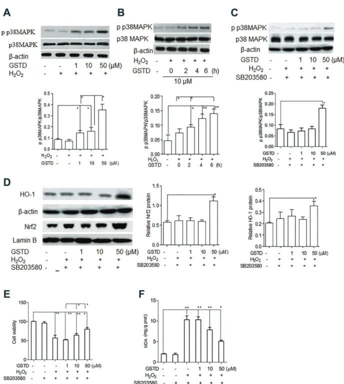
GSTD treatment induced p38 mitogen-activated protein kinase phosphorylation to upregulate the HO-1/Nrf2 pathway
and phosphorylated (p) p38 MAPK were examined. We found that p38 MAPK protein levels in GSTD-treated LSECs exposed to H2O2 were not significantly different from those of control and untreated cells (P40.05, Figure 4A). However, levels of p p38 MAPK were significantly enhanced by GSTD treatment (Po0.05, Po0.01, Figure 4B and C),
suggesting that the p p38 MAPK cascade was asso-ciated with HO-1 and Nrf2 expression. To confirm this,
2 mM SB203580, a specific inhibitor of p38
phospho-rylation, was added to the culture in GSTD-treated LSECs. As shown in Figure 4D, the increase of HO-1 and Nrf2 expression induced by the treatment of 1 and 10 mM GSTD in H2O2-exposed cells was significantly inhibited by the addition of SB2033580 (Po0.05). In the presence
of 2 mM SB203580, H2O2-exposed LSECs treated with
50mM GSTD showed significantly higher HO-1 and Nrf2
expression than those in control and lower dose GSTD (1 and 10 mM) treatment (Po0.05). Incubated with the
inhibitor, 10 and 50 mM GSTD treatment still showed significantly higher viability than those of 1mM and without GSTD treatment (Po0.05, Po0.01, Figure 4E), although
the viability of H2O2-exposed LSECs treated with 1 mM GSTD was not significantly different from those without GSTD treatment (SB203580 plus H2O2). In the presence of inhibitor, MDA content of H2O2-exposed LSECs treated
with 1mM GSTD was not significantly different from those without GSTD treatment (SB203580 plus H2O2), but was significantly higher than those treated with 10mM GSTD. H2O2-exposed LSECs treated with 50mM GSTD in the presence of SB203580 had significantly lower MDA content than those treated with 1 and 10 mM GSTD (Po0.05,
Po0.01, Figure 4F).
Discussion
Oxidative stress is a common pathological mechanism involved in several types of liver disease, including NAFLD, IR, drug- and non-drug-induced liver injury, and hepatic fibrosis (10–12). Because the pharmacological action of GSTD is mainly associated with anti-inflammatory and antioxidant activities, we focused on looking into the antioxidant mechanisms of GSTD treatment in H2O2 -treated LSECs.
Apoptosis in tissues is common under the physiologi-cal condition of stress and disease. In this study, we found that 1 mM H2O2induced apoptosis and reduced viability of LSECs. The results are consistent with a previous study, which reported that cell survival of rat LESCs was remarkably reduced by H2O2(24). Treatment with GSTD reduced H2O2-induced apoptosis and protected cell viability Figure 3.A, Heme oxygenase-1 (HO-1) expression andB, nuclear factor erythroid-related factor 2 (Nrf2) expression in H2O2-exposed liver sinusoidal endothelial cells (LSECs) induced by gastrodin (GSTD) treatment, using Western blot. C, Cell viability.
in a dose-dependent manner. When looking into the mechanism of apoptosis reduction by GSTD treatment, we found that GSTD suppressed H2O2-induced caspase-9 pathway activation and maintained a high Bcl2/Bax ratio. GSTD was previously reported to suppress 1-methyl-4-phenylpyridine (MPP+)-induced caspase-3 pathway
activ-ity in human dopaminergic SH-SY5Y cells (25). In the present study, the caspase-3 pathway was not affected by GSTD treatment or H2O2exposure. The caspase-9 path-way rather than the caspase-3 pathpath-way of LSECs may be sensitive to oxidative stress. H2O2-induced ROS accumula-tion is one of the most common reasons for cell death (26,27). Elevation of MDA levels is commonly used to indicate oxidative injury by ROS species. We found that GSTD treatment reduced ROS generation in H2O2 -exposed LSECs in a dose-dependent manner. Similarly, the MDA content of GSTD-treated cells were significantly lower than untreated cells after H2O2 exposure. It was concluded that GSTD treatment reduced H2O2-induced oxidative injury and protected the viability of LSECs.
HO-1 catalyzes the oxidative cleavage of the a-mesocarbon of Fe-protoporphyrin-IX, yielding equimolar amounts of biliverdin-IXa, free divalent iron, and carbon monoxide (CO) (28). HO-1 can be induced by a variety of stimuli, most of which are linked by their ability to induce oxidative stress (29). HO-1 induction potentially confers protection against oxidative stress in a variety of experi-mental models, such as liver IR secondary to transplanta-tion or hemorrhage/resuscitatransplanta-tion (30,31). The human liver generates comparable amounts of the HO-1-derived products in both Kupffer cells and hepatocytes in the presence of sufficient substrates (32). However, there are no reports regarding their function and expression in LSECs.
After discovering that GSTD treatment provided protec-tion against H2O2-induced oxidative stress, we investigated the effect of GSTD on HO-1 expression in H2O2-exposed LSECs. The results showed that HO-1 expression of H2O2 -exposed LSECs was enhanced by GSTD treatment. When the catalytic function of HO-1 was inhibited by Znpp, the protective role of GSTD on viability became weaker and a higher MDA was observed compared with cells that did not receive Znpp, suggesting that HO-1 plays a key role in the defense of H2O2-induced oxidative stress in LSECs. These data indicate that GSTD exerted anti-oxidative effects on H2O2-exposed LSECs by inducing HO-1 protein expression. Interestingly, we observed that the HO-1 expression of H2O2-exposed LSECs was not statistically different from normal controls. HO-1 protein expression in LSECs seemed unresponsive to H2O2 -induced stress. However, it should be noted that nearly 50% of LSECs were found to be apoptotic after exposure to H2O2. LSECs may be too sensitive to 1 mM H2O2stress with the resulting low cell viability, which can give a misleading result on whether H2O2 stress is able to induce a change in HO-1 protein expression in LSECs.
Among the many mechanisms that regulate HO-1 protein expression, Nrf2 is a typical transcription factor that regulates an array of genes involved in detoxification and antioxidant defense. Recently, upregulation of Nrf2 induced by GSTD was found to alleviate MPP+-induced
oxidative injury in SH-SY5Y cells (33). Nrf2 nuclear translocation and HO-1 protein expression were both induced by GSTD, ameliorating oxidative stress in a mouse model of NAFLD (6). Nrf2 expression was also evaluated. The pattern of detection of Nrf2 expression was similar to that of HO-1, as both were induced by GSTD treatment in H2O2-exposed LSECs. HO-1 upreg-ulation by GSTD may occur at the transcriptional level, requiring Nrf2 regulation.
MAPK cascades have been shown to be involved in HO-1 activation. The synthesis of HO-1 protein and the activation and translocation of Nrf2 were reported to be regulated by p38 (34–36). The p38 cascade may protect Nrf2 from cytosolic degradation by facilitating nuclear translocation of the protein. Several studies showed that multiple inducers of HO-1 also activated protein phos-phorylation-dependent signaling cascades, which ultimately depended on the transcription factors that regulate HO-1 gene expression (37,38). We conducted the experiments designed to determine a possible role of MAPK path-ways in regulating GSTD-induced HO-1 expression. The results showed that p38 was activated by GSTD treatment. The use of a specific inhibitor for p38 pathway, SB203580, confirmed the involvement of p38 in GSTD-induced HO-1 expression, Nrf2 nuclear translocation, and cell viabil-ity protection. In addition to MAPK cascades, it should be noted that GSTD may regulate HO-1 and Nrf2 expression via other pathways. For example, AMPK activation by GSTD was reported to increase the levels of Nrf2 phosphorylation and ameliorate oxidative stress/ proinflammatory response in a mouse model of NAFLD (6). ERK1/2 phosphorylation induced by GSTD was shown to regulate Nrf2 expression and nuclear translo-cation in response to both neurotoxicity in SH-SY5Y cells and rat hippocampal neurons, and motor deficits and oxidative stress in a mouse model of Parkinson’s disease (32,39,40). To fully understand the mechanism of GSTD regulation of HO-1 and Nrf2 expression in H2O2-exposed LESCs, more comprehensive investiga-tion is needed.
Apart from antioxidative protection, GSTD may have prevented H2O2-induced cell death by anti-inflammatory action. Apoptosis of cells treated with 10mM GSTD was significantly lower than those treated with 1mM GSTD in LSECs exposed to H2O2. A more potent protection on cell viability was provided by the treatment with 10mM GSTD. However, HO-1 and p p38 MAPK levels induced by 1 and
10 mM GSTD were not statistically different. Treatment
a more prominent protection of 10 mM vs 1 mM GSTD treatment on cell viability was still observed. Therefore, the stronger protection of cell viability from 10 mM GSTD treatment in H2O2-exposed LSECs may be attributed to more potent anti-inflammatory activity.
In summary, this study demonstrated for thefirst time that GSTD treatment alleviated H2O2-induced oxidative stress in LSECs by reducing ROS, cell apoptosis, and MDA content. GSTD-induced HO-1 and Nrf2 protein expression was involved in protecting H2O2-induced oxidative injury in LSECs, which might be a common protective mechanism for liver oxidative detoxification regulated by p p38 MAPK.
GSTD may be a protective agent against hepatic diseases associated with oxidative stress.
Acknowledgments
We thank the financial support from the National Natural Science Foundation of China (No. 81660113 and 81760119) and Training Program of High-Level Health Technical Talents in Yunnan Province (L-201604 and H-201603). We also thank Sarah Bubeck, Ph.D., from Liwen Bianji, Edanz Editing China (www.liwenbianji.cn/ac), for editing the English of this manuscript.
References
1. Huang Q, Shi J, Gao B, Zhang HY, Fan J, Li XJ, et al. Gastrodin: an ancient Chinese herbal medicine as a source for anti-osteoporosis agents via reducing reactive oxygen species. Bone 2015; 73: 132–144, doi: 10.1016/j.bone. 2014.12.059.
2. Peng Z, Wang S, Chen G, Cai M, Liu R, Deng J, et al. Gastrodin alleviates cerebral ischemic damage in mice by improving anti-oxidant and anti-inflammation activities and inhibiting apoptosis pathway. Neurochem Res 2015; 40: 661–673, doi: 10.1007/s11064-015-1513-5.
3. Kumar H, Kim IS, More SV, Kim BW, Bahk WW, Choi DK. Gastrodin protects apoptotic dopaminergic neurons in a toxin-induced Parkinson’s disease model.Evid Based Com-plement Alternat Med2013; 2013: 514095, doi: 10.1155/ 2013/514095.
4. Hu Y, Li C, Shen W. Gastrodin alleviates memory deficits and reduces neuropathology in a mouse model of Alzheimer’s disease. Neuropathology 2015; 34: 370–377, doi: 10.1111/ neup.12115.
5. Zhang CJ, Zhang JZ, Zheng WH. Protection of gastrodin on vincristine-induced liver injury in rat.J Pract Oncol2012; 27: 521–524.
6. Qu L L, Yu B, Li Z, Jiang WX, Jiang JD, Kong WJ. Gastrodin ameliorates oxidative stress and proinflammatory response in nonalcoholic fatty liver disease through the AMPK/Nrf2 pathway.Phytother Res2016; 30: 402–411, doi: 10.1002/ ptr.5541.
7. Zhao S, Li N, Zhen Y, Ge M, Li Y, Yu B, et al. Protective effect of gastrodin on bile duct ligation-induced hepatic
fibrosis in mice.Food Chem Toxicol2015; 86: 202–207, doi: 10.1016/j.fct.2015.10.010.
8. De Groot H, Rauen U. Ischemia-reperfusion injury: pro-cesses in pathogenetic networks: a review. Transplant Proc2007; 39: 481–484, doi: 10.1016/j.transproceed.2006. 12.012.
9. Cursio R, Colosetti P, Gugenheim J. Autophagy and liver ischemia-reperfusion injury.Biomed Res Int2015: 417590, doi: 10.1155/2015/417590.
10. Poisson J, Lemoinne S, Boulanger C, Durand F, Moreau R, Valla D, et al. Liver sinusoidal endothelial cells: physiology and role in liver diseases.J Hepatol2016; 66: 212–227, doi: 10.1016/j.jhep.2016.07.009.
11. Maslak E, Gregorius A, Chlopicki S. Liver sinusoidal endothelial cells (LSECs) function and NAFLD; NO-based
therapy targeted to the liver.Pharmacol Rep2015; 67: 689– 694, doi: 10.1016/j.pharep.2015.04.010.
12. Peralta C, Jiménez-Castro MB, Gracia-Sancho J. Hepatic ischemia and reperfusion injury: effects on the liver sinusoidal milieu.J Hepatol2013; 59: 1094–1106, doi: 10.1016/j.jhep. 2013.06.017.
13. Xu B, Broome U, Uzunel M, Nava S, Ge X, Kumagai-Braesch M, et al. Capillarization of hepatic sinusoid by liver endothelial cell-reactive autoantibodies in patients with cirrhosis and chronic hepatitis. Am J Pathol 2003; 163: 1275–1289, doi: 10.1016/S0002-9440(10)63487-6. 14. Warren A, Bertolino P, Benseler V, Fraser R, McCaughan
GW, Le Couteur DG. Marked changes of the hepatic sinusoid in a transgenic mouse model of acute immune-mediated hepatitis.J Hepatol2007; 46: 239–246, doi: 10.1016/j.jhep. 2006.08.022.
15. Hu J, Srivastava K, Wieland M, Runge A, Mogler C, Besemfelder E, et al. Endothelial cell-derived angiopoietin-2 controls liver regeneration as a spatiotemporal rheostat.
Science2014; 343: 416–419, doi: 10.1126/science.1244880. 16. Ding BS, Nolan DJ, Butler JM, James D, Babazadeh AO, Rosenwaks Z, et al. Inductive angiocrine signals from sinusoidal endothelium are required for liver regeneration.
Nature2010; 468: 310–315, doi: 10.1038/nature09493. 17. He X, Wang L, Szklarz G, Bi Y, Ma Q. Resveratrol inhibits
paraquat-induced oxidative stress andfibrogenic response by activating the nuclear factor erythroid 2-related factor 2 pathway. J Pharmacol Exp Ther 2012; 342: 81–90, doi: 10.1124/jpet.112.194142.
18. Nguyen T, Nioi P, Pickett CB. The Nrf2-antioxidant response element signaling pathway and its activation by oxidative Stress.J Biol Chem2009; 284: 13291–13295, doi: 10.1074/ jbc.R900010200.
19. Bekyarova G, Tzaneva M, Hristova M. Melatonin protects against burn-induced hepatic oxidative injury by inducing HO-1 via the Nrf2 pathway. Vet Med2015; 60: 621–628, doi: 10.17221/8530-VETMED.
20. Liu CM, Ma JQ, Xie WR, Liu SS, Feng ZJ, Zheng GH, et al. Quercetin protects mouse liver against nickel-induced DNA methylation and inflammation associated with the Nrf2/HO-1 and p38/STAT1/NF-kB pathway.Food Chem Toxicol2015; 82: 19–26, doi: 10.1016/j.fct.2015.05.001.
21. Beyer TA. Impaired liver regeneration in Nrf2 knockout mice.
22. Rao J, Qian X, Li G, Pan X, Zhang F, Zhai Y, et al. ATF3-Mediated NRF2/HO-1 Signaling regulates TLR4 innate immune responses in mouse liver ischemia/reperfusion injury.
Am J Transplant2015; 15: 76–87, doi: 10.1111/ajt.12954. 23. Ge M, Yao W, Yuan D, Zhou S, Chen X, Zhang Y, et al.
Brg1-mediated Nrf2/HO-1 pathway activation alleviates hepatic ischemia-reperfusion injury.Cell Death Dis2017; 8: e2841, doi: 10.1038/cddis.2017.236.
24. Bronk SF, Gores GJ. Acidosis protects against lethal oxidative injury of liver sinusoidal endothelial cells. Hepatol-ogy2010; 14: 150–157, doi: 10.1002/hep.1840140125. 25. Jiang G, Hu Y, Liu L, Cai J, Peng C, Li Q. Gastrodin protects
against MPP(+)-induced oxidative stress by up regulates heme oxygenase-1 expression through p38 MAPK/Nrf2 path-way in human dopaminergic cells.Neurochem Int2014; 75: 79–88, doi: 10.1016/j.neuint.2014.06.003.
26. Brookes PS. Mitochondrial H(+) leak and ROS generation: an odd couple. Free Radic Biol Med 2005; 38: 12–23, doi: 10.1016/j.freeradbiomed.2004.10.016.
27. Rigoulet M, Yoboue ED, Devin A. Mitochondrial ROS generation and its regulation: mechanisms involved in H2O2 signaling. Antioxid Redox Signal 2011; 14: 459–468, doi: 10.1089/ars.2010.3363.
28. Bauer M, Bauer I. Heme oxygenase-1: redox regulation and role in the hepatic response to oxidative stress. Antioxid Redox Signal 2002; 4: 749–758, doi: 10.1089/15230860 2760598891.
29. Haines DD, Lekli I, Teissier P, Bak I, Tosaki A. Role of haeme oxygenase-1 in resolution of oxidative stress-related pathologies: focus on cardiovascular, lung, neurological and kidney disorders. Acta Physiol (Oxf)2012; 204: 487–501, doi: 10.1111/j.1748-1716.2011.02387.x.
30. Shen XD, Ke B, Zhai Y, Gao F, Busuttil RW, Cheng G, et al. Toll-like receptor and heme oxygenase-1 signaling in hepatic ischemia/reperfusion injury.Am J Transplant2015; 5: 1793– 1800, doi: 10.1111/j.1600-6143.2005.00932.x.
31. Rensing H, Bauer I, Peters I, Weni T, Silomon M, Jaeschke H, et al. Role of reactive oxygen species for hepatocellular injury and heme oxygenase-1 gene expression after hemor-rhage and resuscitation. Shock1999; 12: 300–308, doi: 10.1097/00024382-199910000-00009.
32. Qiang L, Niu C, Zhang X, Dong M. Gastrodin and isorhynch-ophylline synergistically inhibit MPP+-induced oxidative stress in SH-SY5Y cells by targeting ERK1/2 and GSK-3b
pathways: involvement of Nrf2 nuclear translocation.
Acs Chem Neurosci 2018; 9: 482–493, doi: 10.1021/ acschemneuro.7b00247.
33. Paxian M, Rensing H, Rickauer A, Schonhofen S, Schmeck J, Pannen BH, et al. Kupffer cells and neutrophils as paracrine regulators of the heme oxygenase-1 gene in hepatocytes after hemorrhagic shock.Shock2001; 15: 438–445, doi: 10.1097/ 00024382-200115060-00005.
34. Zipper LM, Mulcahy RT. Inhibition of ERK and p38 MAPKinases inhibits binding of Nrf2 and induction of GCS genes.Biochem Biophys Res Commun 2000; 278: 484– 492, doi: 10.1006/bbrc.2000.3830.
35. Yong PH, Jeong HG. The coffee diterpene kahweol induces heme oxygenase-1 via the PI3K and p38/Nrf2 pathway to protect human dopaminergic neurons from 6-hydroxydopamine-derived oxidative stress.FEBS Lett2008; 582: 2655–2662, doi: 10.1016/j.febslet.2008.06.045.
36. Yeh CT, Yen GC. Involvement of p38 MAPK and Nrf2 in phenolic acid-induced P-form phenol sulfotransferase expression in human hepatoma HepG2 cells.Carcinogenesis
2006; 27: 1008–1017, doi: 10.1093/carcin/bgi281.
37. Naidu S, Vijayan V, Santoso S, Kietzmann T, Immenschuh S. Inhibition and genetic deficiency of p38 MAPK up-regulates heme oxygenase-1 gene expression via Nrf2. J Immunol
2009; 182: 7048–7057, doi: 10.4049/jimmunol.0900006. 38. Lee SE, Jeong SI, Yang H, Park CS, Jin YH, Park YS.
Fisetin induces Nrf2-mediated HO-1 expression through PKC-d and p38 in human umbilical vein endothelial cells.
J Cell Biochem2011; 112: 2352–2360, doi: 10.1002/jcb.23158. 39. Wang XL, Xing GH, Hong B, Li XM, Zhou Y, Zhang XJ, et al. Gastrodin prevents motor deficits and oxidative stress in the MPTP mouse model of Parkinson’s disease: involvement of ERK1/2-Nrf2 signaling pathway.Life Sci2014; 114: 77–85, doi: 10.1016/j.lfs.2014.08.004.