Ana Isabel Porém Am aral
Dissert at ion present ed t o obtain t he Ph.D degree in Biochem ist ry, Neuroscience
Inst it ut o de Tecnologia Quím ica e Biológica | Universidade Nova de Lisboa
Oeiras,
Met abolism in Prim ary Cult ures
Ana Isabel Porém Amaral
Dissertation presented to obtain the Ph.D degree in
Biochemistry, Neuroscience
Instituto de Tecnologia Química e Biológica | Universidade Nova de Lisboa
Oeiras, September 2011
by Ana Isabel Amaral
Second Edition: October 2011
ITQB-UNL and IBET, Animal Cell Technology Unit
Instituto de Tecnologia Química e Biológica
–
Universidade Nova de Lisboa and
Instituto de Biologia Experimental e Tecnológica
Av. da República
–
EAN, 2780-157 Oeiras, Portugal
Fax: +351 21 442 11 61; Phone: +351 21 446 91 00
http://tca.itqb.unl.pt
http://www.itqb.unl.pt
http://www.ibet.pt
Copyright © 2011 by Ana Isabel Amaral
All Rights Reserved
Amaral, Dr. Paula M. Alves, Prof. Helena Santos.
Supervisors
Dr Paula Marques Alves, Principal Investigator and Head of the Animal Cell Technology Unit at ITQB-UNL/IBET and Executive Director of IBET (Supervisor)
Professor Ursula Sonnewald, Full Professor and Head of the Metabolic Neuroscience Group, Department of Neuroscience, Faculty of Medicine, Norwegian University of Science and Technology (NTNU), Trondheim, Norway (co-supervisor)
Jury
Professor Mary McKenna, Full Professor at the Department of Pediatrics, University of Maryland, School of Medicine; Baltimore – Maryland, USA.
Professor Sebastian Cerdán, Full Professor and Director of the Laboratory of Imaging and Magnetic Resonance Spectrocopy (LISMAR), Instituto de Investigação Biomédica “Alberto Sols”, Universidade Autónoma de Madrid;
This thesis dissertation is the result of more than four years of research at the Animal
Cell Technology Unit of Instituto de Tecnologia Química e Biológica– Universidade Nova de Lisboa and Instituto de Biologia Experimental e Tecnológica (Oeiras, Portugal) under the supervision of Dr. Paula M. Alves and co-supervision of Prof. Ursula Sonnewald.
This thesis aims at contributing with new metabolic flux analysis-based approaches to
improve the investigation and understanding of neural cell metabolism in vitro, with particular emphasis on metabolic responses to pathological insults. Moreover, this thesis
Aos meus pais
Ao meu maninho
This thesis is the product of a long scientific and personal journey at the Animal Cell Technology (ACT) Unit of ITQB-UNL and IBET but also, for shorter periods, at NTNU, Norway, and MIRCen – CEA, France. It was such an enriching period full of challenges, obstacles, joyful moments, personal growth, and exchange of experiences. At this stage I can say that doing research is definitely not easy. However, it is extremely rewarding if we are able to face every challenge with a smile, enthusiasm, motivation, persistence, and hard work, until we finally achieve our goals. Nothing would have been the same without so many extraordinary people that shared this path with me and to whom I wish to acknowledge.
To my supervisor, Dr. Paula Alves, thank you for bringing me to the exciting field of neurochemistry, for your guidance, support, trust, encouragement, energy, and also for being a demanding boss when necessary. Thank you for all the opportunities that allowed me to grow both personally and as a scientist, including the visits abroad, which made total difference in the person I am today. To me, you are a role model of a successful female scientist with great personality who is able to brilliantly manage the broad and excellent ACT unit.
To my co-supervisor, Prof. Ursula Sonnewald, thank you for such a warm hosting in your lab in Trondheim, for inspiring me with your enthusiasm for research, for the encouragement and for all the support both in Norway and via Skype or email whenever I needed.
To Dr. Gilles Bonvento, thank you for receiving me at MIRCen – CEA, in France, for your support and guidance during my stay and during the course of our collaboration project.
To Prof. Manuel Carrondo, for your encouragement, for the example of leadership and for transmitting us that excellence, rigor, hard work and pro-activity are fundamental for success in science.
To Ana Teixeira, thank you for always being there for me, for your friendship, companionship, support, for your guidance, and for being so demanding when we wrote all our papers together. My thesis would have never been possible without your excellent skills and mentoring on MFA. To Pedro Cruz, thank you for all the support and advice and for cheering us up with your visits at the office. I truly admire your enthusiasm for research and your example of entrepreneurship. To Isabel Marcelino, my first scientific mentor at the ACT lab, for your encouragement, enthusiasm and joy at work, for showing me how persistence and hard work are important, and for your friendship.
To my colleagues who contributed directly to the work developed in this thesis: Sanja Martens, Vicente Bernal, Marcos Sousa, Nuno Carinhas, and Joana Tavares, thank you for your hard work and commitment and for making those long working marathons much nicer.
outside the lab. For so many great friends I will keep, you know who you are. It would be impossible to mention all of you, so I will thank you all personally! A special word to the memory of our technician and lab-manager, Nita, a true example of dedication and courage for all of us. To my colleagues at NTNU, Norway, who made my stay in Trondheim even more special and unforgettable, in particular, my dear friends Elvar and Linn – “Tusen takk for alt”. An additional acknowledgement goes to Bjørn Håkonsen and Lars Evje for their significant work contribution to this thesis.
To my colleagues at MIRCen-CEA, France, for such a nice experience, particularly, Carole Escartin, Lydie, Marion, and Marta for all the support. To Mirta and Susanne, my dear friends at “Maison du Portugal”, in Paris.
To all the professors, researchers, students and friends I met in conferences and courses around the world, who inspired me and gave me that extra-motivation about my work. And to all the great people I met at this great institute ITQB who are also part of my PhD experience.
To the financial support provided by Fundação para a Ciência a Tecnologia (SFRH/BD/29666/2006; PTDC/BIO/69407/2006) and to the Clinigene – NoE (LSHB-CT2006-010933). I further acknowledge the Norwegian Research Council for a fellowship that allowed me to perform part of my PhD work at NTNU, Norway.
Aos meus amigos bioquímicos da FCUL, em particular, ao Filipe, Nuno, Tiago, Irina, Catarina, Ana Maria, Raquel, João, e também ao Pedro, pela amizade, por partilharem comigo a minha paixão pela ciência, e por compreenderem tão bem o que é fazer um doutoramento. Tenho a certeza que a ciência portuguesa está em boas mãos com bioquímicos como vocês.
Aos meus amigos de (quase) sempre, Pedro e João, e à Diana, por me tentarem compreender mesmo que não percebam nada de bioquímica, e por continuarem a estar aí, apesar das nossas vidas ocupadas. Vocês são a prova de que os verdadeiros amigos ficam para sempre.
À Sara, pela amizade, por todos os sorrisos e gargalhadas e tantos bons momentos partilhados. Ao Tiago, meu grande e tão especial amigo. Obrigada por tudo aquilo que me tens dado, o carinho, a amizade, e o apoio incondicionais. Por acreditares em mim, por me inspirares, e por teres trazido “novos mundos ao meu mundo”.
Às minhas imensas Amigas Vera, Ana Cristina e Ana Catarina. Por serem quem são, por me acarinharem e mimarem quando eu preciso, pela amizade e apoio incondicional. São o meu orgulho e sabem que a distância entre nós será sempre insignificante, qualquer que seja o lugar do mundo onde estejamos.
Brain energy metabolism results from a complex group of pathways and trafficking
mechanisms between all cellular components in the brain, and importantly provides the
energy for sustaining most brain functions. In recent decades, 13C nuclear magnetic resonance (NMR) spectroscopy and metabolic modelling tools allowed quantifying the
main cerebral metabolic fluxes in vitro and in vivo. These investigations contributed significantly to elucidate neuro-glial metabolic interactions, cerebral metabolic compartmentation and the individual contribution of neurons and astrocytes to brain
energetics. However, many issues in this field remain unclear and/or under debate.
Despite the valuable amount of data generated in cell culture studies involving 13C-labelled substrates and NMR spectroscopy or mass spectrometry, only a few studies
have employed modelling approaches to fully explore the results obtained. Thus, the
main goal of this thesis was to implement novel Metabolic Flux Analysis (MFA)
methodologies combined with information provided by isotopomers of key compounds derived from the metabolism of 13C-labelled precursors, allowing for a more
comprehensive investigation of cell metabolism in cultured brain cells. In addition to
providing a novel in vitro model of ischemia, this work was particularly aimed at
quantifying metabolic fluxes in neurons and in astrocytes and investigating the metabolic
responses of these cells to pathological conditions with high impact on human health,
such as ischemia and hypoglycaemia, by analyzing the changes in the distribution of those fluxes.
Chapter 1 starts by introducing the state of the art in the field of brain energy
metabolism giving particular emphasis on the current topics relevant to the studies in this thesis and on the contribution of certain techniques, such as 13C-NMR spectroscopy
and metabolic modelling, to the current knowledge in the field. A brief introduction on
ischemic episode on the metabolic fluxes of astrocytes is presented in Chapter 2. This
work contributed, in first place, a new in vitro model to mimic ischemia, by cultivating rat astrocytes on Cytodex 3® microcarriers in small-scale bioreactors. The use of bioreactors technology is advantageous as it allows for a tight control and manipulation
of dissolved oxygen levels, a parameter of extreme importance in these studies. By
combining MFA and 13C NMR spectroscopy data, we were able to characterize in detail the metabolic response of astrocytes to oxygen and glucose deprivation. The fast and
transient activation of most metabolic pathways and the parallel reestablishment of
intracellular ATP levels after the insult demonstrate the remarkable capacity of metabolic
adaptation by these cells.
In order to explore the potential of MFA to investigate neuronal metabolism
under different conditions, the work described in Chapter 3 was aimed at characterizing
the effects of glucose deprivation (mimicking brain hypoglycaemia) on neuronal metabolic fluxes. This work provided new evidence on the capacity of neurons to change
their metabolism in the absence of glucose and to metabolize other substrates. The
results suggest that glutamine appears to be an important neuronal fuel during and after hypoglycaemia, and that the pyruvate recycling pathway might be significant for
glutamine oxidation in cerebellar neurons, both under control conditions and, even
more, after hypoglycaemia. These results challenge a number of in vitro studies which
have mainly indicated a predominant astrocytic operation of pyruvate recycling, in
contrast to what had been initially reported in vivo.
Taking into account the complexity of cellular metabolism, and the increasing
availability of techniques generating a larger amount of metabolomics data, more
powerful methodologies have been recently developed. Thus, in Chapter 4, a new model
based on the most recent version of MFA, 13C isotopic transient MFA, was implemented with the aim of estimating the metabolic fluxes in cultured astrocytes in greater detail.
number of fluxes were estimated with high accuracy, including those of parallel and
reversible pathways. In particular, this work suggests that the glutamate/α-ketoglutarate
exchange rate appears to be similar to the TCA cycle flux, a subject that has been highly
controversial and had never been investigated in vitro. This work further allowed
identifying and quantifying the contribution of substrates and metabolic pathways (e.g.
pentose phosphate pathway and catabolism of branched-chain amino acids) to the
isotopic dilution phenomenon typically observed in modelling studies both in vitro and in vivo, reinforcing their importance and the complexity of astrocytic metabolism, even
under physiological conditions.
Chapter 5 describes the work performed in a collaboration project with the
Molecular Imaging Research Centre – Commissariat à L’Énergie Atomique (MIRCen -CEA; France) which aimed at applying MFA to investigate the role of the glial glutamate
transporters, GLAST and GLT-1, in energy metabolism. These studies are relevant not
only to elucidate the role of these proteins in physiological conditions but also in the context of various neurodegenerative diseases involving glutamate excitotoxicity.
Experiments were carried out with the aim of implementing a protocol to down-regulate
the expression of GLAST or GLT-1 in cultured astrocytes. Lentiviral vectors carrying
specific shRNA sequences as well as transfection methods (electroporation and lipofection) using plasmid DNA coding for the same sequences were tested but none was
proven successful. Preliminary data suggests that the viral envelope used led to very low
transduction efficiencies. Therefore, unfortunately, the main aim of this part of the work was not completed and additional studies will be required to generate a good lentiviral
vector and infection protocols that will allow investigating the role of glutamate
transporters in astrocytic metabolism.
Finally, Chapter 6 provides an integrated overview and discussion of the main results of this thesis, highlighting the main findings and the contribution to current
hypoglycaemia by taking advantage of the amount and specificity of the information
provided by MFA methodologies. In addition, the novel modelling tools employed will
be useful for in depth investigations of the responses of brain cells under physiological
O metabolismo energético cerebral resulta de uma complexidade de vias metabólicas e
mecanismos que interligam os diferentes componentes celulares cerebrais, sendo
extremamente importante pois fornece a energia que suporta as diversas funções do cérebro. Nas últimas décadas, estudos de espectroscopia de ressonância magnética
nuclear (RMN) de 13C e ferramentas de modelação metabólica permitiram quantificar os
principais fluxos metabólicos cerebrais, tanto in vitro como in vivo. Estas investigações
contribuíram significativamente para elucidar as interacções metabólicas entre neurónios
e astrócitos, a compartimentação metabólica no cérebro e a contribuição individual de
neurónios e astrócitos para o metabolismo cerebral. No entanto, existem ainda muitos aspectos controversos e/ou pouco compreendidos nesta área.
Diversos estudos in vitro têm permitido obter uma grande e valiosa quantidade
de informação a partir do uso de compostos marcados com 13C e espectroscopia de
RMN e/ou espectrometria de massa. No entanto, poucos estudos utilizaram abordagens
quantitativas que permitem uma caracterização mais aprofundada dos resultados obtidos. Assim, o principal objectivo desta tese foi implementar novas metodologias
baseadas na Análise de Fluxos Metabólicos (AFM), em combinação com informação
obtida através do uso de compostos marcados com 13C e, consequentemente, investigar
com maior detalhe as vias metabólicas em culturas de células de cérebro. Para além da
implementação de um novo modelo in vitro para mimetizar isquémia cerebral, esta tese
pretendeu quantificar, em particular, os fluxos metabólicos de astrócitos e neurónios e investigar as respostas metabólicas destas células a condições patológicas com grande
impacto na saúde humana, como a isquémia e a hipoglicémia, analisando as alterações
na distribuição desses mesmos fluxos.
O Capítulo 1 começa por introduzir o estado da arte na área do metabolismo cerebral, salientando os temas mais relevantes para os estudos desta tese e a contribuição
e as suas vantagens no âmbito dos estudos realizados nesta tese.
No Capítulo 2 apresenta-se o primeiro estudo de AFM que pretendeu investigar
os efeitos de um insulto de isquémia cerebral nos fluxos metabólicos de astrócitos. Em
primeiro lugar, este trabalho contribuiu com um novo modelo in vitro para mimetizar
isquémia, recorrendo ao uso de bioreactores de pequena escala. Estes permitem
controlar e manipular com rigor os níveis de oxigénio dissolvido no meio de cultura, um parâmetro de extrema importância neste tipo de estudos. A combinação de AFM com
dados de espectroscopia de RMN de 13C permitiu caracterizar detalhadamente a
resposta metabólica dos astrócitos à privação simultânea de oxigénio e glucose. Em
particular, salienta-se uma rápida e transiente activação de diferentes vias e o restabelecimento simultâneo dos níveis de ATP intracelulares após o insulto,
demonstrando uma grande resistência e capacidade de adaptação metabólica destas
células.
Com o objectivo de explorar o potencial da AFM na investigação do
metabolismo neuronal em diferentes condições, o trabalho descrito no Capítulo 3
pretendeu caracterizar os efeitos da privação da glucose (hipoglicémia) nos fluxos metabólicos de neurónios em culturas primárias. Este trabalho forneceu evidências
importantes acerca da capacidade de adaptação metabólica destas células na ausência de
glucose e da utilização de outros substratos. Os resultados sugerem que a glutamina é um
substrato neuronal importante durante e após situações de hipoglicémia e que a via de reciclagem do piruvato será significativa para a oxidação de glutamina em neurónios,
tanto em condições fisiológicas como, mais ainda, após um período de hipoglicémia.
Estes resultados contradizem um largo número de estudos in vitro que têm apontado
para uma operação predominante da via de reciclagem do piruvato em astrócitos,
disponibilidade de técnicas que geram cada vez mais dados na área da metabolómica,
novas e mais poderosas metodologias têm sido desenvolvidas. Assim, no Capítulo 4, um
novo modelo baseado na versão mais recente da AFM, AFM transiente isotópica de 13C,
foi implementado com o objectivo estimar com maior detalhe os fluxos metabólicos de astrócitos. Um grande número de fluxos foi estimado com elevada precisão, incluindo
fluxos de vias paralelas e também de vias reversíveis. Em particular, este trabalho fornece,
pela primeira vez, dados obtidos in vitro que apoiam a hipótese de que o fluxo da reacção
de inter-conversão entre glutamato e -cetoglutarato é semelhante ao fluxo do ciclo dos
ácidos tricarboxílicos (TCA), uma questão muito controversa nos estudos de modelação
metabólica in vivo. Esta abordagem permitiu ainda identificar e quantificar a
contribuição de substratos adicionais à glucose e vias metabólicas (ex. aminoácidos de
cadeia ramificada e via das pentoses-fosfatadas) para as diluições isotópicas normalmente observadas nestes estudos, reforçando a sua importância e a complexidade do
metabolismo destas células, mesmo em condições fisiológicas.
O Capítulo 5 descreve o trabalho realizado num projecto de colaboração com o MIRCen-CEA (França) que teve como objectivo aplicar a AFM à investigação do papel
dos dois transportadores de glutamato dos astrócitos, GLAST e GLT-1, no metabolismo
energético. Estes estudos são relevantes, não só para elucidar o papel destas proteínas no
metabolismo dos astrócitos em condições fisiológicas, mas também no contexto de diversas doenças neurodegenerativas que envolvem mecanismos de excitotoxicidade.
Diferentes experiências foram realizadas com o objectivo de implementar um protocolo
para reduzir a expressão do GLAST e GLT-1 em culturas de astrócitos. Vectores lentivirais com sequências específicas de shRNA e métodos de transfecção com DNA
plasmídico para as mesmas sequências (electroporação e lipofecção) foram testados mas
nenhum teve sucesso. Dados preliminares sugerem que o envelope viral utilizado terá
papel dos transportadores de glutamato no metabolismo dos astrócitos.
Finalmente, no Capítulo 6 é feita uma discussão integrada dos principais
resultados da tese, salientando-se as principais conclusões, o contributo para o
conhecimento actual e as perspectivas futuras do trabalho. Em suma, esta tese contribui com novos conhecimentos acerca das respostas metabólicas dos astrócitos a um insulto
de isquémia e dos neurónios a um período de hipoglicémia, tirando partido da
quantidade e especificidade da informação fornecida pelas metodologias de AFM. Estas novas ferramentas serão certamente vantajosas para investigar detalhadamente as
respostas celulares a condições patológicas assim como o efeito de drogas que actuem ao
AI Amaral, AP Teixeira, S Martens, V Bernal, MFQ Sousa, PM Alves (2010)
Metabolic alterations induced by ischemia in primary cultures of astrocytes:
merging
13C NMR spectroscopy and metabolic flux analysis J Neurochem
113(3):735-48.
AI Amaral, AP Teixeira, U Sonnewald,
PM Alves (2011) “
Estimation of
intracellular fluxes in cerebellar neurons after hypoglycemia: importance of the
pyruvate recycling pathway and glutamine oxidation” J N
eurosci Res, 89
(5):700-710.
AI Amaral
*, AP Teixeira*, BI Håkonsen, U Sonnewald, PM Alves (2011) “
A
comprehensive metabolic profile of cultured astrocytes using Isotopic Transient
Metabolic Flux Analysis and
13C-
labelled glucose”
(*equal contribution) Front
Neuroenerg, 3:5 doi 10.3389/fnene.2011.00005
Additional Publications
AI Amaral, AS Coroadinha, O-W.
Merten, PM Alves (2008) “
Improving
retroviral vectors production: Role of carbon sources in lipid biosynthesis”
J
Biotechnol 138 (3-4):57-66
AS Coroadinha, L Gama-Norton,
AI Amaral, H Hauser, PM Alves, PE Cruz
(2010) “Production of Retroviral Vectors” Curr Gene Ther
1;10(6):456-73.
PE Cruz, T Rodrigues, M Carmo, D Wirth,
AI Amaral, PM Alves, AS
Coroadinha (2011)
“
Manufacturing of Retroviruses
” In:
Methods in Molecular
Biology, O-W Merten and M Al-Rubeai (editors) Springer Science
Abbreviation Full Form
2D two-dimensional
3D three-dimensional
3PG 3-phosphoglycerate
AAT aspartate aminotransferase
AGC aspartate-glutamate carrier
ANLS astrocyte-neuron lactate shuttle
ATP adenosine-3-phosphate
BBB blood-brain barrier
BCA bicinchoninic acid
BCAA branched-chain amino acids
cME cytosolic form of malic enzyme
CNS central nervous system
DIV days in vitro
DMEM Dulbecco’s modified Eagle’s medium
DMF N,N-Dimethylformamide
FAD flavin adenine dinucleotide (oxidized form) FADH2 flavin adenine dinucleotide (reduced form)
FBA Flux Balance Analysis
FBS fetal bovine serum
GABA -amino butyric acid
GC-MS gas chromatrography-mass spectrometry
GDH glutamate dehydrogenase
GFAP glial fibrillary acidic protein
GFP green fluorescent protein
GLUT glucose transporter
GS glutamine synthethase
GTP guanosine-3-phosphate
HEK human embryonic kidney
HIF-1 hypoxia-inducible factor 1
Htt Huntingtin
Lac/Glc lactate production rate over glucose consumption rate
LDH lactate dehydrogenase
MAS malate-aspartate shuttle
MCT monocarboxylate transporter
ME malic enzyme
MFA Metabolic Flux Analysis
mHtt mutant Huntingtin
mME mitochondrial form of malic enzyme
MRI magnetic resonance imaging
MS mass-spectrometry
MSNs medium-sized spiny neurons
MSTFA N-Methyl-N-(trimethylsilyl)trifluoroacetamide MTBSTFA N–Methyl-N-(t-Butyldimethylsilyl) trifluoroacetamide NAD+ nicotinamide adenine nucleotide (oxidized)
NADH nicotinamide adenine nucleotide (reduced)
NG2+ nerve/glial antigen 2 - positive
NGS normal goat serum
NMR Nuclear Magnetic Resonance
OGD oxygen and glucose deprivation
PAG phosphate-activated glutaminase
PBS phosphate buffered saline
PC pyruvate carboxylase
PDH pyruvate dehydrogenase
Pen-Strep Penicillin–Streptomycin
PEP phosphoenolpyruvate
PFKFB3 6-phosphofructo-2-kinase/fructose2,6-bisphosphatase, isoform 3
pO2 partial pressure of oxygen
poly-Q poly-glutamine
PPP pentose phosphate pathway
RT-qPCR Reverse Transcriptase - quantitative Polimerase Chain Reaction SDS-PAGE sodium dodecyl sulphate-polyacrylamide gel electrophoresis t-BDMS-Cl t-butyldimethylchlorosilane
Chapter 1
–
Introduction ..………..
..1
Chapter 2
–
Metabolic Flux Analysis of Cultured Astrocytes
–
Effects of Ischemia
………..69
Chapter 3
–
Metabolic Flux Analysis of Cultured Neurons
–
Effects of
Hypoglycaemia .
………
105
Chapter 4
–
Improving Metabolic Flux Estimations in Astrocytes using
13C
Isotopic Transient MFA ……….…
133
Chapter 5
–
RNAi of Glutamate Transporters in Primary Cultures of Astrocytes
……….
. 171
Chapter 6
–
General Discussion and Conclusions ………..
209
C
HAPTER
1
C
ONTENTS1 Introduction ... 3
2 The brain and its cellular populations ... 4
2.1 Neurons ... 5 2.2 Glial Cells ... 5 2.2.1 Astrocytes ... 6
3 In vitro models for neuroscience research ... 8 4 Brain Energy Metabolism: Nutrients and Metabolic Pathways ... 11
4.1 Glucose - the main cerebral energy fuel ... 11 4.1.1 Glucose metabolism in the brain ...12 4.1.1.1 Glycolysis, TCA cycle and Oxidative Phosphorylation ...12 4.1.1.2 Pentose Phosphate Pathway (PPP) ...15 4.1.1.3 Glycogen ...17 4.1.1.4 Cellular redox balance and shuttling of NADH ...18 4.1.1.5 Anaplerotic versus oxidative metabolism ...20 4.2 Glucose vs. Lactate supporting brain activity ... 21 4.2.1 The Astrocyte-Neuron Lactate Shuttle...22 4.2.2 Criticisms to the ANLS and alternative theories ...23 4.2.3 The Redox Switch/Redox Coupling Hypothesis ...24 4.3 Glutamate and glutamine metabolism ... 25 4.3.1 Glutamate-Glutamine cycle ...26 4.3.2 Glutamate/Glutamine Oxidation and Pyruvate Recycling ...28
5 Brain Metabolism in Neuropathologies ... 31
5.1 Cerebral Ischemia and Metabolic Features ... 31
5.2 Brain hypoglycaemia ... 33
6 Tools to Investigate Brain Metabolism ... 34
6.1 13C NMR spectroscopy ... 35
6.2 Mass Spectrometry ... 37 6.3 Metabolic modelling and metabolic flux estimations ... 38 6.3.1 In vitro and ex-vivo studies ...38 6.3.2 In vivo13
C metabolic modelling ...39 6.3.2.1 Modelling assumptions and related controversies ...41 6.3.3 Metabolic Flux Analysis ...43 6.3.3.1 Classical or stoichiometric MFA ...44 6.3.3.2 Isotopic transient 13
C MFA ...46
1
Introduction
The brain is the most complex organ in mammals. It controls most vital functions
although many of the mechanisms underlying its functioning are still unknown.
Moreover, it accounts for only 2% of the body weight, but receives 15% of the cardiac
output (Williams and Leggett 1989) which demonstrates its high energetic demand.
Cerebral metabolism is crucial to provide the energy needed in the numerous processes
sustaining brain activity and, consequently, the disruption of any metabolic-related
mechanism will evidently compromise brain function.
In addition to the profound metabolic alterations known to be associated with
ischemic stroke and hypoglycaemia, it is currently known that many other brain
pathologies, such as neurodegenerative diseases, including Huntington‘s, Alzheimer‘s and Parkinson‘s disease, and psychiatric disorders, like schizophrenia and depression
have a metabolic component. Indeed, changes in metabolic signals detected by imaging
techniques such as magnetic resonance imaging (MRI) and positron emission
tomography (PET) are promising biomarkers for some of these pathologies (Andrews
and Brooks 1998; Coimbra et al. 2006; Mueller et al. 2006; Liepelt et al. 2009).
Research in the last decades has shown that brain energy metabolism is very
complex and compartmentalized due to the highly specialized cellular and sub-cellular
localization of transporters, enzymes and metabolic pathways (reviewed by McKenna et
al. 2006a). Great advances in techniques and methodologies including nuclear magnetic
resonance (NMR) spectroscopy and imaging, PET, mathematical modelling, molecular
biology, microscopy, genomics, proteomics and many others, largely contributed to the
current knowledge in the field. However, many issues remain unclear or under intense
debate. Therefore, research in this field remains an exciting task towards elucidating the
mechanisms underlying brain function under physiological and pathological conditions.
This chapter summarizes the state of the art regarding research on brain energy
metabolic modelling tools. A general overview of the metabolic alterations associated
with hypoglycaemia and ischemia in the brain is also provided, as these were the
pathological conditions mimicked in some studies included in this thesis.
2
The brain and its cellular populations
The brain is constituted mainly by neurons and glial cells. Although presenting distinct
morphologies and specific roles, they strongly depend on close functional interactions
between them and with blood vessels (Figure 1.1).
Figure 1.1 - Interactions between glial cells and neurons in the brain. Different types of glia interact with neurons and the surrounding blood vessels. Oligodendrocytes wrap myelin around axons to speed up neuronal transmission. Astrocytes extend processes that cover > 99% of the cerebrovascular surface and synapses. Microglia keep the brain under surveillance for damage or infection. Reproduced from Allen and Barres (2009) with permission of the publisher.
The blood–brain barrier (BBB), constituted mainly by endothelial cells, enzymes and
environment of the central nervous system (CNS) (Abbott et al. 2006). In addition to
neurons and glia, a new class of cells called pericytes has been recently described in the
brain and is thought to be important in regulating the permeability functions of the BBB
(Armulik et al. 2011).
2.1 Neurons
Neurons were, for more than a century, the only studied cell type in the brain due to
their unique capacity of emitting electrical signals. They are highly specialized cells and
the core components of the nervous system. The number of neurons in the brain varies
dramatically from species to species. It is estimated that the human brain possesses about
100 billion (1011) neurons and 100 trillion (1014) synapses (Williams and Herrup 1988).
Neurons are electrically excitable cells which are able to process and transmit
information using electrical (action potentials) and chemical (neurotransmitters) signals
through a mechanism designated by synaptic transmission (Hof et al. 2004). The shape,
size and neurochemistry determine their specific function. In this respect, three major
classes of neurons can be considered: the inhibitory GABA ( -amino butyric acid)ergic
interneurons that make local contacts, the local excitatory spiny stellate cells in the
cerebral cortex, and the excitatory glutamatergic efferent neurons, such as the cortical
pyramidal neurons (Hof et al. 2004). In addition, other types of neurons localized in
more specialized areas include dopaminergic, cholinergic and serotoninergic neurons.
Still, and despite the heterogeneous distribution among different brain regions, more
than 90% of neurons in the brain are either glutamatergic or GABAergic, according to
the neurotransmitter used for their signalling process, glutamate or GABA, respectively
(Hof et al. 2004). Neurotransmitters play an important role linking energy metabolism of
neurons and astrocytes. This will be further elucidated later in this chapter.
2.2 Glial Cells
Despite the large number of neurons, glial cells occupy the most part of the brain
regions but seems to be correlated with the animal‘s size as the mouse, human and
elephant brain possess approximately 65%, 90% and 97% of glial cells, respectively
(Allen and Barres 2009). Their initial designation of neuroglia (―Nervenkitt‖) was
attributed to Rudolph Virchow in the late 19th century, who described these cells as the
―brain glue‖. They were thought to be mostly connective tissue that filled up the
extracellular space and worked as chemical and physical insulators to support the diverse
neuronal functions (Kimelberg 2004). Only in the beginning of the 20th century did glia
finally lose their passive identity and many crucial and active functions started being
attributed to these cells. In mammals, glial cells are divided into three major groups,
based on their morphology, function and localization in the nervous system: (1)
Schwann cells and oligodendrocytes, the myelinating cells of the peripheral and CNS,
respectively (Nave and Trapp 2008); (2) microglia, the immune cells of the CNS
(Hanisch and Kettenmann 2007); and (3) astrocytes, a diverse cell population with
variable morphology and numerous functions, contacting essentially with all other
cellular elements in the brain (Agulhon et al. 2008; Wang and Bordey 2008) (Figure
1.1). A fourth group, the nerve/glial antigen 2 - positive (NG2+) glia, has been more
recently considered and includes oligodendrocyte and astrocyte progenitor cells as well as
NG2+ cells that persist in the mature brain (Agulhon et al. 2008). Among the different
types of glial cells, astrocytes were those studied in detail in this thesis due to their close
relationship with neurons at the metabolic level, as described below.
2.2.1 Astrocytes
Astrocytes derive their name from the stellate morphology traditionally observed in
histological preparations. However, they are quite heterogeneous among different brain
regions, even at the transcriptome level (Bachoo et al. 2004). Astrocytes are found
throughout the brain and spinal cord and, on the basis of number, surface area, and
volume, are the predominant glial cell type. Protoplasmic astrocytes are the most
common type of astrocytes (Agulhon et al. 2008). Individually, they occupy distinct,
junctions at their boundaries (Bushong et al. 2002). More than 99% of the
cerebrovascular surface is ensheathed by astrocyte processes (Haydon and Carmignoto
2006) and processes from a single astrocyte can envelop approximately 140 000 synapses,
as in the CA1 region of the hippocampus (Bushong et al. 2002).
Astrocytes have, thus, the unique role of dynamic coordination of cerebral
functions. They participate in the regulation of water and ionic homeostasis (Simard and
Nedergaard 2004) and in the maintenance of the BBB (Hawkins and Davis 2005).
Moreover, even though astrocytes do not produce action potentials, they individually
respond to synaptically released neurotransmitters through elevations in intracellular
Ca2+ levels (Agulhon et al. 2008; Schummers et al. 2008). These Ca2+ transients induce
the release of chemical transmitters (―gliotransmitters‖ – ATP, glutamate and D-serine)
allowing them to communicate with neurons. In this way, astrocytes control synaptic
transmission in a process called tripartite-synapse, and regulate local blood flow in
situations of intense neuronal activity (neurovascular-coupling mechanism) (Haydon and
Carmignoto 2006; Iadecola and Nedergaard 2007; Halassa et al. 2009). Nevertheless, it
is still elusive how both excitatory and inhibitory signals provided by the same glial cell
act in concert to regulate neuronal function.
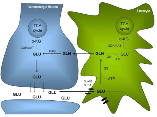
Finally, astrocytes play a key role in brain energy metabolism. They supply energy
substrates to neurons, in a process essential for neurotransmission, and are responsible
for neurotransmitter uptake and recycling, thereby preventing excitotoxicity and
controlling synaptic signals (Hertz and Zielke 2004; Pellerin et al. 2007). Moreover, they
synthesize the main antioxidant molecule in the brain, glutathione (Dringen and
Hirrlinger 2003). The role of both astrocytes and neurons in brain energy metabolism,
in physiological and pathological scenarios, is the basis for the work described in this
thesis and, therefore, will be more thoroughly addressed in the subsequent sections of
3
In vitro models for neuroscience research
The high degree of complexity of the brain makes it difficult to investigate specific
biochemical and cellular mechanisms using in vivo models and, therefore, simple
research models are required. As illustrated in the following sections of this chapter,
cultured brain cells have been crucial to elucidate many aspects of brain energy
metabolism and are still widely used to investigate a number of research questions,
including mechanisms of disease.
In addition to their simplicity, in vitro models are advantageous compared to
animal models since they possess high cellular specificity and are not influenced by the
blood flow component, hormones, immune system, and temperature variations
occurring in vivo (Meloni et al. 2001). The absence of these factors allows, for example,
investigating cell-specific changes in gene/protein expression, determining the metabolic
responses of a particular cell type under different conditions or determining the
mechanism of action of a therapeutic agent (Meloni et al. 2001). In addition, it is easier
and less expensive to perform molecular manipulations, such as antisense
oligonucleotide and gene transfection experiments, in cultured cells. Also, in vitro
models are valuable for drug-screening, enabling to select the most promising
compounds to be tested in vivo, as well as to perform preliminary studies regarding novel
research hypotheses, thereby reducing the number of animals used. This is actually a
crucial aspect in current neuroscience research, due to the stricter rules and definitions
of ethical impact underlying the use of animals, which need to be fully accomplished by
researchers.
One of the oldest in vitro models to study the brain is the organotypic brain slice
culture from the CNS of young rodents (reviewed by Gahwiler et al. 1997). This
preparation continues to differentiate in culture and preserves a level of cellular
organization that closely resembles that observed in situ. Therefore, it has been
it is very technically demanding and its heterogeneous composition and complexity
makes it difficult to investigate cell-specific mechanisms.
Some of these aspects can be overcome by the use of monotypic neural cell
cultures, including both tumour-derived cell lines and primary cultures, which are
simpler models regarding both culture preparation/maintenance and the cellular
features reproduced. Cell lines, such as glioma or neuroblastoma cells (Bouzier et al.
1998; Rae et al. 2003), are much easier to cultivate but their immortalization properties
limit their ability to model non-tumoural tissue. Dissociated primary neural cell cultures
are prepared from neonatal rodent brain, are normally selectively enriched in one
particular cell type and therefore are considered to represent mainly the features of that
cell population (Hertz et al. 1998).
Neural cells can be cultivated in the more classical two-dimensional (2D)
monolayer systems, such as tissue-culture flasks or dishes, but also immobilized in
microcarriers or in gel threads (Alves et al. 1996b; Alves et al. 2000a; Sa Santos et al.
2005) and in the form of three-dimensional (3D) aggregates (Santos et al. 2007),
depending on the purpose of the study. Monolayer 2D cultures have the advantage of
being easily monitored and characterised using microscopy techniques. By cultivating
more than one cell type (e.g. neurons and astrocytes) in the same dish, a 2D co-culture is
obtained (e.g. Waagepetersen et al. 2002). However, these culture approaches are limited
in respect to their spatial environment when compared to 3D aggregates. Aggregating
neural cell cultures or neurospheres are able to reconstitute spontaneously a histotypic
brain architecture to reproduce critical steps of brain development and to reach a high
level of structural and functional maturity (reviewed by Honegger et al. 2011). Even so,
in addition to being much more technically demanding, 3D cultures might have the
possible drawback of nutrient and oxygen transport limitation and accumulation of toxic
byproducts in the centre of aggregates with higher diameters, which might affect cell
advantageous in vitro models to investigate metabolic interactions between different cell
types since they retain some degree of complexity, including cell-cell interactions.
Biotechnological advances in suspension culture systems adequate for neural
cells, namely small scale bioreactors and low shear-stress impellers, have been
advantageous for the implementation of novel in vitro models for neuroscience research,
for example, using primary cultures (e.g. Sa Santos et al. 2005) and stem cells (e.g. Serra
et al. 2009). These systems are hydrodynamically well characterized, allow for a better
homogeneity of the cultures, easy-sampling, and reproducibility between experiments
due to the tight control of culture variables (gas-composition of the culture medium, pH,
temperature, stirring rate). Oxygen and nutrient delivery to the cells is also much more
efficient in stirred systems, as well as its manipulation during culture time. Therefore,
they are particularly well-suited to perform pathological challenges involving the
manipulation of oxygen levels, such as hypoxia (Sa Santos et al. 2005), anoxia (Sa Santos
et al. 2011) or ischemia (Amaral et al. 2010). The use of bioreactors is thus advantageous
when compared to the use of hypoxic incubators/chambers (e.g. Almeida et al. 2002),
that do not enable the tight monitoring of oxygen levels.
Finally, although neural cell cultures have been mainly of rodent origin,
human-derived in vitro models, particularly those based on stem cells have been increasing in
recent decades (reviewed by Gibbons and Dragunow 2010). These are very promising to
investigate cellular, molecular or biochemical mechanisms of human cells in physiology
and disease. While it is important to acknowledge that pre-clinical in vivo studies in
rodents and primates will always be required to validate findings obtained in vitro, before
their translation to humans, all the advances in in vitro neural cell models and culture
systems will certainly continue to make them excellent and privileged models for many
4
Brain Energy Metabolism: Nutrients and Metabolic Pathways
4.1 Glucose - the main cerebral energy fuel
Energy consuming processes in the adult brain account for 25% of the total body
glucose utilization and for 20% of the total oxygen consumed (Magistretti 2004).
However, glucose is not the main energy substrate in all stages of brain development. In
fact, a transitory switch occurs from a combination of glucose, monocarboxylates,
including lactate, and ketone bodies, such as acetoacetate and ß-hydroxybutyrate in the
postnatal period to a predominance of glucose as main fuel in the mature brain (Nehlig
1997; Vannucci and Simpson 2003). The uptake of metabolic substrates from the blood
into the brain is mediated by specific facilitative transporter proteins in endothelial cells
of the BBB and in brain cells. It is the differential developmental expression of glucose
and monocarboxylate transporters (GLUTs and MCTs, respectively) that determines the
maturational increase in glucose utilization (Rafiki et al. 2003; Vannucci and Simpson
2003).
Average cerebral concentrations of glucose range between 0.8–2.3 mM as
estimated using several techniques and show a linear correlation with blood glucose
levels (Gruetter et al. 1998a; Dienel and Cruz 2004; Barros et al. 2007). Although
approximately 96% of endothelial cells are covered by astrocytic end feet, experimental
evidence has shown that approximately equal proportions of glucose are taken up by
neurons and astrocytes (Nehlig et al. 2004), even though this is still a matter of debate.
As shown by autoradiography and PET, the rate of glucose consumption differs between
brain regions, with higher values in grey matter, and also varies with time, with active
areas capturing glucose more avidly (Raichle and Mintun 2006). Although it is
established that oxidative metabolism predominates, as indicated by the cerebral
respiratory coefficient (CO2 production/O2 consumption) of 0.97 (Clarke and Sokoloff
1999), the exact contribution of neurons and astrocytes to this process also remains
activation (e.g. Dienel and Cruz 2008) and that both cell types are able to oxidize lactate
(Zielke et al. 2009). These issues have provided additional sources of discussion with
regard to the main substrate supporting neuronal and astrocytic metabolic activity under
activation and will be further addressed below.
4.1.1 Glucose metabolism in the brain
Cerebral glucose metabolism is similar to that in other tissues but, in the particular case
of the brain, it is almost entirely oxidized to CO2 and water via glycolysis, the
tricarboxylic acid (TCA) cycle and the associated oxidative phosphorylation (Magistretti
2004). Under certain conditions, and depending on the cell type, glucose can be
additionally metabolized in the pentose phosphate pathway (PPP) to a significant extent
(Dringen et al. 2007). Finally, glucose can eventually be stored in astrocytes in the form
of glycogen, the main cerebral energy store (Brown and Ransom 2007). These pathways
are generally described in Figures 1.2 and 1.3 and below.
4.1.1.1 Glycolysis, TCA cycle and Oxidative Phosphorylation
Glycolysis is the pathway responsible for the initial steps of glucose metabolism as it
enters the brain, and it converts glucose into two molecules of pyruvate (Figure 1.2).
Glycolytic activity is thought to be much higher in astrocytes than in neurons (to be
addressed below). This pathway generates a net amount of two ATP molecules. Four
ATP molecules are formed in the two last steps leading to pyruvate formation, the
reactions catalyzed by phosphoglycerate kinase (EC 2.7.2.3) and pyruvate kinase (EC
2.7.1.40), whereas two ATPs are consumed to phosphorylate glucose to
glucose-6-phosphate (by hexokinase; EC 2.7.1.1) and fructose-6-glucose-6-phosphate to
fructose-1,6-bisphosphate (by phosphofructokinase; EC 2.7.1.11), respectively (Magistretti 2004).
Hexokinase and phosphofructokinase both catalyze irreversible reactions, being
important regulation points in carbohydrate metabolism in the brain (McKenna et al.
2006a). This is one of the main reasons why brain cells have a reduced capacity of
the blood. Even so, some authors have provided evidence suggesting the operation of
gluconeogenesis in astrocytes, which are able to produce glycogen from lactate (e.g.
Dringen et al. 1993b; Schmoll et al. 1995; Bernard-Helary et al. 2002).
Anaerobic glycolysis (glucose conversion into lactate) occurs when glucose
utilization is higher than oxygen consumption and, consequently, the fraction of
pyruvate produced from glucose exceeds that oxidized in the TCA cycle (Magistretti
2004). This also represents one of the mechanisms allowing for the maintenance of an
optimal cytoplasmic NAD+/NADH ratio, required for a continuous glycolytic activity
(other mechanisms are described in sub-section 3.1.1.4).
In order for glucose metabolism to proceed via oxidation, it is required that
pyruvate formed in glycolysis is transported into the mitochondria and enters the TCA
cycle. The TCA cycle involves not only the catabolism of energy-rich molecules but also
provides precursors for many intermediates that are utilized in the overall cellular
metabolism. Pyruvate is converted into acetyl-CoA in a reaction catalyzed by the pyruvate
dehydrogenase (PDH) complex localized in the mitochondrial matrix (McKenna et al.
2006a). Acetyl-CoA cannot leave the mitochondrion because of its large size; thus the
PDH complex maintains a positive flow of carbon to the cycle, controlling the rate of
oxidative glucose metabolism.
The oxidation of pyruvate to CO2 in the TCA cycle generates energy-rich
molecules such as GTP, NADH and FADH2. NADH and FADH2 transfer electrons to
oxygen in the electron transport chain, leading to the production of ATP in a process
called oxidative phosphorylation. The mitochondrial NAD+/NADH ratio constitutes
one of the major regulators of the TCA cycle and its value is strongly affected by oxygen
(TCA) cycle. Pyruvate is carried into the mitochondrial matrix for oxidative decarboxylation to acetyl-CoA via the pyruvate dehydrogenase complex (PDH) or for carboxylation to oxaloacetate (OAA) via pyruvate carboxylation (PC; only in astrocytes). Acetyl-CoA (ACoA) is condensed via citrate synthase (1) to citrate (Cit), which is converted to α-ketoglutarate (α-KG) via aconitase (2) and isocitrate dehydrogenase (3). α -ketoglutarate is subsequently decarboxylated via the α-ketoglutarate dehydrogenase complex (4) to succinyl-CoA (Sucsuccinyl-CoA). Succinate (Suc) is formed from succinyl-succinyl-CoA via succinyl-succinyl-CoA synthase (5). Succinate dehydrogenase (6) oxidizes succinate to fumarate (Fum), which is converted into malate (Mal) via fumarase (7). Malate is then oxidized to oxaloacetate via malate dehydrogenase (8) or it can be converted to pyruvate via malic enzyme (ME). NADH and FADH2 are oxidized in the electron transport chain that carries the
electrons through different complexes to O2, which is reduced to H2O at the same time that ADP is
phosphorylated into ATP, in a process called oxidative phosphorylation. Additional abbreviations: DHAP, dihydoxyacetone-phosphate, Isocit, isocitrate; OSuc, oxalosuccinate, PEP, phosphoenolpyruvate; Glu, glutamate; Gln, glutamine.
Oxidation of glucose to CO2 provides the higher yield of ATP per glucose
molecule (34 ATP) and, consequently, is fundamental to support energy-dependent
brain functions. In particular, neurons require a large amount of ATP for recovery of the
ion homeostasis dissipated by excitatory postsynaptic potentials (Attwell and Laughlin
2001). Therefore, it is widely accepted that these cells contribute to a major fraction of
the total brain oxidative metabolism and, consequently, to cerebral ATP synthesis, even
though astrocytes also significantly oxidize glucose (Hertz et al. 2007).
4.1.1.2 Pentose Phosphate Pathway (PPP)
The PPP interconverts sugar phosphates in multiple reactions divided in two branches,
the oxidative and the non-oxidative branch (Figure 1.3). The oxidative part of the PPP is
linked to glycolysis at the level of glucose-6-phosphate and catalyzes its conversion into
ribulose-5-phosphate and CO2. In addition, this branch is responsible for the reduction
of NADP+ into NADPH, the major reducing compound. On the other hand, the
non-oxidative branch interconverts pentose phosphates and phosphorylated aldoses and
ketoses and is connected to glycolysis by their common intermediates
glyceraldehyde-3-phosphate and fructose-6-glyceraldehyde-3-phosphate. It also produces ribose-5-glyceraldehyde-3-phosphates which are
Figure 1.3 - Reactions of the PPP and their connection with glycolysis. For simplicity, only the part of glycolysis which has a link with the PPP is represented. Abbreviations: G6PD. Glucose-6-phosphate dehydrogenase; 6PGL, 6-phosphogluconolactonase; 6PGDH, 6-phosphogluconate dehydrogenase; R5PI, ribulose-5-phosphate isomerase; R5PE,ribolose-5-phosphate epimerase; TK, transketolase; TA, transaldolase; HPI, hexosephosphate isomerase; FBP, fructose-1,6-bisphosphatase; PFK, phosphofructokinase; TIM, triosephosphate isomerase.
The activity of the non-oxidative branch of the PPP in the adult brain appears to
be rather low, being mainly used to support active cellular proliferation during brain
development (Bilger and Nehlig 1992) or for the growth of brain tumours (Spence et al.
1997). Conversely, the oxidative branch predominates in brain cells because it provides
the NADPH required for the regeneration of glutathione from its oxidized form,
glutathione disulfide (GSSG) (Kletzien et al. 1994; Delgado-Esteban et al. 2000; Almeida
et al. 2002). Nevertheless, the significance of the PPP in astrocytes and neurons differs.
Recent findings indicating that the glycolytic enzyme
6-phosphofructo-2-kinase/fructose2,6-bisphosphatase, isoform 3 (PFKFB3) is not active in suspensions of
isolated rat cortical neurons suggested that these cells metabolize glucose mainly through
cultured cells and in vivo. Different authors have shown that neurons use this pathway as
a major antioxidant mechanism in the response to pro-oxidant compounds (Ben-Yoseph
et al. 1996; Garcia-Nogales et al. 2003; Vaughn and Deshmukh 2008) or to stimulation
of glutamate receptors (Delgado-Esteban et al. 2000). Moreover, the over-expression of
PFKFB3 to redirect glucose metabolism from the PPP to glycolysis in these neurons
resulted in depleted glutathione levels, apoptotic cell death and oxidative stress
(Herrero-Mendez et al. 2009), confirming the extreme importance of the PPP in neurons. Even so,
whether neurons use the PPP to compensate for their antioxidant fragility and, at the
same time, inhibit the bioenergetically favourable glycolysis (Bolanos and Almeida 2010),
remains to be elucidated in vivo. Despite exhibiting a high glycolytic rate and a low PPP
basal activity, cultured astrocytes up-regulate this pathway under different conditions.
For example, when subjected to oxidative or nitrosative stress (Ben-Yoseph et al. 1996;
Garcia-Nogales et al. 1999; Bolanos and Almeida 2006), after being exposed to oxygen
and glucose deprivation (Almeida et al. 2002) and when treated with amyloid beta
peptides (Allaman et al. 2010). With regard to the in vivo context, the PPP was shown to
contribute to glucose metabolism after focal brain activation in rats (Cruz et al. 2007)
and was up-regulated after traumatic brain injury in humans (Dusick et al. 2007). All
these findings underline the present importance attributed to the PPP in the metabolic
response of brain cells to a number of pathologies in addition to its physiological
significance.
4.1.1.3 Glycogen
Glucose can additionally be stored in the form of glycogen, which is predominantly
located in astrocytes (Cataldo and Broadwell 1986) and present in the brain at
significant levels (3-6 µmol/g tissue) (Cruz and Dienel 2002; Oz et al. 2007;
Morgenthaler et al. 2008). Glycogen is degraded into glucose-1-phosphate by glycogen
phosphorylase (EC 2.4.1.1) and subsequently enters glycolysis, after being converted into
metabolized to lactate in astrocytes and subsequently exported to fuel neurons and axons
(Dringen et al. 1993a; Wender et al. 2000; Brown et al. 2004; Sickmann et al. 2005;
Tekkok et al. 2005; Pellerin et al. 2007).
Based on its slow turnover under resting conditions (Oz et al. 2003; Oz et al.
2007) and rapid mobilization during an energy crisis or under hypoglycaemia (Choi et al.
2003; Oz et al. 2009), glycogen has been mainly considered an emergency fuel (Gruetter
2003; Dienel et al. 2007 and references therein). Nevertheless, its physiological role has
been progressively reinforced. For instance, glycogenolysis is triggered by neuronal
stimulation, when glucose alone cannot meet the high transient increase in cellular
energy requirements (Swanson et al. 1992; Cruz and Dienel 2002; Brown et al. 2004;
Dienel et al. 2007). Moreover, a ―glycogen shunt‖, i.e., glucose metabolism via glycogen,
is thought to operate in the brain and to contribute to lactate release during activation
(Shulman et al. 2001). More recently, glycogen metabolism was shown to importantly
contribute to long-term memory formation in rats through its conversion into lactate
(Suzuki et al. 2011).
4.1.1.4 Cellular redox balance and shuttling of NADH
The continuous operation of glycolysis and the conversion of lactate into pyruvate via
lactate dehydrogenase involve the reduction of NAD+ into NADH (see Figure 1.2). Thus,
in order to coordinate glycolytic activity with that of the TCA cycle, a low redox state
(NAD+/NADH) needs to be maintained (McKenna et al. 2000a). In addition to the
reaction catalyzed by lactate dehydrogenase (LDH; EC 1.1.1.27) (Figure 1.2), this is
mainly carried out by the malate-aspartate shuttle (MAS) in the brain (McKenna et al.
2006b). Because NADH cannot enter mitochondria, malate is responsible for the
transfer of reducing equivalents from the cytosol into the mitochondria (Berkich et al.
2005; McKenna et al. 2006a). The MAS involves the concerted operation of important
carriers [the aspartate-glutamate carrier (AGC) and the malate-α-ketoglutarate carrier]
and malate dehydrogenase) (Palmieri et al. 2001). This shuttle additionally involves the
irreversible electrogenic exchange of aspartate for glutamate and a proton via the AGC1
carrier (Aralar1), favouring the efflux of aspartate from and entry of glutamate into the
mitochondria (McKenna et al. 2000a).
The real significance of the MAS in each brain cell type is not yet completely
elucidated. Aralar1 and MAS activity appear to be much lower in astrocytes than in
neurons (Ramos et al. 2003; Berkich et al. 2007; Xu et al. 2007) which is consistent with
the enrichment of Aralar1 in neuronal mitochondria (Ramos et al. 2003; Pardo et al.
2011). Despite the presence of Aralar1 mRNA has been demonstrated in acutely isolated
astrocytes from adult mice (Lovatt et al. 2007), it was recently proposed in a quite
controversial study using Aralar-knockout mice that astrocytes do not seem to rely on the
MAS to transfer redox equivalents to mitochondria (Pardo et al. 2011). Rather, Pardo
and colleagues suggested that a neuron-to-astrocyte aspartate efflux may provide a means
to transfer NADH/NAD+ redox potential to astrocyte mitochondria (Pardo et al. 2011).
Considering the lack of strong evidence supporting the existence of the MAS in
astrocytes, the operation of the glycerol-3-phosphate shuttle has alternatively been
proposed to play a similar role in these cells (McKenna et al. 2000a). This shuttle is
based on the concerted action of cytosolic and mitochondrial isoforms of glycerol
3-phosphate dehydrogenase, the former using NAD+/NADH as coenzyme and the latter
using FAD/FADH2 for that purpose. Reducing equivalents are subsequently transferred
to coenzyme Q in the respiratory chain. However, it yields less energy than the MAS due
to the transport of electrons to FAD rather than NAD+ (McKenna et al. 2006b).
Nonetheless, the operation of the glycerol-3-phosphate shuttle remains controversial.
Despite the evidence of glycerol-3-phosphate shuttle activity in cultured astrocytes and
cerebellar neurons (Cammer et al. 1982; McKenna et al. 1993; Atlante et al. 1999), the
cytosolic and mitochondrial isoforms of glycerol-3-phosphate dehydrogenase appear to
be localized in glial and neuronal cells, respectively (Leveille et al. 1980; Nguyen et al.
4.1.1.5 Anaplerotic versus oxidative metabolism
Anaplerosis fuels the TCA cycle with extra carbon units required to the synthesis and
release of TCA cycle intermediates (Sonnewald and Rae 2010). Pyruvate carboxylase
(PC) is the main cerebral anaplerotic enzyme (Patel 1974). It is an ATP-dependent
enzyme which converts pyruvate into oxaloacetate (Wallace et al. 1998). PC was shown
to be primarily, if not exclusively, expressed in astrocytes in vitro and in vivo (Yu et al.
1983; Shank et al. 1985; Kaufman and Driscoll 1993; Shank et al. 1993; Cesar and
Hamprecht 1995). Furthermore, using 13C NMR spectroscopy in monotypic cultures of
astrocytes and neurons it was shown that this pathway occurs in astrocytes and not in
neurons (Sonnewald et al. 1993b; Waagepetersen et al. 2001b). Although malic enzyme
or the combined action of phosphoenolpyruvate carboxykinase (PEPCK, EC 4.1.1.31)
and pyruvate kinase (PK, EC 2.7.1.40) can also fix CO2, their activity in the brain
appears not to be significant (Patel 1974). Even so, Hassel and colleagues have provided
some controversial evidence that neurons can carboxylate, claiming that it occurs
probably via malic enzyme (Hassel 2000).
PC activity is strongly associated with the synthesis and export of glutamine both
in vivo and in vitro (Gamberino et al. 1997; Gruetter et al. 2001; Waagepetersen et al.
2001a). Anaplerosis is important for neurotransmitter metabolism and ammonia
detoxification as it is essential to replenish neuronal TCA cycle and neurotransmitter
pools due to the continuous release of neurotransmitters (e.g. Hertz et al. 1999; Sibson
et al. 2001; Oz et al. 2004; Xu et al. 2004; Zwingmann 2007). In addition, it is important
to compensate for the loss of additional molecules that leave the brain, for example,
lactate, which can occur via the pyruvate recycling pathway (Sonnewald et al,
unpublished data).
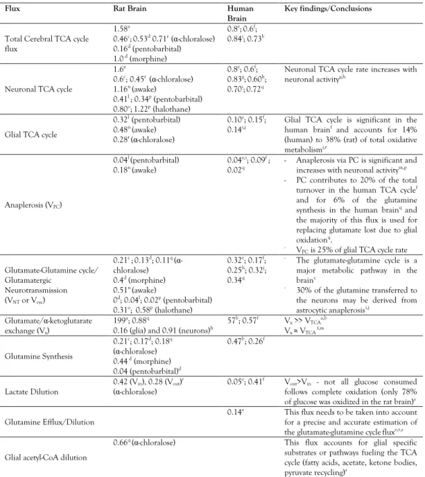
In vivo and in vitro estimations of the different contributions of fluxes through
PC and PDH to the synthesis of different amino acids have been based on the distinct
Leibfritz 2007). However, these estimations might be limited by the appearance of the
same isotopomers arising both through PC and PDH and further complicated by the
equilibration of the label between oxaloacetate and fumarate due to backflow in the
TCA cycle (Merle et al. 1996b, a). Therefore, the range of values reported is rather large
(Table 1.1). The use of different formulas, labelled substrates, and incubation times has
also contributed to this variability (see Zwingmann and Leibfritz 2007 for details). Using
13C NMR spectroscopy and metabolic modelling, PC and PDH fluxes and their
contribution to cerebral oxidative metabolism have been estimated in vivo (see subsection
5.3).
4.2 Glucose vs. Lactate supporting brain activity
Cerebral oxidative metabolism supports brain activity both under resting and activated
conditions. However, glycolytic flux is up-regulated even more than the simultaneous
increase in oxygen consumption upon activation (reviewed by Dienel and Cruz 2004,
2008). These changes suggest an increase in local lactate demand followed by a larger
increase in local lactate production. Lactate transients and increased glucose utilization
are actually the metabolic hallmarks of brain activation detected with functional brain
imaging techniques (Bonvento et al. 2005). Hence, different groups have been trying to
investigate the role of glucose and lactate as substrates supporting synaptic activity and,
at the same time, elucidating the contribution of neurons and astrocytes to cerebral
oxidative metabolism. The different theories proposed to explain these phenomena
remain under a heated debate (Bonvento et al. 2005; Hertz et al. 2007; Simpson et al.
2007; Dienel and Cruz 2008; Mangia et al. 2009; Pellerin 2010). The main controversy


![Figure 1.6 – A - Simplified scheme of labelling patterns in metabolites from [1- 13 C]glucose in neurons and astrocytes](https://thumb-eu.123doks.com/thumbv2/123dok_br/15764121.640096/59.748.88.604.113.671/figure-simplified-labelling-patterns-metabolites-glucose-neurons-astrocytes.webp)