Developmental and paleontological
insights into skull bone homology
and evolution
Rui Alexandre Ferreira Castanhinha
Dissertation presented to obtain the Ph.D degree in
Biology,
Evolutionary Biology
Instituto de Tecnologia Química e Biológica |
Universidade
Nova de Lisboa
Research work coordinated by:
Oeiras,
Declaração/Declaration
Esta dissertação é o resultado do meu próprio trabalho
desenvolvido entre Março de 2010 e Setembro de 2014 nos laboratórios
do Dr. Joaquín Rodríguez-Léon e do Dr. Élio Sucena, no Instituto
Gulbenkian de Ciência em Oeiras, Portugal, no âmbito do Programa
Doutoral Gulbenkian intitulado Programme in Integrative Biomedical
Sciences, do Instituto Gulbenkian de Ciência (edição 2009-2010). Este
trabalho encontra-se em fase de preparação para ser publicado em
revistas da especialidade.
This dissertation is the result of my own research, carried out
between March 2010 and July 2014 in the laboratory of Dr. Joaquín
Rodríguez-Léon and Dr. Élio Sucena, at the Instituto Gulbenkian de
Ciência in Oeiras, Portugal, under the Instituto Gulbenkian de Ciência
PhD programme, entitled Programme in Integrative Biomedical Sciences
(2009-2010 edition). This work is being prepared for publication in
scientific journals.
Apoio Financeiro/Financial Support
O trabalho aqui descrito contou com o apoio financeiro da FCT e
do FSE no âmbito do Quadro Comunitário de Apoio, através da bolsa de
doutoramento SFRH / BD / 51184 / 2010
Inspirações, Ajudas e Empurrões
Joaquin Rodríguez-Léon, Élio Sucena, Gabriel G. Martins, Ricardo
Araújo, António Coutinho, Thiago Carvalho, Patrícia Beldade, José Leal,
Vincent Fernandez, Paul Tafforeau, Elisabeth Dupin, Bárbara Fonseca,
Nicole LeDouarin, Gerard Couly, Marie-Aimée Teillet, Marcelo
Sanchez-Villagra, Dai Koyabu, Torsten, Nelson Nhamutole, Salimo Murrula, Luís
Costa Júnior, Alexandra Tomás, Alexandre Leitão, Vítor Faria, Pedro
Lima, Inês Trancoso, Alexis Hazbún (Perez), Inês Direito, Joana
Rodrigues e Marta Pinto (meninas da histologia), Ânia, Pimpão, Joana
Monteiro, Marco Barros, Christine Koppl, GEAL - Museu da Lourinhã,
ESRF, Jurassic Foundation, e muitos outros.
Aos meus avós Ferreira e Castanhinha,
Summary
The Gallusgallus (chicken) embryo is a central model organism
in evolutionary developmental biology. Its anatomy and developmental
genetics have been extensively studied and many relevant evolutionary
implications have been made so far. However, important questions
regarding the developmental origin of the chicken skull bones are still
unresolved such that no solid homology can be established across
organisms. This precludes evolutionary comparisons between this and
other avian model systems in which skull anatomy has evolved
significantly over the last millions of years. A classical example is the
disputed origin of the frontal bone. Different lineage tracing studies
present dissimilar results. The first hypothesis claims that a population of
cells exclusively derived from neural crest forms this bone. Other authors
advocate for a double ontogenetic contribution from neural crest and
paraxial mesoderm derived cells. In mice the results are unanimous
attributing the origin of the entire frontal bone to cells derived from neural
crest, while the posteriorly contiguous bone (the parietal) is formed
exclusively by paraxial mesoderm derived cells. At the same time the
posterior region of bird's adult skull misses one bone when compared
with other Archosauria and mammals. This absence has been
traditionally interpreted as an evolutionary lost of the interparietal.
Nevertheless, it is not obvious whether the bird's frontal is homologous
to one (frontal), or to a fusion of two skull bones (frontal + parietal). Here,
we present new data from GFP chicken to wt chicken chimeras and a
preliminary interpretation is provided. In addition, embryos from quail,
chicken, duck and crocodile were incubated and stained for bone and
to establish more complete homology relationships between the skull
bones of Aves and Mammalia, shedding new light on our understanding
of the evolution of development of the amniote skull since their last
common ancestor.
We describe a new fossil clutch from the Upper Jurassic of
Portugal. The clutch contains 13 eggs almost without any deformation
ML 1582. We performed propagation phase contrast X-ray synchrotron
microtomography (PPC-SR-µCT) to all individual eggs and found that
inside there were the first Crocodylomorpha embryos ever described. In
addition, we performed a detailed anatomical description comparing the
fossil embryos with PPC-SR-µCT data with embryos from extant
crocodiles (Alligator mississippiensis and Crocodylus niloticus).
Furthermore, we performed a morphometric analysis between using four
different bones in four different species (fossil embryos, Crocodylus
niloticus, Tyto alba, Centrochelys sulcata) and the results confirm a
close relation of the fossil embryos with the Crocodylus niloticus
anatomy.
Combining experimental data with anatomical comparisons
seems to confirm that both paleontology and evolutionary developmental
biology present complementary and independent lines of evidence
towards a better understanding of paleobiology and evolution.
Sumário
O embrião de Gallus gallus (galinha) tornou-se um organismo
modelo central na biologia evolutiva do desenvolvimento. A sua
anatomia e genética do desenvolvimento têm sido extensivamente
estudadas com diversas implicações evolutivas relevantes. No entanto,
questões importantes a respeito da origem do desenvolvimento dos
possam ser estabelecidas relações sólidas de homologia. Isto
impossibilita comparações evolutivas entre este e outros organismos
modelo visto que a anatomia do crânio evoluiu significativamente nos
últimos milhões de anos. Um exemplo clássico é a origem do frontal.
Diferentes estudos de mapeamento de destinos celulares têm
apresentado resultados díspares. Alguns estudos defendem que o
frontal deriva exclusivamente de células da crista neural. Outros autores
defendem que existe uma dupla contribuição da crista neural e de
células da mesoderme paraxial. Em ratinhos os resultados atribuem a
origem de todo o osso frontal a células derivadas da crista neural,
enquanto o osso contíguo (parietal) é formado exclusivamente por
células derivadas da mesoderme paraxial. A região posterior do crânio
da aves adultas não apresenta um osso quando comparada com outros
Archosauria ou mamíferos. Essa ausência tem sido tradicionalmente
interpretada como uma perda evolutiva do interparietal. No entanto, não
é óbvio se o frontal da aves é homólogo a um (frontal) ou a uma fusão
de dois ossos do crânio (frontal + parietal). São aqui apresentado novos
dados resultantes de transplantes de embriões de galinhas GFP em
embriões de galinhas tipo selvagem (wt). Além disso, foram incubados
embriões de codorniz, galinha, pato e crocodilo onde foram corados os
ossos e cartilagem em intervalos regulares. Estas experiências, em
combinação com um exame detalhado do material fóssil publicado,
ajudam a estabelecer relações de homologia mais completas entre os
ossos do crânio de Aves e Mamíferos, o que melhora a compreensão da
evolução do desenvolvimento do crânio amniota desde o seu ancestral
comum mais recente.
Descrevemos ainda uma nova postura de ovos fósseis do
Jurássico Superior de Portugal. A postura (ML 1582) contém 13 ovos
raios-X por contraste de fase usando feixe de sincrotrão (PPC-SR-µCT)
a todos os ovos aqui descritos. Isto permitiu descobrir no seu interior os
primeiros embriões atribuíveis a Crocodylomorpha até agora
conhecidos. Além disso, foi realizada uma descrição anatómica
detalhada comparando os embriões fósseis com dados de PPC-SR-µCT
de embriões de crocodilos actuais (Alligator mississippiensis e
Crocodylus niloticus). Foi ainda realizada uma análise morfométrica
entre quatro ossos diferentes em quatro espécies diferentes (embriões
fósseis, Crocodylus niloticus, Tyto alba, Centrochelys sulcata). Os
resultados confirmam a estreita relação dos embriões fósseis com a
anatomia dos embriões de Crocodylusniloticus.
Uma combinação de dados experimentais alicerçada em
comparações anatómicas clássicas parece confirmar que tanto a
paleontologia como a biologia do desenvolvimento podem apresentar
dados complementares e independentes de forma a melhor se poder
INDEX
INTRODUCTION 1
THE ORIGINS: DEVELOPMENT AND EVOLUTION OF THE SKULL 1 HOMOLOGY: MORE THAN A PROBLEM, A SOLUTION 2 BONE: ANOTHER PROBLEM, A DIFFERENT SOLUTION 6
MELTING THE POT 8
CHAPTER I 11
TOWARDS THE RESOLUTION OF AN EVOLUTIONARY CONUNDRUM: THE AVES
FRONTAL BONE 11
INTRODUCTION 12
THE PALEONTOLOGICAL RECORD 23
RESULTS 26
ALIZARIN-‐RED AND ALCIAN BLUE STAINING 27 CHICKEN (GALLUS GALLUS) 28
QUAIL (COTURNIX COTURNIX) 35
DUCK (ANAS PLATYRHYNCHOS) 38
CROCODILE (ALLIGATOR MISSISSIPPIENSIS AND CROCODYLUS NILOTICUS) 42
MOUSE 45
MICRO-‐CT 48
QUAIL-‐CHICK CHIMERAS 52 CHICK GFP-‐CHICK WT NEURAL CREST TRANSPLANTS 62 NEURAL CREST TRANSPLANTS 62
PARAXIAL MESODERM TRANSPLANTS 75 GRAFTS OF THE PARAXIAL MESODERM AT THE MESENCEPHALIC LEVEL 75
OPT 79
DISCUSSION 82
FRONTAL OSSIFICATION 82
PREFRONTAL/LACRIMAL 89
AUTHORS CONTRIBUTIONS 89
ACKNOWLEDGMENTS 90
THE FIRST FOSSIL CROCODYLOMORPHA EGGS WITH EMBRYOS: A
PRELIMINARY DESCRIPTION 92
INTRODUCTION 93
GEOLOGY 96
RESULTS 98
CLUTCH AND EGGS 98
EGGSHELLS 100
EMBRYO ANATOMY 102
Dermatocranium 104
Dermal ossifications 111
Chondrocranium 111
Splanchnocranium 113
Dermal bones of the jaw 114
Inner ear ossifications 115
Other ossifications 116
Postcrania 116
PROCRUSTES ANALYSIS 121
DISCUSSION 124
AUTHORS CONTRIBUTIONS 126
ACKNOWLEDGMENTS 126
GENERAL DISCUSSION 128
REFERENCES 135
APPENDIX A 147
METHODS 147
WHOLE-‐MOUNT ALIZARIN RED AND ALCIAN BLUE STAINING 147 HISTOLOGICAL TECHNIQUES 150 CRYOPRESERVATION AND SECTIONING 150
HEMATOXYLIN EOSINE STAINING 150
FEULGEN – ROSSENBECK STAINING 151
OPTICAL PROJECTION TOMOGRAPHY (OPT) 153
IMMUNOHISTOCHEMISTRY 154
Protocol 154
IN VIVO MANIPULATION TECHNIQUES 157
Transplants of Neural Crest Cells 157
Transplants of Paraxial Mesoderm Cells 163
MICROSCOPY TECHNIQUES 165
CONFOCAL 165
FLORESCENT STEREO MICROSCOPE 165
COMPUTED TOMOGRAPHY 165 SYNCHROTRON RADIATION-‐BASED MICRO-‐COMPUTED TOMOGRAPHY 165
TOMOGRAPHY DATA PROCESSING 167
APPENDIX B 170
CHAPTER I SUPPLEMENTARY DATA 171
NEURAL CREST 171
MESODERM (FIRST PART) 172
MESODERM (SECOND PART) 173
CHAPTER II SUPPLEMENTARY DATA 175 TABLE SHOWING MEASURES OF EGGS CORRESPONDING TO ML 1582. 175
TABLE SHOWING MEASURES OF APPENDICULAR BONES IN ML 1582 AND CROCODYLUS
Introduction
The origins: development and evolution of the skull
“Nothing was your own except the few cubic centimeters inside
your skull.” George Orwell, 1984.
Our life is housed inside a skull. Everything that we perceive, feel
and think, everything that we say or hear is only possible because our
sensory organs are lodged in a complex structure composed by bones
and cartilages that vary in form and function. Cranial morphology is
extremely variable between different animals and yet each species
usually presents a fixed number of skull bones with a typical topology.
Developmental processes are so robust in shaping each species skull
that many taxonomists use only morphological skull characters when
describing a new taxon. This is particularly true in comparative anatomy
and paleontology given that, additionally, the skull presents a complexity
that is not present in post-cranial skeleton. High complexity implies more
morphological traits that are the basis for taxonomical work.
Furthermore, the skull houses multiple sensory organs and the major
anterior part of the central nervous system. This allows paleobiological
studies to infer, not only morphological traits, but also hypothetical
behaviors of extinct forms. All this stresses the importance of
understanding in detail the origin, development and evolution of probably
the noblest part of the vertebrate skeleton. Still, many questions remain
open regarding how this fascinating structure originated and evolved
Homology: more than a problem, a solution
In any effort to understand the origins of vertebrata, the skull is of
paramount importance. To reconstruct the evolutionary history of any
living form it is critical to find the traits that correctly reflect a true descent
with modification and discard any deceptive similarities that may result
from shared constraints instead of shared ancestrally (convergence).
This is to say that only homologous traits are relevant in constructing
phylogenies. Yet, and as intuitive as it may sound, homology must be
defined in a way that is clear and operational.
The first clear definition of homology was coined by Richard Owen
in 1843, in a time when many concepts and fundamental biological ideas
were yet to be firmly established. The notion of heritability, evolution,
common ancestry and many others were mere hypotheses far from
being tested and even further from being widely accepted by the
scientific community. In this environment Owen, probably the most
influential anatomist of his time, defended a revolutionary idea: the
notion that many (if not all) animal morphological traits were related,
either by form or by function. For the first time the notion of homologous
structure was clearly defined. According to his definition an homologous
trait was “The same organ in different animals under every variety of
form and function” (Owen and Cooper 1843). This definition contrasted
with the term “analogy” that Owen defined at the same time as “a part
which has the same function as another”. To his mind, all this was simply
a corollary of many other concepts that he had been defending all his
life. Owen’s Weltanschauung implied that the body of different animal
groups corresponded to different conceptual archetypes initially
designed by a divine entity. These basic body plans could be compared
and homology established. At that time, Owen imagined that all
along the body axis. In his idealistic morphology, multiple repeated
segments could be shaped and vary, forming different parts of the body
ranging from vertebrae, to limbs or even the different bones of the skull
(see Fig.1). All extant and extinct vertebrates would derive from this ideal
archetype. Here “derive” refers only to variation from an original
prototype and is not an indication of any ancestry relationship.
Fig.1 Vertebrata conceptual archetype as idealized by Owen. The
body of vertebrates was described as having multiple segments that in
combination formed the different elements of the body, from head to tail.
Image adapted from (Owen 1848).
Curiously, Owen never concluded that his archetypes could simply
through evolutionary processes. For him, homologies were indicative of
a divine plan and not of common ancestry. Presently, the expression
“same organ“ in Owen’s original definition is interpreted, not as an
identical structure, but as a trait that is derived from a shared ancestral
structure (Raff 1996). Sameness is now viewed as synonym of ancestry.
Since its origins the term “homologous” has been subject of
extensive debate and different authors use it with dissimilar connotations
(Wagner 2014 and references therein). Actually, Owen (although the first
to propose a clear definition) did not invent the concept. The idea that all
living organisms could be related either by form or by function had being
discussed since Aristotle that recognized dolphins as closely related to
mammals, rather than to fish (Panchen 1999). Many others since
developed multiple ideas about the concept of unity by form. The XVIII
and XIX centuries exploded with new hypotheses and debates over the
nature of the living world. In 1830, Étienne Geoffroy de Saint-Hilaire
defined analogie (referring to homologous structures) as “essential
similarity” or “philosophical similarity” thus defining a concept in all
aspects equivalent to the notion of homology idealized by Owen almost
two decades after. Saint-Hilaire had the opposition of probably the most
important anatomist of his time: Georges Cuvier. The two had publicly
antagonistic views about the natural world and their disagreements were
famous (Rieppel 1994). Cuvier defended the concept that all living forms
were predetermined by the Creator’s mind and no animal or plant could
be viewed as a variation of a typical plan. Each living being was a
perfect match between form and function and without relation, neither
under a shared Bauplan nor subject to modification over time.
The concept of homology has had multiple meanings over
historical time. Here we here use the term “homologous structure” as
variations in their character state” (Wagner 1989). This means that a
particular trait present in a specific organism is only considered
homologous to another trait in another organism if, and only if, this trait
was derived from a common ancestor. Essentially, according to this
definition, identifying a homology between two organisms implies the
existence in the past of the same trait in a common ancestor and the
consequent heritability of that trait. Here the notion of a third element
(the ancestral) that transmitted the trait to the two organisms under
analysis is never a meek hypothesis, rather it is a requirement (and a
result) for establishing any homology. This link of causality is vital to infer
evolutionary history - or “descent with modification” as Darwin defined it -
of organisms, for this is the ultimate goal of evolutionary biology. If not,
any other criterion on how to organize the living world would be a mere
artificial convention as epistemologically valid as any other.
Although agreement over a definition is required for any rational
debate, by itself, it is not sufficient to allow communication. Homology
can be challenging to define but it can be even more difficult to
diagnose. How can one know if a particular common trait is derived from
a shared ancestor and not from shared convergent constrains or even by
mere chance? At this stage, it is important to settle the conditions on
how to identify homologous characteristics.
Classical comparative anatomy use criteria like topology (relative
position), morphology (shape, size and patterning), and some times
even function to find homologous bones and compare the same skeletal
elements between different taxa (Brigandt 2003). Phylogenetic
information should be used with caution given that it can easily create
tautological problems (Raff 1996). Homologies are the fuel to
should pay particular attention when using conclusions from
phylogenetic analysis to infer homologies.
As it is obvious, very rarely paleontologists can use data of
embryological origin, gene expression patterns or molecular networks to
address such homology questions. For example, Rupert Riedl identified
three criteria for the establishment of homologies: position, structure and
transition. The positional criterion tells us if a particular trait is
homologous if is in the same position (e.g. a bone or gene relative
location). The structural criterion says that traits can be presumed
homologous if they share a certain amount of characteristics in common
(e.g. although male testis may present in different parts of the body, they
share structural similarities). The last, transitional criterion, allows
inference of homology if a set of commonly shared historical transitions
can be demonstrated from its origin (Riedl 1978; Raff 1996). Typically,
analyzing the fossil record can help to access the latter criterion. In any
case, all criteria are applicable to both anatomical and molecular
characters. Although usually without any explicit reference, biologists
when referring to homologous traits are recurrently using all (or some of)
these principles.
Bone: another problem, a different solution
Other important terms require a prior definition. One such term,
that is easily (and wrongly) assumed to be well defined is “bone”. Bones
are usually defined as individual parts of the endoskeleton of
vertebrates, but at the same time it is extremely probable that a large
majority of comparative anatomists never asked the question: what is a
bone? The term is usually self-explanatory but it creates uncertainty in
situations such as the skull where high bone fusion rates hinder a clear
assessment of the number of bones present. An even greater confusion
transiently formed during early stages of development and are difficult to
name and relate to final structures in the skull. To prevent any
conceptual misunderstanding, we here use the term bone as an
individual piece of calcified tissue present at any stage of the normal
development of a species. It can be seen as a synonym of “ossification
center” at early stages of development helping to compare transient
structures with other structures present in adult animals for other clades.
In attempting to establish homology between different bones,
paleontology and evolutionary developmental biology (evo-devo) rarely
integrate data from each other. Moreover, although efforts have been
made to standardize ontology (Dahdul et al. 2012), it is common in the
literature to see the same structure named differently or different
structures named with the same term. This lack of interdisciplinary
crosstalk results in misperceptions that preclude further comparisons
between organisms and impairs any wide range analysis regarding their
evolutionary history.
On the other hand, in some cases sound results can be compared
but they suggest contradictory conclusions, creating disagreement over
the identity of certain structures. This is the case for avian digit homology
(Čapek, Metscher, and Müller 2014; Wagner and Gauthier 1999) or
vertebrate cranial bones (Koyabu, Maier, and Sánchez-Villagra 2012). In
other cases the data obtained by different authors using complementary
methods reach opposite conclusions. This is what has been happening
over the last four decades regarding the identity and embryological origin
of some parts of the avian skull (Gross and Hanken 2008a; Evans and
Noden 2006; Couly, Coltey, and Douarin 1993; Couly, Coltey, and
Melting the pot
The need to better understand complex evolutionary patterns
present in modern living forms demands a greater level of integrative
results. Recently, a small but increasing amount of work has been using
both types of analysis to test evolutionary hypotheses and propose new
conclusions. Particularly, when combined, paleontology and evolutionary
developmental biology have shed light onto extremely diverse
evolutionary processes. More than competing, the two disciplines often
complement each other resulting in important advances across diverse
fields such as: developmental plasticity (Standen, Du, and Larsson
2014); bone identity (Botelho et al. 2014; Luo 2011), Bauplan patterning
(Müller et al. 2010), deep time embryological evolution (Chen et al.
2014), cell biology processes (Bomfleur, McLoughlin, and Vajda 2014),
ecological interactions (Topper, Holmer, and Caron 2014) sexual
behavior (Long et al. 2014), and many others (Wilson 2013).
One of the most persistent problems in the evolution of vertebrates
is the establishment of correct homologies between skull bones among a
wide range of animals (Kuratani 2005). This is particularly true for the
tetrapod calvaria. The calvarial bones form the skull roof. The exact list
of bones that can be considered calvarial can vary given that in different
taxa different combinations of bones cover the dorsal surface of the
skull. The bones that can form the skull roof in Amniota are: frontal,
postfrontal, postorbital, preparietal, parietal, postparietal, tabular,
squamosal and supraoccipital.
Anurans have a particular skull anatomy where a long and flat
bone covers the dorsal surface of the head: the frontoparietal. The
frontoparietal complex has been assumed to be homologous to the
frontal and parietal present in labyrinthodonts (Rocek 1988). In that
species of amphibians with some embryological data. Rocek shows that
during development the frontoparietal is formed via the fusion of multiple
centers of ossification. In addition, Rocek demonstrates that not only the
extent of the fusion between the elements that form the frontoparietal
complex differs in various extinct lineages but also that the number of
elements that contribute to this complex differs within anurans.
Collectively, his observations point to the conclusion that the
frontoparietal bone in anurans is probably homologous to the frontal and
parietal bones of other related species.
In amniotes the skull diversity is even greater and the debate over
the skull origin is still alive since Goethe head segmentation hypothesis
was published in 1820 (De Beer 1937). In order to explain the
morphology and origin of the vertebrate skull, Goethe imagined that the
vertebrate head was segmented just like the segments present in the
vertebral column. This idea although originally attributed to Goethe had
been proposed before by many different authors ranging from Oken
(1807), Spix (1815), Bojanus (1819), St. Hilaire (1818), Meckel (1820)
and latter by Owen (1846) (De Beer 1937). In any case, debate was long
but this hypothesis has been losing ground. Gans and Northcutt
published paper in 1983 that settle the new notion that the vertebrate
head is a evolutionary innovation. Ever since this idea has gaining global
acceptance (Gans and Northcutt 1983). These authors based their
hypothesis on the (at that time) new discoveries regarding the
contributions of the anterior neural crest to the formation of the
vertebrate head and on a wide set of fundamental differences between
protochordates and vertebrates. In addition they proposed that a
transition from a protochordate filter feeding lifestyle to a
(protovertebrate) predatory ecology might have been the driving force
Northcutt perspective, the changing of ecology was the driver while the
innovative development of the neural crest was the mechanism that
permitted the appearance of a “new head”.
In this scenario, homology relationships between complex arrays
of bones in the cranium of amniotes are not longer possible to establish
with protochordates axial segments. They represent instead an authentic
evolutionary innovation.
Within recent vertebrates it is interesting to compare divergent taxa
like Synapsida and Sauropsida. These two groups include all extant
Amniota and a rich fossil record. These facts present an opportunity to
combine experimental data with classical comparative anatomical
analysis when trying to reconstruct evolutionary history of living animals.
This thesis will be focused on the importance of finding
complementary data to resolve a particular problem in the bone
homology of the vertebrate skull, particularly of the archosaurian skull.
Over the next chapters we will present two examples, one from
experimental developmental biology and another from a paleobiological
perspective. Chapter one will address the frontal problem in the avian
skull while chapter two will describe a new crocodiliform fossil clutch
Chapter I
Towards the resolution of an evolutionary
Introduction
“Canst thou, O partial sleep, give thy repose
To the wet sea-boy in an hour so rude,
And in the calmest and most stillest night,
With all appliances and means to boot,
Deny it to a king? Then happy low, lie down!
Uneasy lies the head that wears a crown.”
W. Shakespeare. King Henry IV. Part II, 1597.
Gallus gallus (chicken) is a central model organism in comparative
anatomy and evolutionary developmental biology. Its anatomy and
developmental genetics have been extensively studied and many
relevant evolutionary implications have been made so far based on
diverse experimental analysis (Stern 2005 and references therein).
Some examples of unforeseen concepts that have arisen directly from
seminal studies using chicken embryos include: the germ layers
(ectoderm, mesoderm, endoderm) by the paleontologist Pander (Pander
1817a; Pander 1817b) and its latter elaboration by the embryologist von
Baer (von Baer 1828); the neural crest by the anatomist and inventor of
the microtome His (His 1868); and the neural tube, somites and
capillaries by Marcello Malpighi in the XVII century (Malpighi 1672;
Malpighi 1675).
However, important questions regarding the developmental and
evolutionary origin of some chicken skull bones are still unresolved such
that no solid homology can be established across organisms. This
precludes evolutionary comparisons between one of the most widely
used model organisms and other animals in which skull anatomy has
All these questions are even more complex due to the many
different cell movements and migrations occurring during embryogenesis
of the cephalic region. Different lineage tracing studies present
contrasting conclusions, particularly apparent for calvarial bones that lie
in the skull region where the boundary between Neural Crest (NC) and
Paraxial Mesoderm (PM) derived structures is located. A classical
example is the disputed origin of the frontal and parietal bones. It is well
known that the frontal and parietal bones cover a wide part of the
calvarial region of bird skulls where the frontal is usually the largest
bone. Yet, its evolutionary and developmental origins are not well
established.
Using techniques developed by Le Douarin (N. M. Le Douarin
1969) Le Lièvre and Noden and co-workers defined the boundary
between NC and PM derived cells in the mid supraorbital region of the
frontal bone (Le Lièvre 1978; D. M. Noden 1982; D. M. Noden 1984). In
contrast, Couly and colleagues concluded that the NC/PM boundary is
located more posteriorly between the parietal and supraoccipital (Couly,
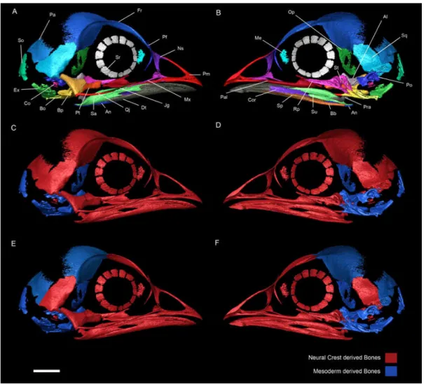
Fig. 2 Right side of a chicken embryo skull at HH40. A, Lateral
view with ossification centers identified with different colors. B, Medial
view with ossification centers identified with different colors. C and D,
respectively lateral and medial views of a chicken skull at HH40 showing
in the embryonic origin of each skull element according to Couly et al
1992, 1993. E and F, respectively lateral and medial views of a chicken
skull at HH40 showing in the embryonic origin of each skull element
Evans and Noden 2006).The left side of the skull was removed including
the left side of unpaired ossification centers. Al, Alisphenoid; An,
Angular; Bb, Basibranchial; Bo, Basioccipital; Bp, Basisphenoid; Co,
Columella; Cor, Coronoid blade of the splenial; Dt, Dentary; Ex,
Exoccipital; Fr, Frontal; Jg, Jugal; Me, Mesethmoid; Mx, Maxilla; Ns,
Nasal; Op, Orbitosphenoid; Pa, Parietal; Pal, Palatine; Pf, Prefrontal;
Pm, Premaxilla; Po, Prootic; Pra, Prearticular; Pt, Pterygoid; Qj,
Quadratojugal; Rp, Rostroparasphenoid (parasphenoid rostrum); Sa,
Surangular; So, Supraoccipital; Sp, Splenial; Sq, Squamosal; Sr,
Sclerotic ring; St, Sella turcica.
Using a pioneering technique, Couly and colleagues were able to
perform unilateral excisions of neural crest from a host chicken embryo
at precise cephalic levels while leaving in situ the neural epithelium. This
procedure was repeated in a quail embryo and the resulting piece was
inserted in the gap created in the chick embryo. Each transplanted piece
of neural fold was about 450 um in length and was always transplanted
from a quail donor to a host chicken embryo of the same stage. Given
the early stages used, the neural crest cells had not started to migrate.
This ensured that tissues, which presented quail cells, were derived from
the neural crest. In addition, these studies presented rigorous
histological sections of the operated embryos at several time points post
operation showing that the neural crest cells were effectively
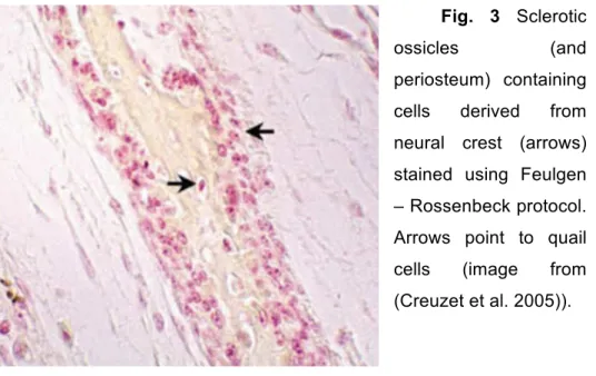
transplanted and contamination was residual. The identity of the cell type
(quail vs chicken) was verified by Feulgen – Rossenbeck staining (Fig.
3). This technique allows a differentiation between the nuclei of quail
cells (which present highly condensed chromatin) and chicken nuclei
Fig. 3 Sclerotic
ossicles (and
periosteum) containing
cells derived from
neural crest (arrows)
stained using Feulgen
– Rossenbeck protocol.
Arrows point to quail
cells (image from
(Creuzet et al. 2005)).
The results showed that in addition to all anterior bones from the
face, cells derived from the donor were present across the whole frontal
and parietal bones. Moreover, these authors also performed the
complementary experiment where they transplanted the paraxial
mesoderm of quail into a chicken host. In these chimaeras they could
not find quail cells in any of these bones. These results strongly
suggested that the frontal and the parietal bones were (at least in quail
and chicken) derived from neural crest cells.
Nevertheless, Evans and Noden criticized these conclusions
arguing that two different problems could lead to misinterpretations of
the results (Evans and Noden 2006). The first argument pertained to the
transplant technique itself. It is known that to produce quail-chick
chimeras an extreme precision is needed. In some cases, at least
theoretically, the transplanted piece of neural fold may carry over some
cells derived from the neural tube, the ectoderm or even of the
mesoderm. If the contamination is from the neural tube or of ectodermal
results and in their interpretation is expected. On the other hand, if the
contamination consists of paraxial mesoderm cells, the identified quail
cells in the posterior region of the frontal and in the parietal bones could
have a different (mesodermal) origin and point to a distinct ontogeny for
those bones. Evans and Noden also noted that these types of
transplants are even more difficult to perform in early stages of
development. For instance, they claimed that “at stage 8 (three somites)
the hindbrain neural folds are vertical and underlying paraxial mesoderm
is tightly adherent and, therefore, difficult to exclude from excised pieces
of neural fold tissue without the use of proteolytic enzymes to separate
epithelial from mesenchymal populations” (Evans and Noden 2006). In
addition, it is argued that the Feulgen staining used to distinguish quail
and chick cells has been shown to be instable in angioblasts therefore
hindering a correct identification of the mesoderm contribution to the
posterior frontal and parietal bones (D. M. Noden 1984; D. M. Noden
1991b).
Specifically, Evans and Noden state that the results obtained by
Couly and colleagues (Couly, Coltey, and Douarin 1993) may be
misleading because they were based “on the presence of cells
containing the quail nuclear marker (Nicole M. Le Douarin 1973) as
identified using Feulgen staining, but did not include assays using the
QH1 antibody to detect quail endothelial cells. Angioblasts are
ubiquitous within early mesodermal populations (D. M. Noden 1984; D.
M. Noden 1991a; Borue and Noden 2004) absent only from notochordal
and prechordal mesoderm (D. M. Noden 1990). Previous studies have
found that the quail nuclear marker is unstable in angioblasts (D. M.
Noden 1984; D. M. Noden 1991a). Therefore, Feulgen-based assays
Trying to circumvent these issues, Evans and Noden infected
chicken embryos with β-galactosidase-encoding, replication-incompetent
retroviruses into the paraxial mesoderm, crest progenitors, and at the
interface between mesodermal and the overlying neural crest. This
produced alternative fate maps to those generated with the quail–chick
grafting technique. These results suggested that the NC/PM boundary
was present at the junction of the supraorbital and calvarial regions of
the frontal bone (Evans and Noden 2006).
A bone is a complex and dynamic organ. It is known that, during
intramembranous ossification, endothelial cells that are always from
mesoderm origin will invade the developing osseous tissue to form blood
vessels (Couly, Coltey, and Douarin 1993). This contradicts critics made
to the quail-chick chimera studies (see Couly et al., 1995 and related
papers) simply because all bones should be invaded by blood vessels
derived from mesoderm cells. Any staining of endothelial cells would
only represent this particular contribution to the bone. On the other hand,
blood vessels are composed of two types of cells: an inner layer of
endothelial cells and an adjacent external layer of pericytes. Cephalic
pericytes are originated anteriorly (forebrain) from neural crest cells and
posteriorly (midbrain and hindbrain) from mesoderm cells. At the border
between the forebrain/midbrain there should be a mixed population of
cells (Etchevers et al. 2001). So any staining of vascular cells would not
be conclusive about the origin of a particular bone. It would be important
to see the exact origin of osteoblasts and osteocytes when performing
fate map studies to understand bone developmental origins.
In addition, Couly and co-workers generated quail-chick mesoderm
transplants that never showed donor cells present in the frontal nor
Until now only two experimental works labeling both neural crest
and mesodermal tissues where publish so far (Couly, Coltey, and
Douarin 1993; Evans and Noden 2006). Thus, it would be also legitimate
to speculate about any possible imprecision in the retroviral infection
studies that could account for the disparate results. Such experiments,
although consider by some as offering finer control over cell labeling
(Gross and Hanken 2008b) can be imprecise given that it is extremely
difficult to control the amount of cells that will be infected. According to
the authors themselves the exact depth of injections was hard to
determined given that the micropipette ejection force was not defined
(Evans and Noden 2006).
Moreover, infections were performed in embryos that range from
Hamburger and Hamilton stages (HH stages) 6 to 11, which in some
cases, depending on the stage they were performed and on the
technique used, resulted in the labeling of mixed cell type populations.
Evans and Noden performed three experiments: 1) injection of retrovirus
into paraxial mesoderm at HH stages 8–9; 2) washing retrovirus over the
surface the embryo, beneath the vitelline membrane, at stages HH 6 to
9.5; and 3) retrovirus injection at the interface between paraxial
mesoderm and overlying neural crest cells at stages HH 10- to 11-
(Evans and Noden 2006). This last experiment was specifically designed
to label both populations of cells. In the same paper the authors
recognize that “each injection typically results in the labeling of some but
not all nearby progenitor cells”. For instance “unintended infection of
crest cells could occur by virus particles reaching the basal surface of
the neural fold or, if they spilled over the surface of the embryo, by
reaching the apical surface of the neural fold” (Evans and Noden 2006).
In addition, in experiment 1, although the infections were performed at
recognize that the virus could remain infectious until crest cells start to
migrate. This is particularly important given that it is known that chicken
cranial NCC migrates via subectodermal streams, immediately dorsal to
mesoderm (Serbedzija, Bronner-Fraser, and Fraser 1992; Lumsden,
Sprawson, and Graham 1991; N. M. Le Douarin and Kalcheim 1982).
Thus cranial NC subectodermal migrating cells could be contaminated
with virus. The fact that the frontal and parietal bones were never
co-labeled only suggests that the domains (either neural crest or mesoderm
or both) are antero-posteriorly separated at early stages of development
(HH 6 to 11-). From experimental procedure 3 where both mesoderm
and neural crest were infected, it is not possible to draw any conclusion
regarding the neural crest:mesoderm origin of these bones. Here, maybe
the most important experiment is number 2 but given the fact that the
authors do not present the amount of positive cases (out of 66
treatments) for a staining of the anterior region of the frontal, it is hard to
extract any conclusion. Moreover, the anterior region of the frontal
should be more compact (if not denser) when compared with the
posterior sheath of bone that forms the posterior region. Any comparison
of a staining should have this into consideration.
Nevertheless it is important to note that in this experiment the
authors did not report any cell in neither the posterior region of the
frontal or in the parietal bones. In any case, it would be interesting to
perform histological cuts in the frontal bone of these embryos and
confirm the complete absence of cells labeled in the posterior region of
the frontal and in the parietal bone.
On the other hand, in mice a three-bone condition is present in the
calvarial region (frontal + parietal + interparietal). The fate map results
are unanimous attributing the origin of the entire frontal bone to cells
parietal) is formed exclusively by paraxial mesoderm derived cells. The
same studies showed that the immediately posterior bone, the
interparietal, is composed medially by a portion derived from NC while its
lateral parts are PM derived (Gross and Hanken 2008a).The medial and
lateral portions have been considered homologous to the postparietal
and tabulars of fossil synapsids respectively (Koyabu, Maier, and
Sánchez-Villagra 2012).
In sum, currently there are two hypotheses regarding the
embryological and evolutionary origin of the calvarial region of the bird
skull. The first hypothesis claims that a population of cells exclusively
derived from neural crest forms the complete frontal and parietal bones
(Couly, Coltey, and Douarin 1993) while other authors advocate for a
double ontogenetic contribution from neural crest and paraxial
mesoderm derived cells (Evans and Noden 2006).
The implications of these two alternative hypotheses go beyond
the avian development realm. As previously mentioned, fundamental
questions regarding skull evolution amongst Archosauria are impacted
differently by these alternatives.
At this point two alternative evolutionary scenarios can be
hypothesized. Hypothesis 1 (H1) corresponds to the classical view
where the frontal and parietal bones in birds are homologous to the
bones with the same name in mammals and to the frontal and parietal of
their last common ancestral. This ancestral should have been a basal
amniote similar to Hylonomus or Protoclepsydrops that lived
approximately 300 Ma in the Carboniferous (Tuinen and Hadly 2004).
The second hypothesis (H2) calls for a revision of the classical view
“frontoparietal”, homologous to the frontal and parietal of mammals. In
addition, this would also imply that the classical parietal bone in birds is
homologous to the mammalian postparietal (medial portion of the
mammalian interparietal) and should also be renamed accordingly.
The two hypotheses entail different and testable predictions.
Namely, throughout ontogeny, different observations are expected. If H1
is correct, one should find a single center of ossification in the frontal,
developing by intramembranous ossification and completely derived
from neural crest cells. If instead H2 is correct, a double center of
ossification developing into the frontal should be observed; both centers
should develop via intramembranous ossification (if not, it could suggest
that one of the centers is an evolutionary novelty rejecting both
hypotheses); the ossification centers should be aligned
antero-posteriorly; have the correct shape (the anterior should be elongated and
the posterior more wide and flat) and the embryological origin should be
neural crest anteriorly and paraxial mesoderm posteriorly (see table 1).
At this point it would be important to consider the fossil record
regarding the ancestral condition in modern Archosauria. Is there any
reason to consider that the extant crocodiles and birds evolved from an
ancestor with a postparietal? And if yes, how can the fossil record help
us test the previous hypotheses?
The Paleontological record
“If there has been a first man he must have been born without
father or mother – which is repugnant to nature. For there could not have
been a first egg to give a beginning to birds, or there should have been a
first bird which gave a beginning to eggs; for a bird comes from an egg”
Aristotle (384–322 BC)
As it is widely known, the posterior region of the bird's adult skull
misses one bone when compared with the most probable ancestor
condition conserved in other Archosauria, namely Alligator
mississippiensis (Klembara 2001). Other closely related Diapsida such
as Euparkeria capensis and most sinapsids (Koyabu, Maier, and
Sánchez-Villagra 2012) do possess a postparietal. The
non-archosaurian archosauriform Euparkeria capensis, found in the Middle
Triassic of South Africa is a very curious fossil in this particular case. It
has been considered as representative of the ancestral pattern to all
Archosauria (Romer 1956) and, not only that, but nearly all phylogenetic
analyses done so far have placed Euparkeria as the closest sister group
of Archosauria (crown group definition) when analyzing
non-archosaurian archosauriforms (Sookias and Butler 2013). In addition to
Euparkeria and some phytosaurs like Machaeroprosopus ((Romer 1956)
although disputed by Nesbitt) present a postparietal. The presence of a
Namely, the basal achosauriforms Proterosuchus fergusi and the
erythrosuchians Erythrosuchus africanus and Shansisuchus
shansisuchus (Nesbitt 2011). All this is suggestive that any possible lost
of this bone could be an autapormophy of Archosauria (exception made
to Alligatormississippiensis (Klembara 2001) (Fig. 4).
Other fossil diapsids also show the presence of a paired
postparietal (also referred as interparietal), for example Araeoscelis,
Protorothyris, Milleretta, Youngina, Petrolacosaurus and even the
anapsid Labidosaurus shows a unpaired interparietal (Koyabu, Maier,
and Sánchez-Villagra 2012; Nesbitt 2011 and references therein).
Fig. 4 Phylogenetic distribution of postparietal in Amniota. Tree
simplified from (Reisz 1997; Nesbitt 2011).
The absence of a postparietal bone (sensu (Koyabu, Maier, and
Sánchez-Villagra 2012)) in birds and in the wide majority of extant
Sauropsida has been traditionally interpreted as an evolutionary loss of
obvious whether the bird's frontal is homologous to one bone (frontal), or
whether it results from a fusion of two skull bones (frontal and parietal).
This doubt makes it difficult to find a definitive terminology for the
calvarial bones of birds and it has been proposed that the frontal bone of
birds should be called “frontoparietal” (Drew M. Noden and Trainor
2005).
Alternatively the postorbital bone is present in almost all members
of Amniota with exception of Aves. This phylogenetic distribution might
also suggest that this bone, rather then being lost simply fused to the
frontal bone and is still present was a transient calvarial center of
ossification (Erdmann 1940).
In this chapter, we will test these competing hypotheses from
different angles using comparative anatomy, developmental studies and
molecular approaches. Embryos from quail, chicken, duck and crocodile
were incubated and stained for bone and cartilage. These experiments,
in combination with a thorough examination of the published fossil
material available, will serve to establish more complete homology
relationships between the skull bones of Aves and Mammalia, shedding
new light onto our understanding of the evolution of development of the
Chicken (Gallus gallus)
We incubated 503 chicken eggs at 38 ºC in a humid environment
and opened 22 eggs at regular 4-hour intervals starting at day 6 plus 23
hours, cleared and stained all embryos with no apparent defect (for
detailed protocol see methods section). The staining performed showed
cartilage (Alcian blue) and bone (Alizarin red). The sequence of
ossification is presented in Table 2. The onset of ossification was only
considered to be positive if any red stain was visible under a binocular
microscope.
The quadratojugal, surangular and angular are the first bones to
appear stained by alizarin red immediately followed by the squamosal at
day 7 plus 19 hours and day 7 plus 23 hours respectively. In contrast,
the epiotic, mesethmoid, vomer, articular and the hyoid apparatus
(except the ceratobranchial) only start ossification at day 11 plus 3
hours.
The premaxilla is stained red just 4 hours after the squamosal and
is then followed by the jugal and pterygoid. Four hours later, at 8 days
plus 11 hours, the palatines start to appear stained and at 8 days plus 15
hours the prootic, maxilla and dentary start to be ossified. The two latter
bones appear to be formed from multiple centers of ossification that will
eventually fuse into one.
Table 2. (next page) Chicken skull bone onset of ossification. Skull
development was monitored over 4 days using Alzarin Red and Alcian
Although it is rare, some bones start to ossify from more than one
center of ossification. For instance, the prefrontal ossifies from the
combination of a larger, dorsally erected center and a ventral needle-like
center. The prefrontal starts to ossify at day 8 + 19 hours but at least
until day 11 + 3 hours it was still possible to observe some embryos with
two unfused centers of ossification. Also, some embryos present a
significant fluctuating asymmetry, displaying one side fused while the
Fig. 5 Chicken embryos (339 and 496) in dorsal view stained with
Alizarin red and Alcian blue, anterior towards right side. A) Dorsal view
of a chicken embryo with 9 days and 3 hours showing two centers of
ossification. B) Magnification of the dashed line square in A. Excpet red
collor all colors were removed. C) Dorsal view of a chicken embryo with
11 days and 3 hours showing the ossification centers completely fused.
D) Magnification of the dashed line square in B. Green dotted line
highlights the contours of the right frontal (double center of ossification).
Frontal
At 8 days plus 19 hours it is already possible to observe two
embryos (out of 16) with a double center of ossification in the frontal.
ends. The posterior center is much more expanded posteriorly and
tapers anteriorly (Fig. 5). These two centers will expand and eventually
fuse in the supraorbital region. During development the fusion of these
two centers occur between the beginning of day 9 (9 days plus 3 hours)
and the start of day 10 (day 10 plus 3 hours, see Fig 6). This fusion
occurs without leaving any scar or suture. After this the frontal continues
to grow thickening at its anterior end and expanding posteriorly to cover
the brain laterally and dorsally.
Fig. 6 Percentage of chicken embryos scored as having 0, 1 or 2 ossification centers in the frontal after alizarin red/alcian blue staining. The X-axis shows times points at which embryos were collected.
Parietal
The first embryos that show some staining of the parietal bone
were collected after 10 days and 7 hours of incubation. At this time point
0% 20% 40% 60% 80% 100%
8D 15h
8D 19h
8D 23H
9D 3h
9D 7h
9D 11h
9D 15h
9D 20h 30m
9D 23h
10D 3h
10D 7h
10D 11h
11D 3h
No ossifica`on 12 15 11 3 8 4 2 1 0 0 0 0 0
Frontal (1 O.C.) 0 0 0 1 1 6 11 17 15 9 13 12 13
5 out of 13 embryos collected presented a faint alizarin red staining in
the parietal. After this point the parietals starts to develop as thin stripes
of bone perpendicular to the antero-posterior axis. Each side of the skull
presents a single center of ossification. These ossification centers
continue to develop in the subsequent days expanding anteriorly in the
direction of the posterior region of the frontal and posteriorly approaching
the anterior border of the supraoccipital. We only observed a single
center of ossification for each parietal bone.
The otoliths, splenials and quadrates start to ossify at day 9 plus 7
hours. After this the next bones to appear are the exoccipital,
orbitosphenoid, opisthotic at day 9 plus 20 hours. The nasal,
parasphenoid and basisphenoid ossify at day 10 plus 3 hours.
The embryos opened at 11 days plus 3 hours presented in addition
some ossification in the basioccipital,
Some authors describe the development of the chick parasphenoid
has having 7 centers of ossification: an anterior rostral
(rostroparasphenoid), a pair below the sella turcica (sellaparasphenoid),
a pair extending out from the dorsal margins of the sella
(alaparasphenoid), and a posterior pair of basitemporals or basicranials
(basiparasphenoid) (Jollie 1957). However, in what concerns birds, other
authors refer five (De Beer 1937) or even three ossification centers
((Parker 1890) cited in (Jollie 1957)). Our results are in agreement with
Jollie regarding the number of ossification centers, but they differ in the
onset. Our embryos showed the ossification centers of the sphenoid
complex at the end of the 9th day, this represents one day earlier the data described before (Jollie 1957). After this point the pattern of
Quail (Coturnix coturnix)
We started by performing a preliminary experimental test where
we incubated 240 eggs and opened every 4 hours to find the interval
where the frontal bone was forming in quail. This preliminary experiment
showed that the frontal starts to ossify around the seventh incubation
day. Then, to increase resolution over the time period where the frontal
is forming we incubated 120 quail eggs. From these, subsets of 20 eggs
were opened every 4 hours between day 7 and 8 days plus 4 hours. The
first embryos to show any ossification in the frontal were sacrificed at 7
days plus 12 hours. Eight hours later all embryos removed from the eggs
presented some degree of staining in the frontal center and no
secondary center formed after this period (Fig. 7 and 8). Two
independent replicas of this experiment were done in parallel and the
results were consistent. In virtually all cases the frontal started to ossify
as a single center. The very few embryos where a putative double center
in the frontal could be hinted were all too faintly stained to allow
interpretation. Moreover, in these cases, the double center was only
present unilaterally. We only scored a double center when it was
conspicuous and bilaterally symmetrical. We used the same criterion in
Fig. 7 Percentage of quail embryos scored as having 0, 1 or 2
ossification centers in the frontal after alizarin red/alcian blue staining.
The X-axis shows time points at which the embryos were collected.
Fig. 8 (next page) Dorsal views of quail embryos with 8 days and 4
hours of incubation stained with Alizarin red. A, C, E, G heads in dorsal
view. B, D, F, H zoom in showing the frontal centers of ossification in
dorsal view. In B, green dotted line surrounds the center of ossification
of the right frontal. 0% 20% 40% 60% 80% 100%
7D + 12h 7D + 16h 7D + 2h0 8D 8D + 4h
No ossifica`on 8 4 0 0 0
Frontal (1 O.C.) 1 5 7 5 9
Duck (Anas platyrhynchos)
In a preliminary test, we incubated 200 eggs and opened
approximately 12 eggs every 8 hours to find the interval where the
frontal bone was forming in the duck. This showed that the frontal bone
was being formed around the beginning of the 10th day of incubation. After this, we incubated 120 eggs opening groups of 12 every 4 hours
starting at day 9 plus 8 hours and ending at day 10 plus 16 hours. This
revealed that the frontal bone in duck is formed by two centers of
ossification as in the chicken. However, the posterior center only
appears almost 12 hours after the anterior one. The first embryos to
show an ossification center in the frontal were collected at 10 days plus
1 hour and the first embryos with the two centers visible were only
collected at 10 days plus 12 hours (Fig. 9 and 10). At 10 days plus 16
hours the percentage of embryos with two centers increased but it was
not possible to continue the tracing of the frontal ossification dynamics
due to lack of embryos. However, in older embryos at 12 days of
Fig. 9 Percentage of duck embryos scored as having 0, 1 or 2
ossification centers in the frontal after alizarin red/alcian blue staining.
The X-axis shows time points at which the embryos were collected. 0%
20% 40% 60% 80% 100%
9D + 20h 10D + 1h 10D + 4h 10D + 8h 10D + 12h
10D + 16h
No ossifica`on 12 4 4 1 1 0
Frontal (1 O.C.) 0 8 10 9 8 5
Fig. 10 Duck embryos stained with Alizarin red. (previous page)
A, right lateral view of an embryo with 10 days and 12 hours of
incubation. B, dorsal view of an embryo with 10 days and 12 hours of
incubation. C, right lateral view of an embryo with 10 days and 16 hours
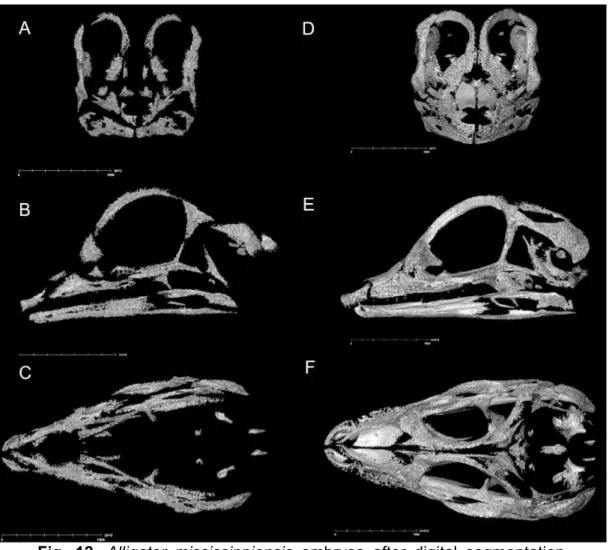
Crocodile (Alligatormississippiensis and Crocodylusniloticus)
The Alligator embryos were staged according to (Ferguson 1987)
and the Crocodylus embryos were staged in days and ranged from 33 to
93 days of incubation. It was not possible to identify a double center of
ossification in the frontal. The same is true for the parietal, postorbital
and supraoccipital.
The frontal bone
starts to ossify
anteroposteriorly. This
long and sheath-like
ossification center tapers
anteriorly and expands
posteriorly to contact the
parietal. The lacrimal
presented two centers of
ossification, one lateral
and another medial to the
lacrimal duct but all other
bones appear to be
formed from only one
center of ossification.
This pattern of
ossification was observed
in both Alligator (Fig. 11
and 12) and Crocodylus
embryos. These results
were manly extracted from synchrotron micro-CT and micro-CT data for
the C. niloticus and A. mississippiensis respectively. Although Alizarin