Healthcare-associated vancomycin resistant
Enterococcus faecium
infections
in the Mansoura University Hospitals intensive care units, Egypt
Dalia Moemen, Doaa Tawfeek, Wafaa Badawy
Department of Medical Microbiology and Immunology, Faculty of Medicine, Mansoura University, Mansoura, Egypt.
Submitted: May 12, 2014; Approved: December 31, 2014.
Abstract
Vancomycin resistantEnterococcus faecium(VREF) ia an emerging and challenging nosocomial pathogen. This study aimed to determine the prevalence, risk factors and clonal relationships be-tween different VREF isolates in the intensive care units (ICUs) of the university hospitals in our geo-graphic location. This prospective study was conducted from July, 2012 until September, 2013 on 781 patients who were admitted to the ICUs of the Mansoura University Hospitals (MUHs), and ful-filled the healthcare-associated infection (HAI) criteria. Susceptibility testing was determined using the disk diffusion method. The clonal relationships were evaluated with pulsed field gel electropho-resis (PFGE). Out of 52E. faeciumisolates, 12 (23.1%) were vancomycin resistant. The significant risk factors for the VREF infections were: transfer to the ICU from a ward, renal failure, an extended ICU stay and use of third-generation cephalosporins, gentamicin, or ciprofloxacin. PFGE with the 12 isolates showed 9 different patterns; 3 belonged to the same pulsotype and another 2 carried a second pulsotypes. The similar pulsotypes isolates were isolated from ICUs of one hospital (EICUs); how-ever, all of the isolates from the other ICUs had different patterns. Infection control policy, in con-junction with antibiotic stewardship, is important to combat VREF transmission in these high-risk patients.
Key words:Enterococcus faecium, ICU, vancomycin, risk factors, PFGE.
Introduction
Enterococciare opportunistic pathogens of the nor-mal humans and aninor-mals intestinal microbiota. The most common Enterococcus species that is involved in noso-comial infections is Enterococcus faecium (E. faecium) (Topet al., 2008; Arias and Murray, 2012).
E. faeciumhas become one of the most important, emerging and challenging nosocomial pathogens (Arias and Murray, 2012). It is a difficult to treat this pathogen due to its intrinsic resistance to cephalosporins, aminogly-cosides, clindamycin and trimethoprim-sulfamethoxazole (Leclercqet al., 2013). Moreover, it has the ability to easily acquire antibiotic resistance genes trough transfer of plas-mids, chromosomal exchange or mutation (Jett et al., 1994).
In addition to this distinct resistance profile, genomic analyses have shown that hospital acquired E. faecium strains have a genetic repertoire that is distinct from that of community associated E. faecium strains that asympto-matically colonize the human gastrointestinal tract (van Schaik et al., 2010). This distinct genetic repertoire in-cludes the enterococcal surface protein, Esp, which is a known virulence determinant (Heikenset al., 2007; 2011). Genomic islands that encode novel metabolic pathways (Heikens et al., 2008), and insertion sequence elements (Leaviset al., 2007). It is now considered that these deter-minants may be adaptive elements that have improved the relative fitness ofE. faeciumsubpopulations in the hospital environment (Willems and van Schaik, 2009). Due to the resistance of multiple antibiotics, the treatment of choice in serious E. faeciuminfections is glycopeptides. However, prudent use of vancomycin is needed as it is associated with DOI: http://dx.doi.org/10.1590/S1517-838246320140403
Send correspondence to D. Moemen. Department of Medical Microbiology and Immunology, Faculty of Medicine, Mansoura University, Egypt. E-mail: [email protected].
an increased risk for vancomycin resistant enterococci (VRE) colonization and infection (Tornieporth et al., 1996).
In the last decades, the number of VRE infections has increased (Graysonet al., 1991; Jones et al., 1995; Rice, 2001). The first VRE isolates were reported in the United Kingdom in the late 1980s (Uttley et al., 1988). In the United States, more than 80% ofE. faeciumisolates from hospitals are vancomycin resistant (Hidronet al., 2008). Vancomycin-resistantE. faecium(VREF) has been associ-ated with outbreaks in hospitals worldwide (Arias and Mur-ray, 2012). The VREF colonization and infection rates have risen steadily, with most cases being caused by the VanA and VanB genotypes, which are the most commonly en-countered glycopeptide resistance forms (Coque et al., 1996; Rice, 2006; Deshpandeet al., 2007).
The clinical impacts of VRE include the limited avai-lability of drugs to treat VRE infections and the ability of VRE to transfer the genetic determinant for vancomycin re-sistance to other Gram-positive pathogens, such as Staphy-lococcus aureus(Schooneveld et al., 2008; Nobleet al., 1992).
VREF is associated with hospital-acquired infections such as urinary tract infections, wounds, bacteremia, endo-carditis and meningitis (Topet al., 2008; Arias and Murray, 2012). Several studies have demonstrated that VRE bacte-raemia patients have a higher mortality rate than those in-fected with vancomycin-susceptibleenterococci(Vergiset al., 2001; Edmondet al., 1996; Bhavnaniet al., 2000).
Information regarding the VRE Infections prevalence in Egypt indicates that there is an increasing VRE infection rate. Ghonaimet al.(2009) reported that VRE isolates con-stituted 20.9% of hospital associated enterococcal infec-tions at the Egyptian National Liver Institute.
To control the rapid spread of multidrug resistant or-ganisms, it is necessary to understand the risk factors for acquiring them. Therefore, the aim of this study was to identify the VREF prevalence and possible risk factors in ICU patients who are at high risk of VREF infection. Addi-tionally, we aimed to identify the clonal relationship be-tween different isolates via pulsed-field gel electrophoresis (PFGE).
Materials and Methods
Patients and methods
Study design
This was a prospective study, which was conducted in nine ICUs in three different MUHs, that were situated within the same geographical region over a 15 months pe-riod from July, 2012 until September, 2013. The ICU bed numbers range from 4 to 27, with a median of 10. They ICUs were categorized as: A. emergency hospital ICUs (EICUs), B. specialized medical hospital ICUs (SMICUs)
and C. children hospital ICUs, which included pediatric (PICU), neonatal ICU (NICU) and surgical ICU (SICU).
Study population
This study included 781 patients (who provided writ-ten consent) that were admitted to different MUHs ICUs and fulfilled the healthcare-associated infection (HAI) cri-teria. (Infections acquired³48 hours after admission that were not present or incubating at the time of hospital admis-sion). The infection types were documented with specific HAIs definitions, which were established by the Centers for Disease Control and Prevention (Horanet al., 2008).
Data collection and definitions
The following data were collected: demographic characteristics, ICU stay duration, transfer to the ICU from a ward, diagnosis at ICU admission, medical conditions that may alter the patient’s immunity (comorbidity) (e.g. diabetes, malignancy, hepatic impairment, renal impair-ment and chronic lung diseases), invasive devices or proce-dures (e.g. surgical procedures, central venous catheter [CVC], peripheral venous catheter [PVC], mechanical ven-tilation, and total parenteral nutrition [TPN], drug therapy (e.g.antibiotics and their durations, prior antibiotic treat-ment, which was defined as any antibiotic treatment during the two weeks preceding ICU admission), immunosup-pressive therapy (which included steroid therapy, cytotoxic chemotherapy or radiotherapy given within one month prior to ICU admission), and neutropenia (less than 500 neutrophils per mm3)
A “case patient” was defined as patient who was in-fected with VREF and was attending a Mansoura Univer-sity ICU during the study period. A “control patient” was defined as a Vancomycin susceptible Enterococcus faecium (VSEF) infected patient who was attending a Mansoura University ICU during the same period. The pa-tients whose initial isolate was susceptible to vancomycin but who subsequently had VREF isolates that were recov-ered were included as case patients.
Microbiologic studies
teicoplanin (30mg), linezolid (30mg), and chloramphenicol (30mg). Glycopeptide antibiotics MICs for: vancomycin and teicoplanin against theE. faeciumisolates were exam-ined with the VITEK 2 system (bioMérieux).
PFGE
The clonal relationships between the vancomycin-resistant strains were studied by evaluating the genomic DNA with PFGE (Antonishynet al., 2000). DNA that was restricted with the SmaI enzyme was separated on an aga-rose gel using a CHEF DR III apparatus (Bio-Rad laborato-ries). The running conditions were 6 V per cm, with pulses ranging from 2 to 15 s for 18 h at 14mC. The DNA banding patterns were visualized under UV light after staining with ethidium bromide (0.5 mg/mL). The similarities between the isolates were determined by visual comparison of the isolate band patterns. The interpretation of the PFGE re-sults was carried out by eye according to the criteria de-scribed by Tenoveret al.(1995).
Statistical analysis
The SPSS software (SPSS Inc., Chicago, IL, USA) was used for data analyseis. Proportions were compared us-ing thec2 test and continuous variables were compared us-ing Student’s t or Mann-Whitney U tests. The results are presented as numbers (percentages) for frequency and as the mean ±standard deviation for quantitative variables. Odds ratios (OR) [95% confidence interval (CI)] were cal-culated for all significant (p < 0.05) qualitative variables in the univariate analyseis.
Results
VREF Prevalence in the ICUs
During the study period, a total of 975 specimens were collected from 782 patients, with clinically suspected HAIs in MUHs ICUs. Out of 52E. faecium isolates de-tected, 12 were VREF, which constituted (12/52) 23.1% of theE. faeciumisolates.
VREF isolates
Twelve VREF single-patient isolates (i.e., 1 isolate per patient) were obtained from the following sites: urine (7 isolates), blood (3 isolates) and wound (2 isolates). A total of 12 infections were documented in 12 patients, including urinary tract infection (7 patients), bacteremia instances (3 patients), and surgical site infection (2 patients). Of these 12 patients, 6 isolates were obtained from the emergency hospital ICUs, 4 were obtained from the ICUs of the spe-cialized medical hospital and 2 were from PICUs.
Antibiotic susceptibility testing of the VREF isolates
Twelve E. faecium isolates were resistant to both glycopeptides antibiotics (vancomycin and teicoplanin) (with a vancomycin MIC³32mg/mL) (VanA phenotype).
All VREF isolates (100%) were resistant to ampicillin, gentamicin, and ciprofloxacin. Most of the isolates (92%) were resistant to erythromycin (only one isolate was sensi-tive), and 5 isolates were resistant to tetracycline (42%). All of the strains were sensitive to linezolid and chloram-phenicol.
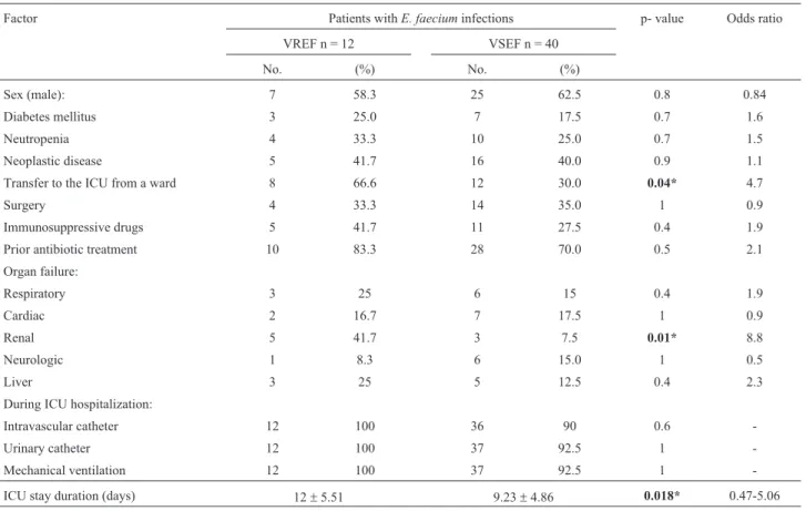
The ICU-acquired VREF Risk factors
Among the 52 E. faecium infected patients, 40 (76.9%) had VSEF infections (control patients), and 12 (23.1%) had VREF infections (case patients). The compar-ative demographic and clinical features for both the case and controls patients are listed in Tables 1 and 2. The risk factors that were significantly associated with the ICU-acquired VREF infections were: transfer to the ICU from a ward (p = 0.04), renal failure (p = 0.01), and longer ICU stay duration (p = 0.018). Additionally, antibiotic use for a long period of time and use of third-generation cepha-losporins, gentamicin, or ciprofloxacin were also associ-ated with VREF infections.
PFGE
The restriction endonuclease patterns obtained with the PFGE following the SmaI treatment for the 12 VREF isolates are presented in Figure 1. In general, the 12 isolates showed 9 different patterns. Three isolates belonged to the same pulsotype and another 2 carried similar pulsotypes. Of interest, the isolates with similar pulsotypes were iso-lated from the ICUs of one hospital (EICUs); however, all of the isolates from the other ICUs had different patterns.
Discussion
fail-ures (P = 0.01) were associated with VREF infections. Intra hospital transfer was associated with VREF colonization or infection in other studies (Tornieporthet al., 1996; Webbet al., 2001). The patients may have acquiredE. faciumduring their ward stay, which later caused infections during their ICU stays. However, our study showed that renal failure was a risk factor for VREF infection acquisition. This is
dif-ferent from other studies in which dialysis was not associ-ated with the VREF infection incidences (Webb et al., 2001; Handwerger et al., 1993; Descheemaeker et al., 2000). Patients who require hemodialysis often have com-plicated illnesses and may receive multiple antibiotic cour-ses, including vancomycin, which places them at greater risk for VREF infection or colonization. Frequent hospital-Table 2- Antibiotic use amongst the study patients during their ICU stays.
Factor Patients withE. faeciuminfections p-value Odds ratio
VREF n = 12 VSEF n = 40
No. (%) No. (%)
Antibiotic treatment duration, days 16±11 5±6 <0.001** 8.09-13.91
Vancomycin use 5 41.7 12 30.0 0.45 1.66
Extended-spectrum penicillin use 7 58.3 19 47.5 0.5 1.6
Fluoroquinolone use 9 75 16 40.0 1 0.8
Carbapenem use 3 25.0 12 30.0 1 0.8
Aminoglycoside use 6 50.0 8 20.0 0.04* 4
First generation cephalosporin use 2 16.7 18 45.0 0.09 0.2
Second generation cephalosporin use 2 16.7 15 37.5 0.3 0.3
Third generation cephalosporin use 6 50.0 6 15.0 0.01* 5.7
*Significant, p-value£0.05; **highly significant, p-value£0.001. Table 1- ICU-acquired VREF infection risk factors.
Factor Patients withE. faeciuminfections p- value Odds ratio
VREF n = 12 VSEF n = 40
No. (%) No. (%)
Sex (male): 7 58.3 25 62.5 0.8 0.84
Diabetes mellitus 3 25.0 7 17.5 0.7 1.6
Neutropenia 4 33.3 10 25.0 0.7 1.5
Neoplastic disease 5 41.7 16 40.0 0.9 1.1
Transfer to the ICU from a ward 8 66.6 12 30.0 0.04* 4.7
Surgery 4 33.3 14 35.0 1 0.9
Immunosuppressive drugs 5 41.7 11 27.5 0.4 1.9
Prior antibiotic treatment 10 83.3 28 70.0 0.5 2.1
Organ failure:
Respiratory 3 25 6 15 0.4 1.9
Cardiac 2 16.7 7 17.5 1 0.9
Renal 5 41.7 3 7.5 0.01* 8.8
Neurologic 1 8.3 6 15.0 1 0.5
Liver 3 25 5 12.5 0.4 2.3
During ICU hospitalization:
Intravascular catheter 12 100 36 90 0.6
-Urinary catheter 12 100 37 92.5 1
-Mechanical ventilation 12 100 37 92.5 1
-ICU stay duration (days) 12±5.51 9.23±4.86 0.018* 0.47-5.06
izations and cross-transmission can also contribute to VREF infection in patients undergoing hemodialysis (Chowet al., 1993; Roghmannet al., 1998). The other risk factor for VREF infection in the ICU was longer ICU stay duration (p = 0.018). This result consistant with most previ-ous studies (Ostrowskyet al., 1999; Warrenet al., 2003; Se et al., 2009; Panet al., 2012; Fridkinet al., 2001). Longer ICU stays can indicate a greater chance of receiving antibi-otics and also a longer exposure time to possible pathogen transmission. Our study showed that patients who had a prior exposure to broad-spectrum antibiotics and for a pro-longed duration were at a higher risk of VREF infections due to the selective pressure from prior antibiotics. Interest-ingly, in this study, vancomycin use was not found to be a risk factor for VREF infection, which is different from other studies (Tornieporthet al., 1996; Fridkinet al., 2001). This can be explained by the theory that in medical settings in which VREF infections are endemic, the patients with VREF colonization may serve as a source for already anti-biotic resistantE. faeciumstrains in patients who have not necessarily received glycopeptide antibiotics (Murray, 2000; Hayden, 2000). According to the PFGE analysis, there were nine pulsotypes that were noted during the study period. The PFGE analysis showed different patterns in the different ICUs of the three hospitals, except for the EICUs in the emergency hospital, in which the 6 VREF isolates showed 3 different patterns. Specifically, 3 isolates be-longed to the same pulsotype and another 2 carried similar pulsotypes, indicating that there was a spread within the ICUs of one hospital (the emergency hospital). Thus, our
data show that all of the strains were not from the same clone, indicating multiple acquisitions of resistant isolates. However, the VREF infections within the ICUs of one hos-pital (the EICUs) may result from cross transmission of prevalent isolates. This shows that spread occurred within the ICUs of an individual hospital but not in the other hospi-tals. This might be because the EICUs have more critical care cases compared with the other ICUs (e.g., care for pa-tients with acute, life-threatening illnesses or injuries), in which VRE infection prevention measures are more impor-tant than in the other ICU type’s. Similarly other studies have documented the spread of VREF clones among hospi-tals (Saderet al., 1994; Fridkinet al., 1998; Nourseet al., 2000; Corsoet al., 2007). The microorganism can be trans-mitted by health care workers in particular via their hands which are most likely the most common mode of noso-comial transmission (Boyceet al., 1994). VREF transmis-sion by way of contaminated medical equipment and health care carriers has been investigated and shown to occur in different hospitals sections especially in the dialysis ward (Kalocheretiset al., 2004). Adherence to infection control precautions by ICU staff members may also affect possible VREF transmission. Other distinct strains may come from intrinsicE. facium, which is selected for under selective pressure. Thus, infection control policy, in conjunction with practices that control antimicrobial use, is important to combat VREF infection transmission in these high-risk pa-tients. Moreover, our data ensure that standard efforts to re-duce cross-transmission might be needed hospital-wide because the VREF rates outside of the ICUs greatly affect the ICU- specific VREF rates.
References
Antonishyn NA, McDonald RR, Chan Eet al.(2000) Evaluation of fluorescence-based amplified fragment length polymor-phism analysis for molecular typing in hospital epidemiol-ogy: comparison with pulsed-field gel electrophoresis for typing strains of vancomycin resistant Enterococcus faecium. J Clin Microbiol 38:4058-4065.
Arias CA, Murray BE (2012) The rise of the Enterococcus: be-yond vancomycin resistance. Nat Rev Microbiol 10:266-278.
Bhavnani SM, Drake JA, Forrest Aet al.(2000) A nationwide, multicenter, case-control study comparing risk factors, treat-ment, and outcome for vancomycin-resistant and -suscepti-ble enterococcal bacteremia. Diagn Microbiol Infect Dis 36:145-158.
Boyce JM, Opal SM, Chow JWet al.(1994) Outbreak of multi-drug-resistantEnterococcus faeciumwith transferable vanB class vancomycin resistance. J Clin Microbiol 32:1148-1153.
Chow JW, Kuritza A, Shlaes DMet al.(1993) Clonal spread of vancomycin- resistant Enterococcus faeciumbetween pa-tients in three hospitals in two states. J Clin Microbiol 31:1609-1611.
Coque TM, Tomayko JF, Ricke SCet al.(1996) Vancomycin-resistant enterococci from nosocomial, community, and ani-Figure 1- SmaI restriction endonuclease patterns obtained by PFGE:
mal sources in the United States. Antimicrob Agents Chemother 40:2605-2609.
Corso AC, Gagetti PS, Rodríguez MMet al.(2007) Molecular ep-idemiology of vancomycin-resistantEnterococcus faecium
in Argentina. Int J Infect Dis 11:69-75.
Descheemaeker P, Ieven M, Chapelle Set al.(2000) Prevalence and molecular epidemiology of glycopeptide-resistant enterococci in Belgian renal dialysis units. J Infect Dis 181:235-241.
Deshpande LM, Fritsche TR, Moet GJet al.(2007) Antimicrobial resistance and molecular epidemiology of vancomycin-resistant enterococci from North America and Europe: a re-port from the SENTRY antimicrobial surveillance program. Diagn Microbiol Infect Dis 58:163-170.
Edmond MB, Ober JF, Dawson JD et al. (1996) Vancomy-cin-resistant enterococcal bacteremia: Natural history and attributable mortality. Clin Infect Dis 23:1234-1239. Fridkin SK, Edwards JR, Courval JMet al.(2001) Intensive Care
Antimicrobial Resistance Epidemiology (ICARE) Project and the National Nosocomial Infections Surveillance (NNIS) System Hospitals. The effect of vancomycin and third-generation cephalosporins on prevalence of vanco-mycin-resistant enterococci in 126 U.S. adult intensive care units. Ann Intern Med 135:175-183.
Fridkin SK, Yokoe DS, Whitney CGet al.(1998) Epidemiology of a dominant clonal strain of vancomycin-resistant
Enterococcus faeciumat separate hospitals in Boston, Mas-sachusetts. J Clin Microbiol 36:965-970.
Ghonaim M, Ghoniem E, Abdulaziz Aet al.(2009) Enterococci in Hospital Associated Infection in the National Liver Insti-tute, Egypt. Egy J Med Microbiol 18:69-79.
Grayson ML, Eliopoulos GM, Wennersten CBet al.(1991) In-creasing resistance to beta-lactam antibiotics among clinical isolates ofEnterococcus faecium: a 22-year review at one institution. Antimicrob Agents Chemother 35:2180-2184. Handwerger S, Raucher B, Altarac Det al.(1993) Nosocomial
outbreak due to Enterococcus faeciumhighly resistant to vancomycin, penicillin, and gentamicin. Clin Infect Dis 16:750-755.
Hayden MK (2000) Insights into the epidemiology and control of infection with vancomycin-resistant enterococci. Clin Infect Dis 31:1058-1065.
Heikens E, Bonten MJ, Willems RJ (2007) Enterococcal surface protein Esp is important for biofilm formation of
Enterococcus faeciumE1162. J Bacteriol 189:8233-8240. Heikens E, Singh KV, Jacques-Palaz KDet al.(2011)
Contribu-tion of the enterococcal surface protein Esp to pathogenesis of Enterococcus faecium endocarditis. Microbes Infect 13:1185-1190.
Heikens E, van Schaik W, Leavis HLet al.(2008) Identification of a novel genomic island specific to hospital-acquired clonal complex 17Enterococcus faeciumisolates. Appl En-viron Microbiol 74:7094-7097.
Hidron AI, Edwards JR, Patel Jet al.(2008) NHSN annual up-date: antimicrobial-resistant pathogens associated with healthcare-associated infections: annual summary of data reported to the national healthcare safety network at the cen-ters for disease control and prevention, 2006-2007. Infect Control Hosp Epidemiol 29:996-1011.
Horan TC, Andrus M, Dudeck MA (2008) CDC/NHSN surveil-lance definition of health care-associated infection and
crite-ria for specific types of infections in the acute care setting. Am J Infect Control 36:309-332.
Institute CaLS (2011) Performance standards for antimicrobial susceptibility testing: 17th informational supplement, M100-S21. Wayne, PA.:Clinical and Laboratory Standards Institute.
Jett BD, Huycke MM, Gilmore MS (1994) Virulence of
enterococci.Clin Microbiol Rev 7:462-478.
Johnson AP, Uttley AH, Woodford Net al.(1990) Resistance to vancomycin and teicoplanin: an emerging clinical problem. Clin Microbiol Rev 3:280-291.
Jones RN, Sader HS, Erwin MEet al.(1995) Emerging multiply resistant enterococci among clinical isolates. I. Prevalence data from 97 medical center surveillance study in the United States. Enterococcus Study Group. Diagn Microbiol Infect Dis 21:85-93.
Kalocheretis P, Baimakou E, Zerbala Set al.(2004) Dissemina-tion of vancomycin-resistant enterococci among haemo-dialysis patients in Athens, Greece. J Antimicrob Chemother 54:1031-1034.
Kapoor L, Randhawa VS, Deb M (2005) Antimicrobial resistance of enterococcal blood isolates at a pediatric care hospital in India. Jpn J Infect Dis 58:101-103.
Kaur N, Chaudhary U, Aggarwal Ret al.(2009) Emergence of VRE and their antimicrobial sensitivity pattern in a tertiary care teaching hospital. J Med Biol Sci 8:26-32.
Leavis HL, Willems RJ, van Wamel WJet al.(2007) Insertion se-quence-driven diversification creates a globally dispersed emerging multiresistant subspecies of E. faecium. PLoS Pathog 3:75-96.
Leclercq R, Canton R, Brown DFet al.(2013) EUCAST expert rules in antimicrobial susceptibility testing. Clin Microbiol Infect 19:141-160.
Mathur P, Kapil A, Chandra Ret al.(2003) Antimicrobial resis-tance in Enterococcus faecalis at a tertiary care centre of northern India. Indian J Med Res 118:25-28.
Murray BE (2000) Vancomycin-resistant enterococcal infections. N Engl J Med 342:710-721.
NNIS (2004) National Nosocomial Infections Surveillance (NNIS) System Report, data summary from January 1992 through June 2004, issued October 2004. Am J Infect Con-trol 32:470-485.
Noble WC, Virani Z, Cree RG (1992) Co-transfer of vancomycin and other resistance genes from Enterococcus faecalis NCTC 12201 toStaphylococcus aureus. FEMS Microbiol Lett 72:195-198.
Nourse C, Byrne C, Kaufmann Met al.(2000) VRE in the Repub-lic of Ireland: clinical significance, characteristics and mo-lecular similarity of isolates. J Hosp Inf 44:288-293. Ostrowsky BE, Venkataraman L, D’Agata EMCet al. (1999)
Vancomycin-Resistant Enterococci in Intensive Care Units: High Frequency of Stool Carriage During a Non-Outbreak Period. Arch Intern Med 159:1467-1472.
Pan SC, Wang JT, Chen YCet al.(2012) Incidence of and risk factors for infection or colonization of vancomycin-resistant enterococci in patients in the intensive care unit. PLoS ONE 7:e47297.
Praharaj I, Sujatha S, Parija SC (2013) Phenotypic & genotypic characterization of vancomycin resistant Enterococcus iso-lates from clinical specimens. Indian J Med Res 138:549-556.
Rice LB (2001) Emergence of vancomycin-resistant enterococci. Emerg Infect Dis 7:183-187.
Rice LB (2006) Antimicrobial resistance in gram-positive bacte-ria. Am J Med 119:11-19. Roghmann MC, Fink JC, Polish L
et al. (1998) Colonization with vancomycin-resistant enterococci in chronic hemodialysis patients. Am J Kidney Dis 32:254-257.
Sader HS, Pfaller MA, Tenover FCet al.(1994) Evaluation and characterization of multiresistant Enterococcus faecium
from 12 U.S. medical centers. J Clin Microbiol 32:2840-2842.
Schooneveld TV, Rupp ME (2010) Control of Gram-positive multidrug-resistant pathogens. In: Lautenbach E, Woeltje KF, Malani PN, editors. Practical healthcare epidemiology. 3rd ed. Chicago, United States: The University of Chicago Press p. 197-208.
Se YB, Chun HJ, Yi HJet al.(2009) Incidence and risk factors of infection caused by vancomycin-resistant enterococcus col-onization in neuro- surgical intensive care unit patients. J Korean Neurosurg Soc 46:123-129.
Taneja N, Rani P, Emmanuel R et al. (2004) Significance of vancomycin resistant enterococci from urinary specimens at a tertiary care centre in northern India. Indian J Med Res 119:72-74.
Tenover FC, Arbeit RD, Goering RVet al.(1995) Interpreting chromosomal DNA restriction patterns produced by pulsed-field gel electrophoresis: criteria for bacterial strain typing. J Clin Microbiol 33:2233-2239.
Top J, Willems R, Bonten M (2008) Emergence of CC17
Enterococcus faecium: from commensal to hospital-adapted pathogen. FEMS Immunol Med Microbiol 52:297-308. Tornieporth NG, Roberts RB, John Jet al.(1996) Risk factors
as-sociated with vancomycin-resistant Enterococcus faecium
infection or colonization in 145 matched case patients and control patients. Clin Infect Dis 23:767-772.
Tornieporth NG, Roberts RB, John Jet al.(1996) Risk factors as-sociated with vancomycin-resistant Enterococcus faecium
infection or colonization in 145 matched case patients and control patients. Clin Infect Dis 23:767-772.
Uttley AH, Collins CH, Naidoo J et al. (1988) Vancomycin-resistant enterococci. Lancet 1:57-58.
van Schaik W, Top J, Riley DRet al.(2010) Pyrosequencing-based comparative genome analysis of the nosocomial pathogenEnterococcus faeciumand identification of a large transferable pathogenicity island. BMC Genomics 11:239. Vergis EN, Hayden MK, Chow JWet al.(2001) Determinants of
vancomycin resistance and mortality rates in enterococcal bacteremia. A prospective multicenter study. Ann Intern Med 135:484-492.
Warren DK, Kollef MH, Seiler Set al.(2003) The Epidemiology of Vancomycin-Resistant Enterococcus Colonization in a Medical Intensive Care Unit Infect Control Hosp Epidemiol 24:257-263.
Webb M, Riley LW, Roberts RB (2001) Cost of hospitalization for and risk factors associated with vancomycin-resistant
Enterococcus faeciuminfection and colonization. Clin In-fect Dis 33:445-452.
Willems RJ, van Schaik W (2009) Transition ofEnterococcus faeciumfrom commensal organism to nosocomial pathogen. Future Microbiol 4:1125-1135.
Associate Editor: Roxane Maria Fontes Piazza