Trypanosoma cruzi
: Genetic Structure of Populations and
Relevance of Genetic Variability to the Pathogenesis of Chagas
Disease
Andréa M Macedo, Carlos R Machado, Riva P Oliveira*, Sérgio DJ Pena/
+Departamento de Bioquímica e Imunologia, Universidade Federal de Minas Gerais, Caixa Postal 486, 30161-970 Belo Horizonte, MG, Brasil *Joslin Diabetes Center and Department of Pathology, Harvard Medical School, Boston, MA, US
Chagas disease, caused by the protozoan Trypanosoma cruzi, has a variable clinical course, ranging from symptomless infection to severe chronic disease with cardiovascular or gastrointestinal involvement or, occasion-ally, overwhelming acute episodes. The factors influencing this clinical variability have not been elucidated, but it is likely that the genetic variability of both the host and the parasite are of importance. In this work we review the the genetic structure of T. cruzi populations and analyze the importance of genetic variation of the parasite in the pathogenesis of the disease under the light of the histotropic-clonal model.
Key words: Trypanosoma cruzi - genetic variability - mismatch repair - population structure - pathogenesis - Chagas disease
Chagas disease is a chronic illness caused by the pro-tozoan Trypanosoma cruzi afflicting over 18 million people in the three Americas, from Southern Argentine to the Southern United States. T. cruzi is transmitted to humans by the feces of infected hematophagus triatomine bugs, blood transfusion, congenitally from infected mothers, or more infrequently by organ transplantation, contaminated food or laboratory accidents (Lambrecht 1965, Prata, 2001). Under natural conditions the life cycle of T. cruzi al-ternates between an invertebrate host, one of more than 130 hemiptera from the family Reduviidae, and a verte-brate host that can be any one of more than 100 mamma-lian species from different orders (Lambrecht 1965, Zeledon & Rabinovich 1981). The geographical distribution of the triatomines and the vertebrate hosts associated with the insects’ preferences for specific blood sources define two transmission cycles of T. cruzi: a sylvatic cycle involving different species of triatomid bugs and wild animals and a domestic/peridomestic cycle in which household animals and human beings act as reservoirs.
The human disease has a very variable clinical pre-sentation. Following the parasite infection, there is a short acute phase characterized by an abundant parasitemia, but frequently very mild and unspecific symptoms that difficult the recognition of the contagion. In less than 5% of the cases, fatal neurological and/or cardiac complica-tions of the acute phase can be observed. The infection then proceeds to a chronic phase, with scarce parasitemia and an unpredictable clinical course that ranges from an absence of symptoms to severe disease with cardiovas-cular and/or gastrointestinal involvement (Prata 2001). The
+Corresponding author. Fax: +55-31-3227.3792. E-mail:
spena@dcc.ufmg.br Received 17 December 2003 Accepted 7 January 2004
heart pathology is characterized by chronic myocarditis that frequently leads to cardiomegaly, congestive heart failure and arrhythmias (Tanowitz 1992). In the digestive forms of the disease, dilatation of the esophagus and/or colon (megaesophagus and megacolon, respectively) may be observed in advanced stages (Lopes et al. 1989, Adad et al. 1991). Pathologically, both the heart and digestive tract exhibit focal lymphocytic infiltrates (Brener 1992), with scarring and loss of myocardial cells in the cardiopa-thy (Palacius-Pru et al. 1989) and neuronal degeneration with denervation in megaesophagus and megacolon (Köeberle, 1968, Adad et al. 1991).
Geographical variations in the prevalence of clinical forms and morbidity of Chagas disease have been re-ported. In Brazil, the asymptomatic or indeterminate form is the most common (60-70%), followed by the cardiac and digestive forms (20-30% and 8-10%, respectively); the cardio-digestive form is less frequent. However in the Central Brazil and in Chile, the digestive form of Chagas disease predominates, while it is practically non-existent in Venezuela and Central America (Luquetti et al. 1986, Dias 1992). The reason for this geographical heterogene-ity and why different patients develop different clinical forms remain unexplained. It certainly cannot be explained by major genetic differences between the human popula-tions that inhabit these areas. Thus, it is generally be-lieved that they are primarily determined by genetic varia-tion of T. cruzi. However,a possible role for environmen-tal, nutritional, and immunological aspects of the host cannot be discarded.
mark-ers capable of establishing a link between the variability of the parasite and the clinical form of the disease. The failure of these endeavors, associated with the scarcity of parasites in the blood and tissues of chronic symptom-atic patients led to the “autoimmune phase”. In this sec-ond period the pathogenetic importance of the parasite was relegated to a second plane, except as providing cross-reacting epitopes capable of unleashing autoim-mune reactions (Schmunis 1987, Brener 1987, Kalil & Cunha-Neto 1996). The discovery of more sensible tech-niques for T. cruzi detection rescued the primary role of the parasite in the pathogenesis of Chagas disease. We are now experiencing the third wave, the “genomic phase”, in which the illness is seen as the product of the interac-tion of two highly variable and coevolving genomes: the parasite genome and the human genome.
THE GENOMIC PHASE
The genomic phase of the pathogenesis of Chagas disease started in the early 90’s when the presence of T. cruzi in chronic infected tissues was revealed by applica-tion of powerfulimmunohistochemical techniques devel-oped in the past two decades (Higuchi et al. 1993). How-ever, it was the application of the sensitive polymerase chain reaction (PCR) that provided definitive molecular evidence that clearly tied tissue lesions to the presence of the parasite (Jones et al. 1993, Brandariz et al. 1995, Vago et al. 1996a). These new studies highlighted the pri-mary role of T. cruzi in the pathogenesis of Chagas dis-ease and set the stage for establishing the notion that genomic variation of T. cruzi might influence the course of the disease.
THE GENETIC VARIABILITY OF T. CRUZI
Different studies including biological, biochemical and molecular ones have demonstrated that T. cruzi is a very heterogeneous taxon (reviewed by Macedo & Pena 1998, Macedo et al. 2001, Devera et al. 2003). In the present review we will recapitulate some of these studies, with emphasis in those that have contributed to a better un-derstanding of the T. cruzi population structure and its role for the pathogenesis of Chagas disease.
ZYMODEMES
The first experimental method that demonstrated the extent of genetic diversity of T. cruzi was the analysis of electrophoretic variants of cellular enzymes (isoenzymes). Based on the isozymic variability of six loci Miles et al.
(1977, 1978, 1980) proposed the existence of three isoen-zymatic clusters (zymodemes Z1, Z2, and Z3) of T. cruzi.
Epidemiological studies demonstrated that Z1 and Z3 were associated with the sylvatic cycle and Z2 with the domes-tic transmission cycle (Miles et al. 1978, 1980, Barret et al. 1980). Carneiro et al. (1990) identified four isoenzymatic groups (ZA, ZB, ZC, and ZD) when analyzing eight isoen-zyme loci. Subsequent studies using 15 isoenisoen-zyme loci predictably evidenced much higher genetic diversity, and 43 genotypes were proposed (Tibayrenc et al. 1986, Tibayrenc & Ayala 1988). Numerical taxonomic analysis of these data later suggested the existence of two major highly heterogeneous phylogenetic lineages of T. cruzi
that differed in several biological properties (Tibayrenc
1995). More recently, Brisse et al. (2000) identified five lower phylogenetic subdivisions within one of the lin-eages and proposed the existence of six principal discrete
T. cruzi groups (so-called lineages I, IIa, IIb, IIc, IId, IIe).
SCHIZODEMES
The first studies of DNA polymorphism in T. cruzi
were published in 1980 by Morel et al. who reported on restriction fragment length polymorphism (RFLP) typing of kDNA minicircles. Parasite populations displaying iden-tical or very similar kDNA minicircle restriction patterns were called schizodemes. kDNA RFLP analyses unrav-eled an unexpected amount of genetic diversity in T. cruzi, although some correlation was seen with the isozyme clas-sification, demonstrating that linkage disequilibrium ex-tended even between different (nuclear and mitochon-drial) genome compartments (Tibayrenc & Ayala 1987). Together with cloning experiments, kDNA restriction analysis demonstrated for the first time that single strains of T. cruzi could contain two or more distinct clones (Mo-rel et al. 1980). The existence of these multiclonal T. cruzi
strains was confirmed later by several groups using dif-ferent techniques (Deane et al. 1984, Marques-Araújo & Chiari, 1988, Carneiro et al.1990, Macedo et al.1992, Oliveira et al. 1998, 1999).
LOW STRINGENCY SINGLE SPECIFIC PRIMER (LSSP)-PCR
An alternative and more sensitive way to study the polymorphism of the kDNA is the LSSP-PCR method (Vago et al. 1996a). In this technique a previously purified DNA amplicon is subjected to PCR using a single specific primer under very low stringency conditions. The primer hybrid-izes specifically to its complementary region and also nonspecifically to multiple sites within the fragment, in a sequence-dependent manner, thus producing a highly complex set of reaction products that can be resolved by electrophoresis to generate ‘gene signatures’ (Pena et al. 1994, Pena & Simpson, 1996). LSSP-PCR has been suc-cessfully applied to the ~ 330 bp variable DNA fragment of T. cruzi kDNA minicircles to produce the ‘kDNA signa-tures’, allowing for the first time to profile parasites present in the tissues of chronically infected patients (Vago et al. 1996b). The initial idea of the differential tissue tropism playing fundamental importance in determining the patho-genesis of Chagas disease, proposed as early as 1911 (Vianna 1911) was revitalized when it was shown that para-sites presenting different genetic profiles could be found in distinct tissues (esophagus and heart) of the same pa-tient (Vago et al. 2000). Moreover, in a study of doubly infected mice, we found a strong correlation between the strain profile (JG or Col1.7G2) and the type of lesion in the heart and rectal tissues (Andrade et al. 1999).
RANDOM AMPLIFIED POLYMORPHIC DNA (RAPD) ANALYSIS
banding patterns were evidenced for different T. cruzi
populations, although no correlation with clinical aspects has been found (Oliveira 1997). However, when we com-pare the RAPD profiles of strains isolated from patients in the chronic phase of the disease, with isolates from pa-tients in the acute phase, and with T. cruzi populations obtained from sylvatic vectors and mammals, a clear in-creasing gradient in variability is observed (Table I). These results are compatible with the idea that not all T. cruzi
strains are capable of establishing effective infections in
Homo sapiens and that humans thus function as a filter in selecting more adapted sub-populations (Macedo & Pena 1998). Even in the domestic environment, the probability of acquiring Chagas disease from one bite of an infected bug has been estimated to be only about one in a thou-sand (Prata 2001).
RAPD patterns have being used in numerical taxonomy studies. A strong correlation has been observed between RAPD profiles and zymodemes (Steindel et al. 1993, Tibayrenc et al. 1993). Moreover, RAPD profiling was also utilized for corroborating the T. cruzi taxon division in two major lineages (Tibayrenc 1995, Souto et al. 1996).
MINI EXON POLYMORPHISM
The mRNA of trypanosomes is unique in that most, if not all, mature species bear an identical 35-bp splicer leader (SL) at their 5’-termini (Borst & Greaves 1986). This SL is encoded in blocks of tandemly repeated units, the mini-exon genes (ME). Comparative alignment of ME from dif-ferent T. cruzi strains showed the presence of highly con-served sequences regions of 39 bp (the exons), similar regions of 73 bp with more than 98% of identity (corre-sponding to the introns) and divergent intergenic regions (less than 59% of similarity) (Murthy et al. 1992, Souto et al. 1996) (Fig. 1). By using a multilocal PCR strategy it was possible to cluster T. cruzi strains into two groups ac-cording to the amplification products. Strains that yielded 300 bp products were designated as belonging to group 1 and those that yielded 350 bp products were designated as belonging to group 2 (Souto et al. 1996). Subsequent analysis of mini-exon gene sequences of Z3 strains rec-ognized a sub-clade inside of group 2 characterized by a ~ 50 bp insertion/deletion in the non-transcribed spacer region of the mini-exon (Fernandes et al. 1998). New sets of primers were designed to amplify the Z3 strains (Fernandes et al. 2001).
rDNA POLYMORPHISMS
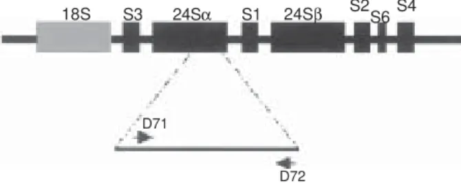
In trypanosomes, the ribosomal cistron exhibits a sin-gular organization. Both the small subunit (SSU) and the large subunit (LSU) are considerably larger than the typi-cal eukaryotic rRNA species. Particularly interesting is the LSU that contains two-high-molecular weight RNAs (24Sα/24Sβ) and six low-molecular-weight RNA (S1-S6) components (Leon et al. 1978, Hernandez et al. 1990, Arruda et al. 1990). The rDNA cistron is constituted of repeated sequences in which the coding regions of the SSU and the LSU are separated by two internal transcribed spacers. The ITS 1 separates the coding region of the 18S subunit and the 5.8 S rDNA, and the ITS2 separates 5.8 S rDNA sequence from the 24S rDNA (Fig. 2). Comparison between the 24Sα rRNA gene of T. cruzi and other trypanosomatids showed high homology except for dis-crete regions. The most divergent domain (D7) is a seg-ment of about 100 bp located at the 3’ end of T. cruzi gene (Arruda et al. 1990, Souto & Zingales 1993). Initially, PCR amplification of a sequence from the D7 domain revealed a dimorphism when DNA from different T. cruzi strains was used (Souto & Zingales 1993). Three different groups or lineages of T. cruzi strains could be detected. Strains from lineage 1 gave a 125 bp product and another group of strains (lineage 2) gave a 110 bp product. A third group presenting both PCR products and denoted group 1/2 was later detected (Souto et al. 1996).
Fig. 1: schematic representation of the Trypanosoma cruzi mini-exon (ME) locus. The ME locus is organized as tandem repeated units in which the exon region are conserved throughout the kinetoplastida (black boxes). Intron regions are indicated by the grey boxes and the non-transcribed spacer is represented by the black connecting line. ME typing is based on a multiplex poly-merase chain reaction using an exon-specific primer (right arrow) and lineage-specific primers located in the spacer region.
TABLE I
Analysis of genetic variability of 58 Trypanosoma cruzi strains by randon amplified polymorphic DNA according to their origin
Number Average Average number Average
of number of of shared bands in proportion of
Sample strains bands per track different strains (± s.d.) shared bands
Strains isolated from non-human sources 11 45.7 ± 7.8 10.2 ± 8.9 22%
Strains isolated from patients with acute Chagas disease 11 55.0 ± 10.3 21.5 ± 13.9 39% Strains isolated from patients with asymptomatic chronic 14 55.4 ± 6.7 28.1 ± 16.8 51% Chagas disease
The phylogenetic relationship of strains from the rDNA group 1, 2, and 1/2 is still under intense investiga-tion. Studies on RAPD and mini-exon profiling had shown that the strains of the D7 rDNA group 1/2 were more ge-netically related to the rDNA lineage 1 than rDNA lineage 2 (Souto et al. 1996). However Zingales et al. (1999) se-quenced both alleles of the D7 rDNA from a strain of group1/2 and while the alignment of the sequence type 1 placed this strain among lineage 1 strains, the sequence type 2 did not allow the clear position of the strain in the tree. In fact, depending on the algorithm used group 1/2 in some tree seemed to be more related to lineage 1 and in the other trees to lineage 2.
Additionally, when comparing 18S and D7-24Sα rDNA sequences, Kawashita et al. (2001) found that T. cruzi could be organized into four riboclades. The riboclade 1 (rDNA group 1) could be separated from the others, but the riboclade 2 (containing the strains rDNA group 2) proved to be a sister group of the two additional riboclades 3 and 4, which grouped representative strains from zymodeme Z3 and rDNA group 1/2.
More recently, extra D7 PCR products of 117/119 bp (Pimenta 2002) or 120/130 pb (Brisse et al. 2001) were found. Brisse et al. (2001) combine data from 18S, D7 re-gion of 24Sa rDNA and mini-exon data to reinforce the their hypothesis of a subdivision of the T. cruzi into six lineages (I, IIa, IIb, IIc, IId, IIe).
KARYOTYPING
The chromatin of T. cruzi is poorly condensed during mitosis and distinct chromosomes cannot be visualized by microscopic methods. The development of the pulsed-field gel electrophoresis (PFGE) technique made possible the identification of 20 chromosomal bands ranging from 450 Kb to 3500 Kb for CL Brener, the model clone of the T. cruzi genome initiative (Santos et al. 1997, Porcile et al. 2003). However, the intensities of different chromosomal bands vary in ethidium bromide stained gels, indicating that the chromosome number may be possibly much higher than 20 (Santos et al. 1997).
Annealing studies with several DNA probes show
hybridization signals in pairs, presumably in homologous chromosomes, which in many cases differ in size. In fact extensive chromosomal size polymorphism has been re-vealed among different strains and even between clones of the same strain of T. cruzi (Henriksson et al. 1995, Alves et al.1996). This polymorphism can be seen in most trypanosomatids and has been attributed to expansion/ contraction of tandem repeats (Dujardin et al. 1995). More-over, differences in ploidy for one or more chromosomes (aneuploidy) have been also observed (reviewed by Herinksson et al. 1996).
Attempts have been made to reconstruct the evolu-tionary history of T. cruzi by using the absolute chromo-somal size difference index (aCSDI) as a genetic distance among the isolates. This method assumes that the ge-nomic distance between two organisms is the sum of the size differences between their homologous. The phenetic trees obtained had defined initially two (Henriksson et al. 2002) and later three (Pedroso et al. 2003) major clusters in
T. cruzi encompassing respectively the strains from D7 rDNA group 1, 2 and 1/2.
MICROSATELLITE ANALYSES
The disclosure of the T. cruzi microsatellites (poly-morphic tandem repeats of 2-6 bases long simple motifs) has provided a new tool for the analysis of the population structure of the parasite (Oliveira et al. 1998). These DNA markers are extremely polymorphic and are dispersed throughout the parasite’s nuclear genome. The microsat-ellite analysis of different T. cruzi strains and clones con-stituted a simple screening test to estimate if a T. cruzi
strain is a monoclonal or a multiclonal population (Oliveira et al. l998, 1999, Macedo et al. 2001). By using microsatellite analyses it was demonstrated that the percentage of multiclonal (or aneuplody) populations decrease progres-sively as we compare strains isolated from the sylvatic cycle with those isolated from man, confirming the previ-ous idea of a human filter selecting more adapted strains (Table II- Oliveira et al. l998, 1999, Macedo et al. 2001).
The T. cruzi microsatellite profile also has been used for phylogenetic investigations. In initial studies involv-ing 53 T. cruzi populations isolated from different geo-graphical areas, the tree topology indicated clustering of the strains in three major branches. The first two of these branches contained respectively the strains belonging to D7 rDNA group 1/mini-exon group 1 and those of D7 rDNA group 2/mini-exon group 2. The third branch was less homogeneous and enclosed strains from the D7 rDNA
Fig. 2: schematic organization of Trypanosoma cruzi rDNA cis-tron. The small subunit (SSU) is indicated by a grey box and the large subunit (LSU) by black boxes. The rDNA cistron is constituted of repeated sequences in which the coding regions of the SSU and the LSU are separated by two internal transcribed spacers (ITS). The ITS 1 separates the coding region of the 18S subunit and the 5.8 S rDNA, and the ITS2 separates 5.8 S rDNA sequences from the 24S rDNA. rDNA typing is performed using primers D71 and D72 that amplifies a dimorphic region at the 3’ end of the 24Sα rRNA gene.
TABLE II
Proportion of Trypanosoma cruzi strains with more than two microsatellite peaks according to their origin
Total Strains with more
Sample number than two peaks
Strains isolated from 40 23 (58%)
non-human sources
Strains isolated from patients 79 8 (10%) with chronic Chagas disease
Total 119 21 (18%)
18S S3 24Sα S1 24Sβ S2S6S4
D71
group 1/2 together with some strains presenting D7 rDNA type 2, but mini-exon type 1(Oliveira et al. 1999, Macedo et al. 2001). It was proposed that strains presenting D7 rDNA type 2 could be split into two distant subgroups with different origins, and that one of these subgroups could be more related to the strains from group 1/2 (Souto et al. 1996, Oliveira et al. 1998, Macedo et al. 2001). Further work using microsatellite analysis demonstrated that the third cluster may correspond to hybrid T. cruzi strains (Augusto-Pinto et al. manuscript in preparation).
OTHER GENE POLYMORPHISMS
Using two different nuclear genes (trypanothione re-ductase - TR and dihydrofolate rere-ductase-thymidylate synthase - DHFR-TS) and a region of the mitochondrial DNA partially encompassing the maxicircle-encoded genes (cytochrome oxidase subunit II - COII and NADH dehydrogenase subunit 1 - ND1) Machado and Ayala (2001) proposed the division of T. cruzi strains into four different clades, named A, B, C, and D. Clade A included all DNA sequences from strains belonging zymodeme 1 (Z1). However, the DNA sequences obtained from strains of Z2 and Z3 were not monophyletic and were clustered into two (mitochondrial data) or three (nuclear data) dis-tinct clades (B, C, and D).
Augusto-Pinto et al. (2003) cloned and sequenced the mismatch repair mutS homologue 2 (MSH2) of T. cruzi,
which proved to be polymorphic. Examination of 13 dif-ferent strains identified three clusters of sequences that could be used for dividing the taxon into three distinct clades (A, B, and C). It was shown that strains belonging to clade A correspond to those presenting D7 rDNA 2, mini-exon 2, and zymodene Z1 (that were initially named as group 2 by Souto et al. 1996, but latter renamed T. cruzi
I, see about). Similarly strains belonging to clade C corre-spond to those strains sharing D7 rDNA 1, mini-exon 1, zymodeme Z2 and initially refereed as group 1 by Souto et al. (1996) and latter T. cruzi II. Into the third clade B were grouped the hybrid strains such as those presenting D7 rDNA 1/2 or zymodeme Z3.
THE MAJOR LINEAGES IN T. CRUZI
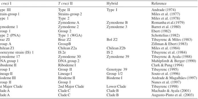
As pointed out previously, T. cruzi is a very polymor-phic species and the overall population structure is far from being completely understood. Until the end of 80’s, the prevalent idea was that T. cruzi strains could not be grouped into few discrete groups that might represent natural taxa (Morel et al. 1980, Tibayrenc et al. 1986). In-stead, a multiclonal population structure was proposed, with the different clones evolving from a very ancient ancestral, basically by clonal reproduction (Tibayrenc et al. 1986, Zhang et al. 1988). However the identification of a strong correlation between different DNA markers such as mini-exon, D7-24S rDNA, RAPD and microsatellites generated a consensus about the existence of two major phylogenetic lineages within the taxon (Tibayrenc 1995, Souto et al. 1996, Nunes et al. 1997, Zingales et al. 1998). The basic dichotomy of the species was further corre-lated with a plethora of epidemiological, biochemical, bio-logical and molecular markers (Fernandes et al. 1998, Oliveira et al. 1998, Macedo et al. 2001, Henriksson et al.
2002, reviewed by Momen 1999, Zingales et al. 1999, and Buscaglia & Di Noia 2003), but the nomenclature of the major groups became very confusing. In 1999, in an effort to homogenize the nomenclature, the two major lineages were renamed T. cruzi I and T. cruzi II (Satellite Meeting 1999, Table III). At the time, putative hybrid strains such as those belonging to D7- rDNA group 1/2 (Souto et al. 1996), clonet 39 (Tibayrenc 1995) and Z3 could not be unequivocal classified as T. cruzi I or T. cruzi II. Thus it was established that these strains should remain identi-fied as T. cruzi without any other specification, until new insights could clarify the phylogenetic relationship of the group (Satellite Meeting 1999).
IS T. CRUZI A SINGLE SPECIES?
One important question that arises is the significance of the division of T. cruzi into discrete major lines. Should be T. cruzi I and T. cruzi II considerate as separated sub-species or even different sub-species?
The modern concept of a species is that of a popula-tion that is simultaneously a reproductive community, an ecological unit and a genetic unit. Obviously, basically clonal asexual species such as T. cruzi cannot be defined in reproductive terms and thus the concept of genetic unity, in the sense of a shared genome, becomes critical. The question here is how much intraspecific genomic vari-ability can be allowed before westart splitting the species into different ones. In attending this question Briones et al. (1999) analyzed the rDNA cistron and found that T. cruzi I and II constitute monophyletic groups with a ge-netic distance compatible with a coalescence time of 88 million years before present. This genetic distance is com-parable to that separating different Leishmania species. They proposed also that T. cruzi emerged as a species over 150 million years ago and that the major groups re-mained separated from each other during the insulation of South America (from 100 to 38 MYR ago), thus favor-ing their cryptic speciation. Accordfavor-ing to this view, T. cruzi I was autochthonous to South America and co-evolved with marsupials and edentates, whereas T. cruzi
II co-evolved in North American placental mammals (Briones et al. 1999).
II is apparently more associated with primates and par-ticularly with human infections while T. cruzi I, which seems to be more adapted to marsupials, is only occa-sionally isolated from humans and even then in regions where T. cruzi is enzootic. Until now all parasites isolated from soropositive individuals from endemic regions in Brazil belong to T. cruzi II major lineage. (Fernandes et al. 1998, 1999, Zingales et al. 1998, reviewed by Zingales et al. 1999). Recent unpublished results obtained in our labo-ratory have shown that all parasites typed directly from affected heart and/or esophagus tissues of Brazilian pa-tients with clinical forms of the disease are from the T. cruzi II lineage (J Freitas, AM Macedo, manuscript in prepa-ration).
THE ORIGIN OF THE NATURAL HYBRID STRAINS
The occurrence of genetic exchange with homologous recombination has just recently been demonstrated for T. cruzi (Stothard et al. 1999, Gaunt et al. 2003). However, in the last two decades evidence for some kind of genetic exchange in T. cruzi was brought to light (reviewed by Tait 1980, Gibson & Stevens 1999). Most of the evidence was based on the identification of putative natural hybrid strains by isoenzyme analyses (Bogliolo et al. 1996, Higo et al. 2000), RAPD (Carrasco et al. 1996), karyotyping and/ or gene sequencing (Sturn et al. 2003).
What is the origin of the natural hybrid populations detected in T. cruzi? An interesting observation is that most of them, if not all, (see below) correspond to those strains that could not be appropriately classified into T. cruzi I or T. cruzi II major lines (satellite Meeting 1999, Momen 1999). The list of such unclassifieable T. cruzi
populations includes strains presenting both alleles for the locus D7 24Sα rDNA (group 1/2 - Souto et al. 1996), strains from clonet 39 (Tibayrenc 1995, Tibayrenc & Ayala 1998), strains belonging to zymodeme Z3 (Fernandes et al. 1998, 1999) and strains showing discrepant alleles for different loci, for example, D7 type 2 miniexon type 1 (Souto et al. 1996). There were no putative hybrids among the strains clearly classified as T. cruzi I or T. cruzi II, except for CL Brener. Another possibly hybrid is the Tulauhén strain. Here, the complication is that there is more than one strain circulating among different laboratories with the same name; some of them are genuinely T. cruzi I, but others are T. cruzi II or hybrids (Souto et al. 1996, Brisse et al. 2001, Augusto-Pinto et al. 2003). CL Brener, the clone chosen for the T. cruzi genome project, was initially clas-sified as T. cruzi II, on the bases of D7 and mini-exon gene typing (Souto et al. 1996). However, several lines of evi-dence have demonstrated that CL Brener is in fact a hy-brid strain presenting characteristics from both major lines and being genetically much more closer to those strains belonging to D7 rDNA group 1/2 than to T. cruzi II (Brisse et al. 2001, Pedroso et al. 2003, Augusto et al. 2003).
More recently there was an effort to classify the hy-brid strains into the T. cruzi I or T. cruzi II major lineages. However this turned out to be a more difficult task than initially expected, since depending on the polymorphic marker used, the hybrid strains seemed to be closer re-lated to one or other major lineage. As an example we can cite the strains belonging to the D7 group 1/2 (Souto et al.
1996). As mentioned before, studies on RAPD and mini-exon profiling showed that these strains seemed to be genetically closer to T. cruzi II than T. cruzi I. However, sequence analyses of the alleles of D7 or 18S rDNAs put the 1/2 strains together with T. cruzi I or T. cruzi II de-pending on the algorithm used (Zingales et al.1999, Kawashita et al. 2001). In order to better accommodate the hybrid strains within T. cruzi II, Brisse et al. (2000, 2001) proposed a subdivision of this major lineage into five lower subgroups (named IIa, IIb, IIc, IId, and IIe). However this strategy did not take into account the fact that depending on the characteristic analyzed the hybrids seem to be more related to the T. cruzi I or even very different from both major lines (Souto et al. 1996, Zingales et al. 1999, Machado & Ayala 2001, 2002, Augusto-Pinto et al. 2003). An alter-native viewpoint now emerging is that the hybrid strains could constitute a third major lineage on T. cruzi taxon, maybe the T. cruzi III lineage (Machado & Ayala 2001, 2002, Augusto-Pinto et al. manuscript in preparation).
Gaunt et al. (2003) have shown evidence that the hy-bridization of T. cruzi strains occurs only with nuclear genome without mitochondrial fusion, so if the natural hybrid strains have the same evolutionary lineage the hybrids will also have the same type of mitochondrial DNA. Machado and Ayala (2001, 2002) demonstrated that the mitochondrial clade B is constituted by at least two Z3 strains and other hybrid strains from isoenzyme type 39 and 43. Recently, Augusto-Pinto et al. (manuscript in preparation) showed that all strains presenting hybrid molecular markers in their nuclear genomes have a spe-cific mitochondrial genome distinct from both the T. cruzi
I and T. cruzi II major lineages. These results suggest that the hybrid T. cruzi strains might represent a truly diverg-ing phylogenetic lineage of T. cruzi, conceivably origi-nating from an ancient recombination event between T. cruzi I and T. cruzi II strains, rather than from multiple recent hybridization episodes.
CLONAL POPULATION STRUCTURE VERSUS SEXUAL REPRODUCTION IN T. CRUZI
Analyses of several genetic loci among different para-site populations have demonstrated that T. cruzi popula-tions present drastic departures from Hardy-Weinberg expectations and strong linkage disequilibrium (reviewed in Ayala 1993). These findings have been interpreted as suggestive that T. cruzi has a clonal population structure in which sexual reproduction is rare or absent. In this perspective each clone represents an independent lineage that divides by binary division and evolves by mutation only from an ancient ancestral (Tibayrenc et al. 1986, Zhang et a. 1988).
as great as 54% in degenerate sites (third positions in codons, where any of the four bases can be found in the DNA without changing the amino acid in the correspon-dent protein). In T. cruzi however, microsatellite data show an excess of homozygosity (Oliveira et al. 1998). Indeed, a recentstudy on gene sequencing did not detect any sig-nificant divergence of the two alleles in strains belonging to T. cruzi I or T. cruzi II lineages (Machado & Ayala 2001). Contrasting with the asexual rotifers, allele diver-gence in T. cruzi was only observed in nuclear genes of some strains presenting hybrid isoenzyme specimens and was better attributed to hybridization events rather than a signal of ancient asexuality. Therefore it has been pro-posed that one or more of four different possible mecha-nisms, automixing, sexuality (gene exchange), gene con-version and mitotic recombination, could be involved in decreasing the T. cruzi allele divergence (Machado & Ayala 2001).
Until now there is no evidence for automixing in T. cruzi. Conversely, Gaunt et al. (2003), have demonstrated that genetic exchange can occur in the parasite. Working with two strains of T. cruzi I carrying different drug-resis-tance markers, they were able to recover six double-drug-resistant progeny clones from the mammalian stage of the life cycle. The hybrids showed a fusion of parental geno-types with loss of alleles and homologous recombina-tion. Isoenzyme and karyotype analyses have confirmed that the putative hybrid clones really shared characters from both parental populations.
As point out above, there are many evidences for natu-ral occurrence of hybrid strains in T. cruzi, but in relation to the “Meselson effect”, the hybrid strains do not ex-plain the high homozygosity observed in T. cruzi. Instead
initially the presence of hybrids would be expected to increase the divergence between the alleles in the same locus. Of course, a meaningful question is which is the frequency of the “recombination events” between strains from the T. cruzi I and T. cruzi II lineages, since excess homozygosity could result from either clonality or from aneuploidy with subsequent allele loss (i.e. from hemi-zygosity, which is experimentally indistinguishable from homozygosity). We cannot rule out the possibility that cell fusion followed by allele loss may be a common fea-ture in these strains. In this case T. cruzi would not have a typical asexual reproduction and the absence of the “Meselson effect” could be explained. However, this pos-sibility seems a priori unlikely, and other mechanisms should be entertained.
Homogenizing mechanisms like mitotic recombination or genetic conversion could limit the “Meselson effect”, decreasing the genetic diversity within and between indi-viduals. Schon and Martens (2003) showed that the an-cient asexual ostracod species Darwinula stevensoni
shows very low nucleotide divergence in three nuclear regions. Likelihood permutation tests confirmed the pres-ence of gene conversion in the multi-copy internal tran-scribed spacer sequence, but rejected rare or cryptic forms of sex as a general explanation for the low genetic diver-sity in the organism (Schon & Martens 2003).
The human Y chromosome is another example of ge-netic conversion as homogenizing mechanism (Skaletsky et al. 2003). It presents a male-specific region (MSY), which is a mosaic of heterochromatic sequences and three classes of euchromatic sequences: X-transposed, X-degenerate and ampliconic. There are evidences that the X-degener-ate and ampliconic sequences were transmitted exclusively
TABLE III
Biological and molecular markers used for characterization of Trypanosoma cruzi sub-division a
T. cruzi I T cruzi II Hybrid Reference
Type III Type II Type I Andrade (1974)
Strain-group 1 Strains-group 2 Miles et al. (1977)
Type 1 Type 2 Miles et al. (1978)
Zymodeme A Zymodeme B Romanha et al.(1979)
Zymodeme 1 Zymodeme 2 Zymodeme 3 Barret et al. (1980)
Group 1 Group 2 Ebert (1982)
Type 2 (PNA) Type 1 (WGA) Schottelius (1982)
Braz ZI Braz Z2 Bol Z2 Tibayrenc & Miles (1983)
Group A GroupB Zillman & Ebert (1983)
Chilean Z1 Chilean Z2a Chilean Z2b Miles et al. (1984)
Isoenzyme strain (IS) 1 IS 2e IS 2 Tibayrenc et al. (1984)
Zymodeme 17 Zymodeme 30 Zymodeme 39 Tibayrenc & Ayala (1988)
DNA group 1 DNA group 2 Muhlpfordt & Berger (1990)
Ribodeme II Ribodeme I Clark & Pung (1994)
Group I Group II Genotype 39 Tibayrenc (1995)
Lineage II Lineage I Group 1/2 Souto et al. (1996)
Biodeme III Biodeme II Biodeme I Andrade & Magalhães (1997)
Group II Group I Nunes et al. (1997)
1st Major Clade 2nd Major Clade Lower Clade Tibayrenc (1998)
Clade A Clade C Clade B Machado & Ayala (2001)
Clade A Clade C Clade B Augusto-Pinto et al. (2003)
through the male germline, and neither participated in meiotic crossing over with a homologous counterpart. In the Y chromosome the shutting down of X-Y crossing over during evolution triggered a monotonic decline in gene function. This model is corroborated by the pres-ence, in the MSY’s X-degenerate sequences, of decayed, intron-bearing pseudogenes. However, in the ampliconic regions, which present functional genes, there is an abun-dant Y-Y gene conversion as a homogenizing mechanism that permitted this region to overcome the problem of sexual absence. This process might provide a mechanism for conserving gene functions across evolutionary time in the absence of crossing over (Skaletsky et al. 2003).
It remains to be established if genetic conversion and/ or mitotic recombination are important phenomena for homogenization of gene sequences in T. cruzi. In this aspect, it was shown that T. cruzi cells present atypical high resistance to ionizing radiation (IR), which could be associated with recombination-based DNA repair (Takeda et al. 1986, Amato-Neto et al. 1996).
MECHANISM OF T. CRUZI GENETIC VARIATION?
Another intriguing characteristic of T. cruzi is how it achieves remarkable heterogeneity among the strains and even clones, paradoxically maintaining at same time high levels of individual homozigosity. One possibility that is unlikely, but cannot be discarded, is that the homozigosity is artefactual and in reality represents hemizigosity oc-curring in aneuploid clones. Ignoring this possibility for the moment, and working within the paradigm of a clonal structure for the T. cruzi populations, one could then ex-pect that mutation and DNA repair would play a role.
The function of mismatch repair (MMR) in decreasing the mutation rate is well understood (reviewed by Hsieh 2001). It is clear that the loss of this mechanism plays a determinant role in some types of human cancers (Peltomaki 2003). However, pathogenic microorganisms presenting defective MMR have been isolated in a pro-portion much higher than initially expected and it has been argued that an increased mutation rate could permit greater host adaptability for these organisms (LeClerc et al. 1996). As already discussed above, the sequence of the T. cruzi MSH2 homologue gene identified tree haplogroups:
A (T. cruzi I), B (hybrid strains) and C (T. cruzi II) (Augusto-Pinto et al. 2003). The MMR efficiency was fur-ther investigated among strains belonging to each haplogroup by analyzing two phenotypes: microsatellite instability (in the presence of hydrogen peroxide) and cisplatin resistance. Microsatellite instability was ob-served only for strains belonging to haplogroups B and C, but not in haplogroup A. Furthermore cells from haplogroups B and C were considerably more resistant to cisplatin treatment, a characteristic known to be conferred by deficiency of mismatch repair in eukaryotic cells (Fujieda et al. 1998). Altogether, these data suggest that strains belonging to haplogroups B and C may have de-creased mismatch repair ability at stress conditions and consequently higher mutations rates when compared with those belonging to haplogroup A (Augusto-Pinto et al. 2003). Up to now the mutation rates for each of the T. cruzi
major lineages are unknown, but it is noteworthy that
haplogroups B and C, which apparently have decreased mismatch repair efficiency, are involved in most cases of human infection (Buscaglia & Di Noia 2003).
CLINICAL IMPLICATIONS OF T. CRUZI GENETIC VA-RIABILITY
REFERENCES
Adad SJ, Andrade DCS, Lopes ER 1991. Contribuição ao estudo da anatomia patológica do megaesôfago chagásico. Rev Inst Med Trop São Paulo 33: 443-450.
Alves AM, de Almeida DF, von Kruger WM 1996. Genomic variation in Trypanosoma cruzi clonal cultures. Parasitol Res 82: 410-415.
Amato Neto V, Pasternak J, Matsubara L, Hammerschlak N, Carignani FL 1996. Trials for the use of gamma rays in the prevention of Trypanosoma cruzi transfusion infection. Rev Soc Bras Med Trop 29: 613-614.
Andrade LO, Machado CRS, Chiari E, Pena SDJ, Macedo AM 1999. Genetic variability of Trypanosoma cruzi implica-tions for the pathogenesis of Chagas disease. Mol Bioch Parasitol 100: 163-172.
Andrade SG 1974. Caracterização de cepas de Trypanosoma cruzi isoladas no Recôncavo Baiano. Ver Patol Trop 3: 65-121.
Andrade SG, Magalhães JB 1997. Biodemes and zymodemes of Trypanosoma cruzi strains: correlations with clinical data and experimental pathology. Rev Soc Bras Med Trop 30: 27-35.
Arruda MV, Reinach FC, Colli W, Zingales B 1990. Sequence of 24S ribosomal RNA gene and characterization of a corre-sponding pseudogene from Trypanosoma cruzi. Mol Biochem Parasitol 40: 35-42.
Augusto-Pinto L, Pimenta JR, Teixeira SMR, Fernandes O, Chiari E, Pena SDJ, Macedo AM, Machado CR 2003. Multilocus genotype data provide evidences that hybrid Trypanosoma cruzi strains constitute a third major lineage. Int J Parasitol in press.
Ayala FJ 1993. Trypanosoma and Leishmania have clonal popu-lation structures of epidemiological significance. Biol Res 26: 47-63.
Barret TV, Hoff RH, Mott KE, Miles MA, Godfrey DG, Teixeira R, Almeida de Souza JA, Sherlock IA 1980. Epidemiologi-cal aspects of tree Trypanosoma cruzi: two types of parsite populations presenting distinct characteristics. PAHO Sci-entific Pub 347: 11-21.
Bogliolo AR, Lauria-Pires L, Gibson WC 1996. Polymorphisms in Trypanosoma cruzi: evidence of genetic recombination. Acta Trop 61: 31-40.
Borst P, Greaves D 1986. Discontinuous transcription and an-tigenic variation in trypanosomes. Annu Rev Biochem 55: 701-732.
Brandariz S, Schijman A, Vigliano C, Arteman P, Viotti R, Beldjord C, Levin MJ 1995. Detection of parasite DNA in Chagas’ heart disease. Lancet 346: 1370-1371.
Brener Z 1987. Pathogenesis and immunopathology of chronic Chagas disease. Mem Inst Oswaldo Cruz 82: 205-212. Brener Z 1992. Immune Response and Immunopathology in
Trypanosoma cruzi Infection Chagas Disease (American Trypanosomiasis): its Impact on Transfusion and Clinical Medicine, ISBT Brazil’92, São Paulo, p. 31-47.
Briones MR, Souto RP, Stolf BS, Zingales 1999. The evolution of the two Trypanosoma cruzi subgroups inferred from rRNA genes can be correlated with the interchange of Ameri-can mammalian faunas in the cenozoic and has implica-tions to pathogenicity and host specificity. Mol Biochem Parasotol 104: 219-232.
Brisse S, Dujardin JC, Tibayrenc M 2000. Identification of six Trypanosoma cruzi lineages by sequence-characterised amplified region markers. Mol Biochem Parasitol 111: 95-105.
Brisse S, Verhoef J, Tibayrenc M 2001. Characterisation of large and small subunit rRNA and mini-exon genes further
supports the distinction of six Trypanosoma cruzi lineages. Int J Parasitol 31: 1218-26.
Buscaglia CA, Di Noia JM 2003. Trypanosoma cruzi clonal diversity and the epidemiology of Chagas disease. Mic-robes Infect 5: 419-427.
Carneiro M, Chiari E, Gonçalves AM, Pereira AAS, Morel C, Romanha AJ 1990. Changes in the isoenzyme and kineto-plast DNA patterns of Trypanosoma cruzi strains induced by maintenance in mice. Acta Trop 47: 35-45.
Carrasco HJ, Frame IA, Valente SA, Miles MA 1996. Genetic exchange as a possible source of genomic diversity in syl-vatic populations of Trypanosoma cruzi. Am J Trop Med Hyg 54: 418-424.
Clark CG, Pung OJ 1994. Host specificity of ribosomal DNA variation in sylvatic Trypanosoma cruzi from North America. Mol Biochem Parasitol 66: 175-179.
Deane MP, Sousa MA, Pereira NP, Gonçalves AM, Momen H, Morel C 1984. Trypanosoma cruzi: inoculation schedules and re-isolation methods selected individual strains from doubly infected mice, as demonstrated by schizodeme and zymodeme analyses. J Protozool 31: 276-280.
Devera R, Fernandes O, Coura JR 2003. Should Trypanosoma cruzi be called “cruzi” complex? A review of the parasite diversity and the potential of selecting population after in vitro culturing and mice infection. Mem Inst Oswaldo Cruz 98: 1-12.
Dias JCP 1992. Epidemiology of Chagas disease. In S Wendel, Z Brener, M S Camargo, A Rassi (eds), Chagas Disease (American Trypanosomiasis): its Impact on Transfusion and Clinical Medicine, ISBTBrazil’92, São Paulo, p. 49-80. Dias Neto E, Steindel M, Passos LK, de Souza CP, Rollinson
D, Katz N, Romanha AJ, Pena SD, Simpson AJ 1993. The use of RAPDs for the study of the genetic diversity of Schistosoma mansoni and Trypanosoma cruzi. EXS 67: 339-345.
Dujardin JC, Dujardin JP, Tibayrenc M, Timperman G, De Doncker S, Jacquet D, Arevalo J, Llanos-Cuentas A, Guerra H, Bermudez H 1995. Karyotype plasticity in neotropical Leishmania: an index for measuring genomic distance among L. (V.) peruviana and L. (V.) braziliensis populations. Para-sitology 110: 21-30.
Ebert F 1982. The identication of two main-groups of Trypano-soma cruzi stocks from Brazil by their isoenzyme patterns of isoelectrofocusing. Tropenmed Parasitol 33: 140-146. Fernandes O, Mangia RH, Lisboa CV, Pinho AP, Morel CM,
Zingales B, Campbell DA, Jansen AM 1999. The complex-ity of the sylvatic cycle of Trypanosoma cruzi in Rio de Janeiro state (Brazil) revealed by the non-transcribed spacer of the mini-exon gene. Parasitology 118: 161-166. Fernandes O, Santos SS, Cupolillo E, Mendonça B, Derre R,
Junqueira ACV, Santos LC, Sturm NR, Naiff RD, Barrett TB, Campbell DA, Coura JR 2001. A mini-exon multiplex polymerase chain reaction to distinguish the major groups of Trypanosoma cruzi and T rangeli in the Brazilian Ama-zon. Trans R Soc Trop Med Hyg 95: 97-99.
Fernandes O, Sturm NR, Derre R, Campbell DA 1998. The mini-exon gene: a genetic marker for zymodeme III of Try-panosoma cruzi. Mol Biochem Parasitol 95: 129-133. Fujieda S, Tanaka N, Sunaga H, Noda I, Sugimoto C, Tsuzuki
H, Saito H 1998. Expression of hMSH2 correlates with in vitro chemosensitivity to CDDP cytotoxicity in oral and oropharyngeal carcinoma. Cancer Lett 132: 37-44. Gaunt MW, Yeo M, Frame IA, Stothard JR, Carrasco HJ,
Tay-lor MC, Mena SS, Veazey P, Miles GA, Acosta N, de Arias AR, Miles MA 2003. Mechanism of genetic exchange in American trypanosomes. Nature 421: 936-939.
trypa-nosomatidae. Adv Parasitol 43: 1-46
Henriksson J, Dujardin JC, Barnabe C, Brisse S, Timperman G, Venegas J, Pettersson U, Tibayrenc M, Solari A 2002. Chro-mosomal size variation in Trypanosoma cruzi is mainly progressive and is evolutionarily informative. Parasitol-ogy 124: 277-286.
Henriksson J, Porcel B, Rydaker M, Ruiz A, Sabaj V, Galanti N, Cazzulo JJ, Frasch AC, Pettersson U 1995. Chromo-some specific markers reveal conserved linkage groups in spite of extensive chromosomal size variation in Trypano-soma cruzi. Mol Biochem Parasitol 73: 63-74.
Henriksson J, Solari A, Rydaker M, Sousa OE, Pettersson U 1996. Karyotype variability in Trypanosoma rangeli. Para-sitology 112: 385-391.
Hernandez R, Rios P, Valdes AM, Pinero D R 1990. Primary structure of Trypanosoma cruzi small-subunit RNA coding region: comparison with other trypanosomatids. Mol Biochem Parasitol 41: 207-212.
Higo H, Yanagi T, Matta V, Agatsuma, Cruz-Reyes A, Uyema N, Monroy C, Kanbara H, Tada I 2000. Genetic structure of Trypanosoma cruzi in american continents: special em-phasis on sexual reproduction in Central America. Parasi-tology 121:403-408.
Higuchi ML, Gutierrez PS, Aiello VD, Palomino S, Bocchi E, Kalil J, Bellotti G, Pileggi F 1993. Correlation between Try-panosoma cruzi parasitism and myocardial inflammatory infiltrate in human chronic chagasic myocarditis: light mi-croscopy and immunohistochemical findings. Cardiovasc Pathol 2: 101-105.
Hsieh P 2001. Molecular mechanisms of DNA mismatch repair. Mutat Res 486: 71-87.
Jones EM, Colley DJ, Tostes S, Lopes ER, Vnencak-Jones CL, McCurley TL 1993. Amplification of a Trypanosoma cruzi DNA sequence from inflammatory lesions in human chagasic cardiomyopathy. Am J Trop Med Hyg 48: 348-357. Kalil J, Cunha-Neto E 1996. Autoimmunity in Chagas disease
cardiomyopathy: fulfilling the criteria at last? Parasitol Today 12: 396-399.
Kawashita SY, Sanson GF, Fernandes O, Zingales B, Briones MR 2001. Maximum-likelihood divergence date estimates based on rRNA gene sequences suggest two scenarios of Trypanosoma cruzi intraspecific evolution. Mol Biol Evol 18: 2250-2259.
Köeberle F 1968. Chagas disease and Chagas syndromes: the pathology of American trypanosomiasis. Adv Parasitol 6: 63-116.
Lambrech FL 1965. Biological variations in trypanosomes and their relation to the epidemiology of Chagas’ disease. Rev Inst Med Trop São Paulo 7: 346-352.
LeClerc JE, Li B, Payne WL, Cebula TA 1996. High mutation frequencies among Escherichia coli and Salmonella patho-gens. Science 274: 1208-1211.
Leon W, Foutz DL, Manning J 1978. Sequence arrangement of 16S and 26S rRNA genes in the pathogenic haemoflagellate Leishmania donovani. Nucleic Acids Res 5: 491-504. Lopes ER, Rocha A, Meneses AC, Lopes MAB, Fatureto MC,
Lopes GP, Chapadeiro E 1989. Prevalência de megas em necrópsias realizadas no Triângulo Mineiro no período de 1954 a 1988. Rev Soc Bras Med Trop 22: 211-215. Luquetti AO, Miles MA, Rassi A, de Rezende JM, de Souza
AA, Povoa MM, Rodrigues I 1986. Trypanosoma cruzi: zymodemes associated with acute and chronic Chagas’ dis-ease in central Brazil. Trans R Soc Trop Med Hyg 80: 462-470.
Macedo AM, Martins MS, Chiari E, Pena SDJ 1992. DNA finger-printing of Trypanosoma cruzi: a new tool for
char-acterization of strains and clones. Mol Biochem Parasitol 55: 147-154.
Macedo AM, Pena SDJ 1998. Genetic variability of Trypano-soma cruzi: implications for the pathogenesis of Chagas disease. Parasitol Today 14:119-123.
Macedo AM, Pimenta JR, Aguiar RS, Melo AI, Chiari E, Zingales B, Pena SD, Oliveira RP AM 2001. Usefulness of Microsatellite Typing in Population Genetic Studies of Try-panosoma cruzi. Mem Inst Oswaldo Cruz 96: 407-413. Machado CA, Ayala FJ 2001. Nucleotide sequences provide
evidence of genetic exchange among distantly related lin-eages of Trypanosoma cruzi. Proc Natl Acad Sci USA 98: 7396-7401
Machado CA, Ayala FJ 2002. Sequence variation in the dihydrofolate reductase-thymidylate synthase (DHFR-TS) and trypanothione reductase (TR) genes of Trypanosoma cruzi. Mol Biochem Parasitol 121: 33-47.
Marques-Araújo, Chiari E 1988. Caracterização biológica de clones das cepas Y, CL e MR de Trypanosoma cruzi em camun-dongos C3H isogênicos. Mem Inst Oswaldo Cruz 83: 175-181.
Miles MA, Apt BW, Widmer G, Povoa MM, Schofield CJ 1984. Isoenzyme heterogeneity and numerical taxonomy of Trypanosoma cruzi stocks from Chile. Trans R Soc Trop Med Hyg 78: 526-535.
Miles MA, Lanhan SM, de Souza AA, Povoa DG 1980. Fur-ther enzymic characters of Trypanosoma cruzi and their evaluation for strain identification. Trans R Soc Trop Med Hyg 74: 221-242.
Miles MA, Souza AA, Povoa M, Shaw JJ, Lainson R, Toye PJ 1978. Isozymic heterogeneity of Trypanosoma cruzi in the first autochthonous patients with Chagas’ disease in Ama-zonian Brazil. Nature 272: 819-821.
Miles MA, Toye PJ, Oswald SC, Godfrey DG et al 1977. The identification by isoenzyme patterns of two distinct strain groups of Trypanosoma cruzi, circulating independently in a rural area of Brazil. Trans R Soc Trop Med Hyg 71: 217-225.
Momen H 1999. Taxonomy of Trypanosoma cruzi: a commen-tary on characterization and nomenclature. Mem Inst Oswaldo Cruz 94: 181-184.
Morel C, Chiari E, Camargo EP, Mattei DM, Romanha AJ, Simpson L 1980. Strains and clones of Trypanosoma cruzi can be characterized by pattern of restriction endonuclease products of kinetoplast DNA minicircles. Proc Natl Acad Sci USA 77: 6810-6814.
Muhlpfordt H, Berger J 1990. Characterization and grouping of Trypanosoma cruzi stocks by DNA base-specific fluo-rochromes and discriminate analysis. Parasitol Res 76: 319-325.
Murthy VK, Dibbern KM, Campbell DA 1992. PCR amplifi-cation of mini-exon genes differentiates Trypanosoma cruzi from Trypanosoma rangeli. Mol Cell Probes 6: 237-243. Nunes LR, de Carvalho MR, Buck GA 1997. Trypanosoma
cruzi strains partition into two groups based on the struc-ture and function of the spliced leader RNA and rRNA gene promoters. Mol Biochem Parasitol 86: 211-224.
Oliveira RP 1997. Estudos Genéticos e Evolucionários do Try-panosoma cruzi, PhD Thesis, Universidade Federal de Minas Gerais, Belo Horizonte.
Oliveira RP, Broude NE, Macedo AM, Cantor CR, Smith CL, Pena SDJ 1998. Probing the genetic population structure of Trypanosoma cruzi with polymorphic microsatellites. Proc Natl Acad Sci USA 95: 3776-3780.
Oliveira RP, Melo AIR, Macedo AM, Chiari E, Pena SDJ 1999. The population structure of Trypanosoma cruzi: expanded analysis of 54 strains using eight polymorphic CA-repeat microsatellites. Mem Inst Oswaldo Cruz 94: 65-70. Palacius-Pru E, Carrasco H, Scorza C, Espinoza R 1989.
Ultra-structural characteristics of different stages of human chagasic myocarditis. Am J Trop Med Hyg 41: 29-40. Pedroso A, Cupolillo E, Zingales B 2003. Evaluation of
Trypa-nosoma cruzi hybrid stocks based on chromosomal size variation. Mol Biochem Parasitol 129: 79-90.
Peltomaki P 2003. Role of DNA mismatch repair defects in the pathogenesis of human cancer. J Clin Oncol 21: 1174-1179. Pena, SDJ, Simpson AJG 1996. Sequence-specific gene signa-tures produced by LSSP-PCR. In U Landegren, Labora-tory Protocols for Mutation Detection, Oxford University Press, Oxford, p. 42-47.
Pena SD, Barreto G, Vago AR, De Marco L, Reinach FC, Dias Neto E, Simpson AJ 1994. Sequence-specific “gene signa-tures” can be obtained by PCR with single specific primers at low stringency. Proc Natl Acad Sci USA 91: 1946-1949. Pimenta JR 2002. Análises Filogenéticas Populacionais em T. cruzi a Partir de Estudos de Microssatélites Polimórficos de DNA, PhD Thesis, Universidade Federal de Minas Gerais, Belo Horizonte.
Porcile PE, Santos MR, Souza RT, Verbisck NV, Brandao A, Urmenyi T, Silva R, Rondinelli E, Lorenzi H, Levin MJ, Degrave W, Franco da Silveira J 2003. A refined molecular karyotype for the reference strain of the Trypanosoma cruzi genome project (clone CL Brener) by assignment of chro-mosome markers. Gene 308: 53-65.
Prata A 2001. Clinical and epidemiological aspects of Chagas disease. Lancet Infec Dis 1: 92-100.
Romanha AJ, Pereira da Silva AA, Chiari E, Kilgour, V 1979. Isoenzyme patterns of cultured Trypanosoma cruzi: changes after prolonged subculture. Comp Biochem Physiol 62B: 139-142.
Santos, FR, Pandya, A, Tyler-Smith, C, Pena, SDJ, Schanfield, M, Leonard, WR, Osipova, L, Crawford, MH, Mitchell, RJ 1999. The central Siberian origin for native American chromosomes. Am J Hum Genet 64: 619-628.
Santos MR, Cano MI, Schijman A, Lorenzi H, Vazquez M, Levin MJ, Ramirez JL, Brandão A, Degrave WM, da Silveira JF 1997. The Trypanosoma cruzi genome project: nuclear karyotype and gene mapping of clone CL Brener. Mem Inst Oswaldo Cruz 92: 821-828.
Satellite Meeting 1999. Recommendations from a satellite me-eting. Mem Inst Oswaldo Cruz 94: 429-432.
Schmuñis, GA 1987. Autoimmunity in Chagas’ disease. Mem Inst Oswaldo Cruz 82: 287-310.
Schon I, Martens K 2003. No slave to sex. Proc R Soc Lond B Biol Sci 270: 827-833.
Schottelius J 1982. The identification by lecitins of two strain groups of Trypanosoma cruzi. Z Parasitenk 68: 147-154. Skaletsky H, Kuroda-Kawaguchi T, Minx PJ, Cordum HS,
Hillier L, Brown LG, Repping S, Pyntikova T, Ali J, Bieri T, Chinwalla A, Delehaunty A, Delehaunty K, Du H, Fewell G, Fulton L, Fulton R, Graves T, Hou SF, Latrielle P, Leonard S, Mardis E, Maupin R, McPherson J, Miner T, Nash W, Nguyen C, Ozersky P, Pepin K, Rock S, Rohlfing T, Scott K, Schultz B, Strong C, Tin-Wollam A, Yang SP, Waterston RH, Wilson RK, Rozen S, Page DC 2003. The male-specific region of the human Y chromosome is a mo-saic of discrete sequence classes. Nature 423: 825-837. Souto RP, Zingales B 1993. Sensitive detection and strain
clas-sification of Trypanosoma cruzi by amplification of a ribo-somal RNA sequence. Mol Biochem Parasitol 62: 45-52. Souto RP, Fernandes O, Macedo AM, Campbell, Zingales B
1996. DNA markers define two major phylogenetic lin-eages of Trypanosoma cruzi. Mol Biochem Parasitol 83: 141-152.
Steindel M, Dias Neto E, Menezes C, Romanha A, Simpson AJG 1993. Random amplified polymorphic DNA analysis of Trypanosoma cruzi strains. Mol Biochem Parasitol 60: 71-80.
Stothard J, Frame I, Miles M 1999. Genetic diversity and ge-netic exchange in Trypanosoma cruzi: dual drug-resistant “progeny” from episomal transformants. Mem Inst Os-waldo Cruz 94: 189-193.
Sturm NR, Vargas NS, Westenberger SJ, Zingales B, Campbell DA 2003. Evidence for multiple hybrid groups in Trypano-soma cruzi. Int J Parasitol 33: 269-279.
Tait A 1980. Evidence for diploidy and mating in trypano-somes. Nature 287: 536-538.
Takeda GK, Campos R, Kieffer J, Moreira AA, Amato Neto V, Castilho VL, Pinto PL, Duarte MI 1986. Ação de raios gama sobre formas sanguícolas de Trypanosoma cruzi. Estudo experimental em camundongos. Rev Inst Med Trop Sao Paulo 28: 15-18.
Tanowitz HB 1992. Chagas disease. Clin Microbiol 5: 400-419. Tibayrenc M 1995. Population genetics of parasitic protozoa
and other microorganisms. Adv Parasitol 36: 47-115. Tibayrenc M 1998. Genetic epidemiology of parasitic
proto-zoa and other infectious agents: the need for an integrated approach. Int J Parasitol 28: 85-104.
Tibayrenc M, Ayala FJ 1987. High correlation between isoen-zyme classification and kinetoplast DNA variability in Try-panosoma cruzi. C R Acad Sci III 304: 89-92.
Tibayrenc M, Ayala F 1988. Isoenzyme variability of Trypa-nosoma cruzi, the agent of Chagas’ disease: genetic, taxo-nomic and epidemiological significance. Evolution 42: 277-292.
Tibayrenc M, Miles MA 1983. A genetic comparison of Brazil-ian and BolivBrazil-ian zymodemes of Trypanosoma cruzi. Trans R Soc Trop Med Hyg 77: 76-83.
Tibayrenc M, Echalar L, Dujardin, P Poch O, Desjeux P 1984. The microdistribution of isoenzymic strains of Trypano-soma cruzi in Southern Bolivia: new isoenzyme profiles and futher arguments against Mendelian sexuality. Trans R Soc Trop Med Hyg 78: 519-525.
Tibayrenc M, Neubauer K, Barnabé C, Guerrini F, Skarecky D, Ayala F 1993. Genetic characterization of six parasitic pro-tozoa: parity between random primer DNA typing and multilocus enzyme electrophoresis. Proc Natl Acad Sci USA 90: 1335-1339.
Tibayrenc M, Ward P, Moya A, Ayala FJ 1986. Natural popu-lations of Trypanosoma cruzi, the agent of Chagas disease, have a complex multiclonal structure. Proc Natl Acad Sci USA 83: 115-119.
Vago AR, Andrade LO, Leite AA, d’Avila Reis D, Macedo AM, Adad SJ, Tostes S Jr, Moreira MC, Filho GB, Pena SD 2000. Genetic characterization of Trypanosoma cruzi di-rectly from tissues of patients with chronic Chagas disease: differential distribution of genetic types into diverse or-gans. Am J Hum Pathol 156: 1805-1809.
Vago AR, Macedo AM, Adad SJ, Reis DA, Corrêa-Oliveira R 1996a. PCR detection of Trypanosoma cruzi DNA in esoph-ageal tissues of patients with chronic Chagas disease. Lan-cet 348: 891-892.
Vago AR, Macedo AM, Oliveira RP, Andrade LO, Chiari E, Galvão LMC, Simpson AJG, Tostes S, Pena SDJ 1996b. Kinetoplast DNA signatures of Trypanosoma cruzi strains obtained directly from infected tissues. Am J Pathol 149: 2153-2159.
patológica da moléstia de Chagas. Mem Inst Oswaldo Cruz 3: 276-293.
Welch DM, Meselson M 2000. Evidence for the evolution of Bdelloid rotifers without sexual reproduction or genetic exchange. Science 288: 1211-1215.
Zeledon R, Rabinovich JE 1981. Chagas’ disease: an ecological appraisal with special emphasis on its insect vectors. Annu Rev Entomol 26: 101-33.
Zhang Q, Tibayrenc M, Ayala FJ 1988. Linkage disequilibrium in natural populations of Trypanosoma cruzi (Flagellate), the agent of Chagas’ disease. J Parasito l 35: 81-85. Zillman U, Ebert F 1983. The characterization of Trypanosoma
cruzi stocks by starch gel electrophoresis, comparison of results with those of isoeletric focusing. Tropemmed Parasitol 34: 84-88.
Zingales B, Souto RP, Mangia RH, Lisboa CV, Campbell DA, Coura JR, Jansen A, Fernandes O 1998. Molecular epide-miology of American trypanosomiasis in Brazil based on dimorphisms of rRNA and mini-exon gene sequences. Int J Parasitol 28: 105-112.