Arq Neuropsiquiatr 2002;60(3-B):730-733
CEREBRAL TRYPANOSOMIASIS AND AIDS
Apio Claudio Martins Antunes
1, Felipe Martins de Lima Cecchini
2,
Fernando von Bock Bolli
2, Patricia Polanczyk de Oliveira
2,
Ricardo Gurgel Rebouças
2, Thais Lampert Monte
3, Daniele Fricke
4ABSTRACT - A 36 year-old black female, complaining of headache of one month’s duration presented with nausea, vomiting, somnolence, short memory problems, loss of weight, and no fever history. Smoker, intravenous drugs abuser, promiscuous lifestyle. Physical examination: left homonimous hemianopsia, left hemiparesis, no papilledema, diffuse hyperreflexia, slowness of movements. Brain CT scan: tumor-like lesion in the splenium of the corpus calosum, measuring 3.5 x 1.4 cm, with heterogeneous enhancing pattern, sugesting a primary CNS tumor. Due to the possibility of CNS infection, a lumbar puncture disclosed an opening pressure of 380 mmH20; 11 white cells (lymphocytes); glucose 18 mg/dl (serum glucose 73 mg/dl); proteins 139 mg/dl; presence of Trypanosoma parasites. Serum Elisa-HIV tests turned out to be positive. Treatment with benznidazole dramatically improved clinical and radiographic picture, but the patient died 6 weeks later because of respiratory failure. T. cruzi infection of the CNS is a rare disease, but we have an increasing number of cases in HIV immunecompromised patients. Diagnosis by direct observation of CSF is uncommon, and most of the cases are diagnosed by pathological examination. It is a highly lethal disease, even when properly diagnosed and treated. This article intends to include cerebral trypanosomiasis in the differential diagnosis of intracranial space-occupying lesions, especially in immunecompromised patients from endemic regions.
KEY WORDS: trypanosomiasis, trypanosoma cruzi, AIDS, benznidazole, nifurtimox.
Tripanossomíase cerebral e SIDA
RESUMO - Uma mulher negra de 36 anos, procurou a emergência do hospital com quadro de cefaléia holocraniana há 1 mês. Evoluiu com náuseas e vômitos, sonolência e diminuição da memória, associadas a emagrecimento, sem história de febre. Tabagista, usuária de drogas endovenosas, história de promiscuidade sexual. Exame físico: hemianopsia homômima esquerda, hemiparesia esquerda, sem papiledema, hiperreflexia profunda difusa e lentificação dos movimentos. A TC de crânio mostou lesão expansiva no esplênio do corpo caloso com 3,5 x 1,4 cm, com impregnação heterogênea pelo contraste, sugestiva de tumor primário do SNC. Por causa da possibilidade de neuroinfecção, foi realizada uma punção lombar que revelou pressão de abertura de 380 mmH2O; 11 leucócitos (mononucleares), glicose 18 mg/dl (glicemia 73mg/dl), proteínas 139 mg/dl, presença de parasitas com características de Trypanossoma. Testes para HIV foram reagentes. Iniciado tratamento com benzonidazol, a paciente apresentou melhora neurológica e tomográfica. Faleceu 6 semanas após, por insuficiência respiratória. A infecção por T. cruzi no SNC é entidade rara, observando-se número crescente de casos em pacientes imunocomprometidos por infecção pelo HIV. O diagnóstico por exame direto no líquor é incomum, em sua maioria sendo os casos diagnosticados por exame anátomo-patológico (biópsia ou necrópsia). É doença de alta letalidade, mesmo com diagnóstico e tratamento adequados. É necesário incluir a tripanossomíase cerebral no diagnóstico diferencial das patologias expansivas intracranianas, principalmente em pacientes imunodeprimidos e provenientes de áreas endêmicas.
PALAVRAS-CHAVE: tripanossomíase, tripanosoma cruzi, SIDA, benzonidazol, nifurtimox.
Hospital de Clínicas de Porto Alegre, Porto Alegre RS, Brasil; 1Professor-Adjunto de Neurocirurgia, Doutor, Faculdade de Medicina da
Universidade Federal do Rio Gande do Sul (UFRGS); 2Residente em Neurocirurgia; 3Médica Neurologista, Mestre em Clínica Médica,
UFRGS, 4Residente em Neurologia.
Received 13 March 2002. received in final form 13 April 2002. Accepted 20 April 2002.
Dr. Apio Claudio Martins Antunes - Rua Luciana de Abreu 471/308 - 90570-060 Porto Alegre RS - Brasil. E-mail: [email protected]
The problem of a patient with an intracranial mass
lesion and acquired immunodeficiency syndrome
(AIDS) imposes an enormous list of diagnostic
possi-bilities. The management of this particular situation
is sometimes a real challenge. Cases of patients
pre-viously infected with T. cruzi presenting with
chaga-sic meningoencephalitis have been reported since
1990
1. We present a patient with AIDS and
Arq Neuropsiquiatr 2002;60(3-B) 731
CASE
A 36-year-old black female was admitted complaining of severe headache non-amenable to common analgesics, and progressive left hemiparesis for one month. She also had a history of general malaise, vomitting and significant weight loss (10 kg in two months). She had no allergies and was not using medication on chronic basis, but there was history of smoking and intravenous drug abuse. She had a promiscuous life, with nine pregnancies and one abortion, and had been submitted to blood transfusion. The patient was oriented, but mildly somnolent, vital signs were stable, and she had disseminated hypocromic skin lesions. Neurologic examination disclosed a left homoni-mous hemianopsia, mild left hemiparesis, diffuse hyper-reflexia and normal fundoscopy. Her movements were slow with normal coordination. She was unable to walk with-out someone’s help.
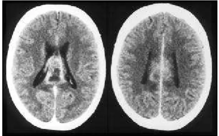
Blood tests showed leukopenia (2730 leukocytes, 6% lymphocytes), hematocrit was 27.42%. HIV tests were per-formed (ELISA). Brain CT disclosed a contrast-enhancing tumor-like lesion in the splenium of the corpus calosum, measuring 3.5 x 1.4 cm, with heterogeneous enhancing pat-tern (Fig 1).
Due to the possibility of central nervous system (CNS) infection, the patient was then submitted to a lumbar puncture. CSF was clear, pressure was 380 mmH2O, and the examination showed 11 white cells/mm3 (lymphocytes)
glucose 18 mg/dL (blood glucose 73 mg/dL), proteins 139 mg/dl and the presence of parasites with the characteristics of Trypanosomatidae family (Fig 2). HIV blood tests were positive, with a CD4 count of 8/mm3. Serologic tests for
Chagas’ disease were all positive. No sign of chronic infec-tion was found. Treatment with benznidazole 5 mg/kg was initiated. The patient showed clinical and radiographic im-provement (Fig 3), and a new lumbar puncture performed 20 days later disclosed clear CSF with no parasites. In spite of the good response to phamacological therapy with benznidazole, the patient died 40 days later due to pul-monary complications.
DISCUSSION
American trypanosomiasis affects 16-18 million
people and some 100 million, i.e. about 25% of the
population of Latin America, is at risk of acquiring
Chagas’ disease
2. The disease exists only in the
Ame-rican Continent, and is caused by Trypanosoma cruzi,
a flagellated protozoan parasite, transmitted to
hu-mans by haematophagous triatomine insects.
Trans-mission of T. cruzi may also occur through blood
trans-fusion, especially in individuals who receive repeated
transfusions
3.
In the acute phase, the disease is usually silent.
Only 35% of patients show symptoms
4like: fever,
general malaise, headache, edema,
lymphadeno-pathy and hepatosplenomegaly
5. Myocarditis,
mos-Fig 1. CT scan: lesion in the splenium of the corpus calosum with heterogenous enhancing pattern.
Fig 2. CSF examination: T. cruzi parasites indentified on direct examination.
732 Arq Neuropsiquiatr 2002;60(3-B)
tly seen as an EKG alteration, may happen.
Someti-mes the clinical presentation takes the form of a
me-ningoencephalitis, a severe condition that occurs
spe-cially in infants and children younger than three
years-old, commonly associated with acute myocarditis.
Al-though CNS involvement is rare, the presence of the
parasite in the CNS has been proved in 1953 by
Fre-itas et al.
6. Hoff et al.
7showed that 72% of patients
investigated in the acute phase had T. cruzi in the CSF.
The chronic phase has three main presentations:
(a) indeterminate, (b) cardiac and (c) digestive
8. The
indeterminate type is the most common one: the
patient has positive test for Chagas’ disease but is
asymptomatic. Progressive deterioration of the
myo-cardium leading to cardiomegaly and cardiac failure
is the hallmark of the cardiac form and involvement
of the bowel and esophagus is what characterizes
the digestive form (megacolon and megaesophagus)
and is caused by a loss of ganglion cells of the
auto-nomic nervous system in the gut
9.
The acquired immunodeficiency syndrome has
changed the profile of many diseases in the past two
decades and Chagas’ disease is no exception to the
rule. Until 1969, reactivation of chronic Chagas’
dise-ase had been recorded only experimentally, when a
case of a patient with lymphatic leukemia was
repor-ted
10afterwards. Reactivation due to AIDS has been
reported since 1990
1. The disease tends to present
as a severe form of meningoencephalitis,
myocardi-tis, or both: it is a highly lethal disease and only a
few cases of cure have been reported.
Chagasic meningoencephalitis may be the first
presentation of a patient with AIDS. Some patients
are found to have low CD4 counts, as demonstrated
by Pagano et al. in the analysis of 8 cases (CD4 counts
ranging from 126 to 63 cell/mm3)11 . Imaging of
patients with chagasic meningoencephalitis usually
presents as a cerebral mass, often called cerebral
tumor-like American Trypanosomiasis. Ring and
no-dular enhancing patterns are the most frequent
non-specific findings, and a differential diagnosis with
toxoplasmosis or lymphoma are highly suggested.
The lesions appear mostly in the supratentorial white
matter
9, but masses have been reported everywhere
in the CNS, including infratentorial region and basal
ganglia. The serological tests for Chagas’ disease are
usually positive, but may be negative and
insuffi-cient for the diagnosis, which is made by
identifica-tion of the parasite in the CSF or more frequently by
histological examination of the brain tissue. There
are no sufficient data to evaluate the sensitivity of
the CSF examination. When the disease is strongly
suspected on clinical grounds and when
appropri-ate tests fail to stablish the etiology, a brain biopsy
shoud be performed without delay.
There is usually an inflamatory process and
neu-ronal damage
10. Microglial nodules and, less
frequen-tly, perivascular mononuclear cuffings are the main
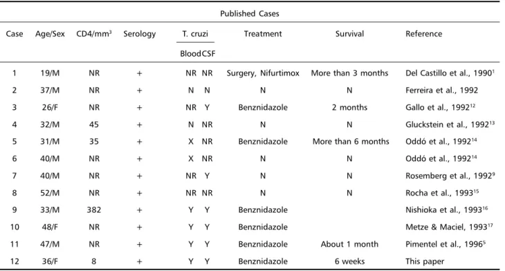
Table 1. Published cases of Trypanosoma cruzi meningoencephalitis in AIDS patients. Modified from Veronesi8.
Published Cases
Case Age/Sex CD4/mm3 Serology T. cruzi Treatment Survival Reference
BloodCSF
1 19/M NR + NR NR Surgery, Nifurtimox More than 3 months Del Castillo et al., 19901
2 37/M NR + N N N N Ferreira et al., 1992
3 26/F NR + NR Y Benznidazole 2 months Gallo et al., 199212
4 32/M 45 + N NR N N Gluckstein et al., 199213
5 31/M 35 + X NR Benznidazole More than 6 months Oddó et al., 199214
6 40/M NR + X NR N N Oddó et al., 199214
7 40/M NR + NR Y N N Rosemberg et al., 19929
8 52/M NR + NR NR N N Rocha et al., 199315
9 33/M 382 + Y Y Benznidazole Nishioka et al., 199316
10 48/F NR + Y Y Benznidazole Metze & Maciel, 199317
11 47/M NR + Y Y Benznidazole About 1 month Pimentel et al., 19965
Arq Neuropsiquiatr 2002;60(3-B) 733
pathologic findings. In several cases the lesions have
a necrotic character. Parasites are seen either in glial
cells, macrophages and neurons or may be free in
the parenchyma, where they tend to form
pseudo-cysts. Observation of the coexistence of nuclei and
kinetoplasts shows the presence of the intracellular
amastigote form of the prasasite.
Reactivation of chronic Chagas’ disease due to
AIDS is uncommon and there have been a few
re-ports since 1990 (Table 1).
Specific treatment has been based on three
op-tions: benznidazole (5 mg/kg/day for 60 days), or
nifurtimox (8 to 10 mg/kg/day for 120 days in four
divided doses; 15 to 20 mg/kg/day in four divided
doses for children 1 to 10 years old). Nifurtimox is
effective in treatment of acute Chagas’ disease,
re-ducing its severity, but ineffective in the chronic
stages of the disease
18. When therapy is complete,
over 80% of patients are cured.
In conclusion, cerebral trypanosomiasis must be
included in the diferential diagnosis of the
intra-cranial lesions in an immunosupressed patient,
espe-cially if the patient comes from an endemic area.
REFERENCES
1. Del Castillo M, Mendoza G, Oviedo J, et al. AIDS and Chagas’ disease with central nervous system tumor-like lesion. Am J Med 1990;88:693-694. 2. World Health Organization. Control of Chagas’ disease: report of the WHO Expert Committee. Weekly Epidemiological Record, No. 1/2, 3/10 January 1997.
3. Leiguarda R, Roncoroni A, Taratuto AL, et al. Acute CNS infection by Trypanosoma cruzi (Chagas’ disease) in immunosuppressed patients. Neurology 1990;40:850-851.
4. Teixeira, MGL. C. Doença de Chagas: estudos da forma aguda inaparente. Tese, Universidade Federal do Rio de Janeiro, 1977.
5. Pimentel PCA, Handfas BW, Carmignani M. Trypanosoma cruzi men-ingoencephalitis in AIDS mimicking cerebral metastase. Arq Neuropsiquiatr 1996;54:102-106.
6. Pedreira de Freitas JL, Lion MF, Tartari JTA. Resultados de umm investigação sobre moléstia de Chagas realizada no município de Marília e outros, com estudo de dois casos agudos da doença. Rev Hosp Clin São Paulo 1953;8:81-92
7. Hoff R, Teixeira RS, Carvalho JS, Mott KE. Trypanosoma cruzi in the cerebrospinal fluid during the acute stage of Chagas’s disease. N Engl J Med 1978; 298: 604-606.
8. Ferreira MS, Lopes ER, Chapadeiro E, Dias JCP, Ostermayer AL. Doença de Chagas. In Veronesi R., Focaccia R (eds). Tratado de Infectologia. São Paulo: Atheneu, 1996:1175-1213.
9. Rosemberg S, Chaves CJ, Higushi ML, et al. Fatal meningoencephalitis cause by a reactivation of Trypanosoma cruzi infection in a patient with Sida. Neurology, 1992;42:640-642.
10. Spina-França A, Mattosinho-França LC. American trypanosomiasis (Chagas’ disease). In Vinken PJ, Bruyn GW (eds). Handbook of clinical neurology, vol 35: infections of the nervous system. Amsterdam: Elsevier/North Holland, 1978:85-114.
11. Pagano MA, Segura MJ, Di Lorenzo GA, et al. Cerebral tumor-like American trypanosomiasis in acquired immunodeficiency syndrome. Ann Neurol 1999;45:403-6.
12. Tracy JW, Webster LT,. Drugs used in the chemotherapy of protozoal infections: trypanosomiasis, leishmaniasis, amebiasis, giardiasis, tri-chomoniasis and other protozoal infections. In Gilman GA. Goodman & Gilman’s the pharmacological basis of therapeutics. 9.Ed, New York: McGraw-Hill,1996:998-999
13. Gallo P, Fabião OM Neto, Suarez JMM, Borba RP. Acute central ner-vous system infection by Trypanosoma cruzi and AIDS. Arq Neuropsiquiatr 1992;50:375-377.
14. Gluckstein D, Ciferri F, Ruskin J. Chagas’ disease: another cause of cerebral mass in the acquired immunodeficiency syndrome. Am J Med 1992;92:429-432.
15. Oddó D, Casanova M, Acuña G, et al. Acute Chagas disease (Trypa-nosomiasis americana) in acquired immunodeficiency syndrome: re-port of two cases. Human Pathol 1992;23:41-44.
16. Rocha A, Ferreira MS, Nishikosha SA, et al. Trypanosoma cruzi men-ingoencephalitis and myocarditis in a patient with acquired immuno-deficiency syndrome. Rev Inst Med Trop São Paulo 1993;35:205-208. 17. Nishioka AS, Ferreira MS, Rocha A, et al. Reactivation of Chagas
dis-ease successfully treated with benznidazole in a patient with acquired immunodeficiency syndrome. Memórias do Instituto Oswaldo Cruz 1993;88:493-496.