www.microbialcell.com
ABSTRACT
The baker’s yeast Saccharomyces cerevisiae has been extensively
explored for our understanding of fundamental cell biology processes highly
conserved in the eukaryotic kingdom. In this context, they have proven
inval-uable in the study of complex mechanisms such as those involved in a variety
of human disorders. Here, we first provide a brief historical perspective on
the emergence of yeast as an experimental model and on how the field
evolved to exploit the potential of the model for tackling the intricacies of
various human diseases. In particular, we focus on existing yeast models of
the molecular underpinnings of Parkinson’s disease (PD), focusing primarily
on the central role of protein quality control systems. Finally, we compile and
discuss the major discoveries derived from these studies, highlighting their
far-reaching impact on the elucidation of PD-associated mechanisms as well
as in the identification of candidate therapeutic targets and compounds with
therapeutic potential.
From the baker to the bedside: yeast models of
Parkinson's disease
Regina Menezes
1,2, Sandra Tenreiro
3,5, Diana Macedo
2, Cláudia N. Santos
1,2, Tiago Fleming Outeiro
4,5,6,*
1Instituto de Biologia Experimental e Tecnológica, Apartado 12, Oeiras 2781-901, Portugal.
2
Instituto de Tecnologia Química e Biológica António Xavier, Av. da República, 2780-157 Oeiras, Universidade Nova de Lisboa, Portu-gal.
3
Instituto de Medicina Molecular, Av. Prof. Egas Moniz, Lisboa 1649-028, Portugal.
4
Instituto de Fisiologia, Faculdade de Medicina da Universidade de Lisboa, Lisboa 1649-028, Portugal.
5
CEDOC – Chronic Diseases Research Center, Faculdade de Ciências Médicas, Universidade Nova de Lisboa, Campo dos Mártires da Pátria, 130, Lisboa 1169-056, Portugal.
6
Department of NeuroDegeneration and Restorative Research, University Medical Center Göttingen, Waldweg 33, Göttingen 37073, Germany.
* Corresponding Author: Prof. Dr. Tiago Fleming Outeiro, Department of NeuroDegeneration and Restorative Research, University Medical Center Gottingen, Waldweg 33; 37073 Gottingen, Germany; Tel: +49 551 39 13544; Fax: +49 551 39 22693; E-mail: [email protected]
INTRODUCTION
The unicellular eukaryote
Saccharomyces cerevisiae has
been extensively used as an industrial microorganism. The
first records indicating its use in fermentation processes to
produce alcoholic beverages and to leaven bread date back
to ancient Egypt, over 5,000 years ago [1, 2]. Ever since, S.
cerevisiae has been used for making bread, being therefore
also frequently referred as baker’s yeast.
The first uses of S. cerevisiae as an experimental model
organism date back to the mid-thirties of the 20
thcentury
[3], and its consequent establishment as a robust model
system in diverse areas of biology was largely fueled by its
unique features. They include short generation time, easy
handling which is further simplified by its nonpathogenic
nature, inexpensive culture conditions, and, most
im-portantly, its amenability for genetic manipulation. Being
very versatile for biological and genetic studies, these
at-tributes placed yeast at the forefront for the development
of countless genetic tools to address major biological
is-sues. Hence, for almost a century, S. cerevisiae has served
as a remarkable experimental model for several seminal
discoveries in biology, revealing important aspects of
mi-crobiology and biochemistry. For example, studies using
yeast have contributed to the elucidation of fundamental
cellular mechanisms involved in DNA replication,
recombi-nation and repair [4], in RNA metabolism [5], and in cell
doi: 10.15698/mic2015.08.219 Received originally: 19.05.2015; in revised form: 10.06.2015, Accepted 11.06.2015 Published 27.07.2015.
Keywords: protein misfolding, neurodegeneration, alpha-synuclein, Parkinson’s disease, synucleinopathies.
Abbreviations:
BiFC – bimolecular fluorescence complementation,
CPs- cyclic peptides, ER - endoplasmic reticulum, FRAP - fluorescence recovery after photobleaching,
MYTH - membrane yeast two-hybrid, PD - Parkinson’s disease,
division and cell cycle progression [6].
The discovery of the high-degree of evolutionary
con-servation of disease genes and of fundamental biological
processes among eukaryotes, combined with the power of
yeast genetics, has brought S. cerevisiae from the baker to
the bedside, as a model organism with an unprecedented
potential to decipher the intricacies of devastating human
pathologies such as Parkinson’s disease (PD), as well as to
help in the identification of molecular targets and lead
molecules with therapeutic potential.
In this review, we summarize the impact of yeast
mod-els on the current knowledge and understanding of the
molecular underpinnings of PD. We first discuss the most
relevant findings in the yeast field and the extent to which
they have paved the way for the use of S. cerevisiae in
bi-omedical research. We then briefly review the main
as-pects of PD, emphasizing the molecular players and
path-ways governing disease pathology. Finally, we cover the
most important yeast models generated thus far, and
dis-cuss their contribution to the elucidation of PD-related
mechanisms, as well as to the identification of molecular
targets and compounds with therapeutic potential.
THE POWER OF YEAST GENETICS
The peculiar life cycle of S. cerevisiae constitutes, in itself,
an invitation for performing genetic studies. In the wild, it
can be found in both haploid and diploid forms that
repro-duce vegetatively, by budding. In nutrient-poor
environ-ments, a condition easily mimicked experimentally, diploid
cells undergo meiosis and sporulate, yielding a progeny of
four haploid cells. Hence, under controlled laboratory
con-ditions, sporulation of a particular diploid cell allows the
generation of different combinations of genotypes with
desired genetic traits. Additionally, the life cycle of budding
yeast also greatly simplifies the study of lethal mutations in
heterozygous diploids as well as recessive mutations in
haploid cells [7, 8].
Yeast research has definitely won a place in history
af-ter the demonstration that yeast strains with a mutation in
LEU2 locus, therefore unable to grow in media depleted of
the amino acid leucine, can be transformed with a chimeric
ColE1 plasmid encoding the wild type (WT) yeast
LEU2
gene, and that this sequence can integrate into the yeast
chromosome restoring leucine prototrophy [9]. The
dis-covery of the amenability of yeast cells for transformation
opened new avenues for manipulation of yeast genome,
allowing insertion or deletion of genes to generate
recom-binant strains. The high efficiency of the transformation
process, aided by a very effective homologous
recombina-tion system, has provided yeast geneticists a tremendous
flexibility in experimental design, which is currently
incre-mented by the availability of a large collection of
recombi-nation-based Gateway vectors [7, 10-12].
S. cerevisiae was also the host organism for the
devel-opment of pioneering approaches to investigate the
inter-action between biomolecules. Taking advantage of the
bi-modular nature of the yeast transcription factor Gal4,
re-searchers generated a novel genetic system to study
pro-tein-protein interactions in which two known proteins are
separately fused to the DNA-binding and transcriptional
activation domains of Gal4 [13]. The principle of the
meth-odology relies on the premise that the interaction between
proteins reconstitutes a functional Gal4, which in turn
acti-vates expression of reporter genes. After its original
de-scription, a number of “variations on the theme” has been
described to allow the study of DNA-protein (one-hybrid),
RNA-protein (RNA-based three-hybrid) and small
molecule-protein interactions (ligand-based three-hybrid), as well as
to identify mutations, peptides or small molecules that
dissociate macromolecular interactions – the reverse
n-hybrid systems [14]. Additionally, a split-ubiquitin
mem-brane-based two-hybrid assay (also referred as membrane
yeast two-hybrid - MYTH) was designed to overcome the
limitation of the original system on the assessment of
pro-tein interactions forced to occur within the nucleus, for
membrane-embedded proteins [15, 16]. The fact that
nov-el hits for a target protein, from both yeast and other
or-ganisms, can be identified in the screening of libraries,
without any prior bias or knowledge of their identity, is the
most powerful application of these techniques.
Another tool allowing the study of protein interactions
in vivo is the bimolecular fluorescence complementation
(BiFC). This method, originally developed in mammalian
cells [17, 18], has been efficiently used in yeast to visualize
protein interactions with minimal perturbation of the
nor-mal cellular environment [19, 20]. It is based on the
princi-ple that two fragments of a fluorescent protein are each
fused to target proteins. The reassembly of these
non-fluorescent fragments into a non-fluorescent complex is
medi-ated by the interaction between the target proteins,
thereby constituting a powerful tool to resolve spatial and
temporal aspects of many molecular interactions [21].
The genetic features highlighted have distinguished
yeast as a versatile model organism. As such, S. cerevisiae
was the first eukaryotic organism to have its genome fully
sequenced [22]. Thus, the completion of the yeast genome
in 1996 represented a landmark achievement in the history
of eukaryotic biology. This has been providing, in the
course of the last two decades, a wealth of information
allowing the development of several biological resources
such as the yeast gene deletion strains [23], the
tetracy-cline (tet)-repressible [24] and heat-inducible shutoff set of
strains [25] to generate conditional mutants of essential
genes, the GFP- [26] and TAP-tagged [27] collection of
strains, collections of other protein tags, and collections
engineered for protein overexpression [28-30]. A
compen-dium of existing tools and resources available for the yeast
research community is provided by Tenreiro and Outeiro
[31] and by Duina and coauthors [7].
scrutinize the huge amount of data generated towards
building a comprehensive model of eukaryotic cell
func-tioning [7, 8, 32]. Compilations of genetic and biological
data from these analyses, including information regarding
predicted orthologues in humans, are easily accessible at
the comprehensively annotated
Saccharomyces Genome
Database online resource (http://www.yeastgenome.org/)
[33] and other public databases [7, 31, 34].
The power of yeast genetics has fueled all the
achievements discussed, rendering
S. cerevisiae as the
best-understood eukaryotic organism. A surprising, but
delightful finding, emerging from the
cumulative
knowledge on yeast genomics and biology, was the
unpre-dictable gene homology and functional conservation of key
fundamental cellular processes between yeasts and higher
eukaryotes [4, 5, 8].
Importantly, the
S. cerevisiae genome encodes nearly
1000 genes which are members of orthologous gene
fami-lies related to human disease, representing about 20% of
the total yeast genes [35]. The mammalian orthologues of
most of these genes are functional in yeast and
comple-ment the respective yeast deletion mutant. In line with the
evolutionary conservation of disease genes in eukaryotes,
it has been extensively shown that several
disease-associated cellular pathways are also highly conserved
from yeast to humans [11, 34, 36], enabling the modeling
of specific disease aspects in this model organism. Protein
quality control systems [37], vesicular trafficking and
secre-tion [38], autophagic pathways [39], the unfolded protein
response [40], and mitochondrial biogenesis and
metabo-lism [41] are among the conserved cellular mechanisms,
allowing the study of fundamental mechanisms associated
with neurodegenerative diseases, such as PD, in yeast cell
models [11].
Modeling particular molecular aspects of human
dis-eases in yeast models can be achieved using distinct
strat-egies, depending on the presence or absence, of
disease-genes orthologues in the yeast genome [42]. If the disease-genes of
interest have yeast counterparts, a unique opportunity to
directly study their function is offered, either through their
deletion or overexpression. In a more physiological context,
human wild type (WT) and mutant alleles of favorite genes
can be heterologously expressed in the respective yeast
mutant backgrounds, since they are capable of replacing
the function of the endogenous yeast gene product.
Oth-erwise, if the disease-associated genes do not have a yeast
orthologue, a functional analysis can still be conveniently
performed via heterologous expression in WT strains [31,
43] to provide paradigms of their function on cellular
phys-iology and metabolism. Insights into the function of most
PD-associated genes have been obtained using the latter
strategy. However, the study of endogenous yeast proteins
has been also providing clues on the role of key PD players,
as discussed in detail bellow.
Once comprehensively validated as reliable
experi-mental model systems to recapitulate specific aspects of
human diseases, “humanized” yeasts constitute powerful
toolboxes for high-throughput screenings of genes that
may constitute therapeutic targets, and as robust primary
drug-screening platforms to filter for cytoprotective
com-pounds [31, 44].
Despite several singularities and idiosyncrasies of yeast
cells, their simplicity can be turned from a limitation into
an asset, by virtue of all the attributes mentioned above.
Thus, S. cerevisiae is uniquely suited for the task of
assist-ing our understatassist-ing of the cellular mechanisms underlyassist-ing
human diseases as well as in the search for novel
molecu-lar targets and compounds prone for therapeutic
interven-tion.
PARKINSON'S DISEASE AND THE KEY GENETIC PLAYERS
PD, described by James Parkinson in 1817 [45] and then
further refined by Jean-Martin Charcot [46], is one of the
most common neurodegenerative disorders. Currently, it is
estimated that there are 4 million diagnosed PD patients
worldwide. However, it is estimated that 7 to 10 million
people live with this devastating chronic disease (data from
the Parkinson’s Disease Foundation). The typical motor
symptoms of PD include tremor at rest, bradykinesia,
stiff-ness, and postural instability [47]. These symptoms are
caused by the progressive degeneration of nigrostriatal
dopaminergic neurons from the
substantia nigra pars
compacta of the brain. Nevertheless, PD is currently
con-sidered as a whole-brain disorder, affecting multiple brain
areas and presenting a broad variety of symptoms [48].
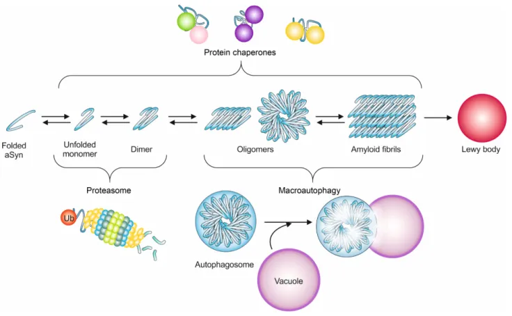
The histopathological hallmark of PD, and other
synucle-inopathies, is the appearance of proteinaceous
intraneu-ronal cytoplasmic inclusions termed as Lewy bodies (LB)
and Lewy neurites [49]. These insoluble aggregates are
predominantly composed of alpha-synuclein (aSyn) [49], a
protein encoded by the SNCA gene, and are decorated with
components of protein quality control systems such as
molecular chaperones, and proteasomal and lysosomal
subunits [50].
The SNCA gene was the first genetic locus to be
associ-ated with familial forms of PD, and was later also
implicat-ed in sporadic cases of the disease [51]. It encodes aSyn, a
protein whose function remains unclear, but that has been
proposed to be linked to diverse functions ranging from
transcriptional regulation [52-57], mitochondrial
homeo-stasis [58] and vesicle trafficking [59-61], possibly
regulat-ing dopamine neurotransmission, synaptic function and
synaptic plasticity [62-65].
Genetic alterations in the
SNCA gene linked to PD
in-clude duplication or triplication of the SNCA locus [66-68],
as well as missense mutations A30P [69], E46K [70], H50Q
[71, 72], G51D [73], A53T [51, 74, 75], and A53E [76],
caus-ing autosomal dominant forms of the disease. The precise
effect of each of these mutations is unclear, but they seem
to affect the interaction of aSyn with membranes [77-82],
and to alter the propensity of the protein to aggregate, at
least in vitro [81, 83-89].
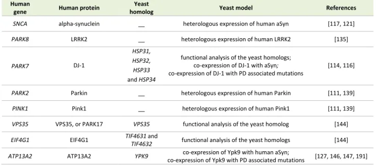
TABLE 1. Yeast models of PD.
Human
gene Human protein
Yeast
homolog Yeast model References
SNCA alpha-synuclein __ heterologous expression of human aSyn [117, 121]
PARK8 LRRK2 __ heterologous expression of human LRRK2 [135]
PARK7 DJ-1
HSP31, HSP32,
HSP33 and HSP34
functional analysis of the yeast homologs; co-expression of DJ-1 with aSyn; co-expression of DJ-1 with PD associated mutations
[114, 116]
PARK2 Parkin __ heterologous expression of human Parkin [111, 139]
PINK1 Pink1 __ heterologous expression of human Pink1 [111, 139]
VPS35 VPS35, or PARK17 VPS35 functional analysis of the yeast homolog [144]
EIF4G1 EIF4G1 TIF4631 and
TIF4632 functional analysis of the yeast homologs [144]
ATP13A2 ATP13A2 YPK9 co-expression of Ypk9 with human aSyn;
co-expression of Ypk9 with PD associated mutations [127, 146, 147, 191]
leucine-rich repeat kinase 2 LRRK2 (PARK8), the E3
ubiqui-tin-ligase Parkin (PARK2), the mitochondrial PTEN-induced
putative kinase 1 Pink1 (PARK6), the oxidation-sensitive
chaperone DJ-1 (PARK7), and the lysosomal ATPase
ATP13A2 (PARK9) [90]. Additionally, several genes are
known to be associated with an increased risk of
develop-ing PD, such as the vacuolar protein sortdevelop-ing 35 homolog
VPS35 (PARK17), the ubiquitin carboxyl-terminal esterase
L1 UCH-L1 (PARK5), the translation initiation factor
4-gamma 1 EIF4G1 (PARK18), and beta-glucocerebrosidase
(GBA) [90]. A comprehensive list of PD genetic risk factors,
including the six risk loci associated with proximal gene
expression or DNA methylation recently identified in
large-scale meta-analysis of genome-wide association studies,
are available online at ‘PDGene’ (www.pdgene.org) [91, 92].
Mutations in LRRK2, encoding a 2527-amino acid
cyto-solic kinase, are the most frequent genetic cause of PD [93,
94]. The role of this kinase has been associated with
bio-logical processes such as endocytosis of synaptic vesicles
[95, 96], autophagy [97], and neurite outgrowth [98]. The
recent discovery of LRRK2 interactions with members of
the dynamin superfamily of large GTPases, by yeast
two-hybrid analyses, implicates its function in the regulation of
membrane dynamics relevant for endocytosis and
mito-chondrial morphology [99]. In line with these findings,
LRRK2 appears to modulate the cellular protection against
oxidative insults in a mechanism that is dependent on
en-docytosis and mitochondrial function [100]. LRRK2 was also
shown to interact with aSyn [101, 102], possibly mediating
its phosphorylation on S129 [103]. The pathogenic
mecha-nisms triggered by mutant versions of LRRK2 are still
un-clear [94]. However, it is speculated that mutations may
affect its interactions with other proteins [90], possibly also
with aSyn.
Parkin homozygous mutations are the most frequent
cause of juvenile PD. Parkin has been associated with
vari-ous cellular pathways but special importance has been
given to its role in mitochondrial quality control, where it
participates in common pathways with Pink1 to regulate
the formation of mitochondrial-derived vesicles and
mi-tophagy [104-110]. Indeed, the MVD pathway has been
referred as the primary defense mechanism against
mito-chondrial damage. Mitophagy only plays a role once MVDs
are overwhelmed. Parkin-assisted Pink1 translocation into
mitochondria is associated with autophagy of damaged
mitochondria [107, 109], and was further supported by
yeast studies on the modulation of mitochondrial
degrada-tion upon oxidative injury and chronological aging [111].
The DJ-1 protein, the most extensively studied member
of the DJ-1 superfamily, is a multifunctional protein
associ-ated with numerous cellular functions including oxidative
stress responses [112]. The cellular mechanisms by which
mutations in DJ-1 cause PD are still unclear, but DJ-1 may
act as a redox-dependent chaperone preventing aSyn
ag-gregation [113]. DJ-1 overexpression confers protection
against neurodegeneration in model organisms, in a
mech-anism that is dependent on protein-protein interactions
between DJ-1 and disease proteins [114, 115]. This
sug-gests that the direct interaction between DJ-1 and its
tar-gets constitutes the basis for the neuroprotective effect of
DJ-1. Further insights into putative roles of members of the
DJ-1 superfamily were obtained in yeast, where Hsp31-like
chaperones may serve as regulators of autophagy [116].
Finally, it is important to stress that most of the genes
associated with PD encode proteins whose functions
ap-pear to be related to mitochondrial function, membrane
trafficking and protein quality control systems,
underscor-ing the importance of these mechanisms in the
pathophys-iology of PD.
MODELLING PD IN YEAST
dis-ease. This also led to the development of various cell and
animal models that are widely used. Among these, yeast
models have proven extremely useful to dissect the basic
molecular mechanisms associated with PD and other
synu-cleinopathies (Table 1). These models are based either on
the heterologous expression of the human genes, or by
studying the function and pathological role of the yeast
counterparts, when these are represented in the yeast
genome (Fig. 1).
aSyn
The first yeast model of PD was based on the heterologous
expression of human aSyn [117]. Since then, many other
studies used this approach to investigate the molecular
mechanisms underlying aSyn toxicity and to identify novel
compounds of therapeutic interest, as described below.
The expression of aSyn in yeast results in
dose-dependent cytotoxicity [117], as observed in other model
systems. This is also in line with the identification of
famili-al forms of PD associated with duplications [67] and
tripli-cations [66] of the aSyn locus. In addition, expression of
aSyn in yeast also resulted in the formation of intracellular
inclusions. The nature of these inclusions was matter of
debate, as it was observed that aSyn leads to the
accumu-lation of vesicles [118-120], raising doubts on whether the
inclusions observed were indeed aggregated aSyn.
Howev-er, amyloid-like aggregates of aSyn seem to be also formed
in yeast cells, as some inclusions are positively stained by
thioflavin S [121] or thioflavin T [122]. More recently, the
formation of large oligomeric species of aSyn in yeast cells
was also demonstrated using both sucrose gradients and
size exclusion chromatography [123].
The various yeast models based on the heterologous
expression of aSyn present different phenotypes,
depend-ing on the expression system, given that this affects the
level of aSyn expression [117, 124, 125]. This feature has
been also explored according to the objectives of the
stud-ies.
The use of multicopy plasmids revealed that yeast cells
reduce the average plasmid copy number in order to
re-duce aSyn expression and toxicity [117]. To avoid this,
in-sertions of the aSyn coding sequence in the yeast genome
enabled more stable expression, and the levels of toxicity
could be manipulated by varying the number of copies of
the aSyn cDNA inserted in the genome [125, 126]. The use
of a galactose-inducible promoter provided additional
con-trol for the synchronous induction of expression of aSyn,
avoiding the negative pressure during routine cell
manipu-lations.
Using these various expression systems, several genetic
modifiers (enhancers and suppressors) of aSyn toxicity
were identified in genetic screenings in yeast [124, 125].
In other studies, yeast cells expressing different levels
of aSyn, hence displaying different levels of cytotoxicity,
revealed the involvement of multiple cellular pathways in
the toxicity [117, 119, 125-129]. In turn, this facilitated the
identification of synergistic effects between different
ge-netic suppressors of toxicity [126, 127]. Overall, these
stud-ies greatly expanded our understanding of the cellular
pathways affected by aSyn. Among these, ER-to-Golgi
traf-ficking appeared to be significantly disrupted [125]. Also,
mitochondrial stress was identified as an early signature of
aSyn toxicity [126]. Indeed, the aSyn expression system
controlled by the yeast
MET25
promoter [121] enabled the
verification that aSyn cytotoxicity requires functional
mito-chondria [130]. Moreover, mitomito-chondria has been pointed
out as the site of enhanced ROS production in response to
Pmr1-dependent Ca
2+overload leading to cellular death in
yeast, flies and worms aSyn models [131].
Other effects of aSyn overexpression in yeast are
im-pairment of proteasome activity [117], accumulation of
cytoplasmic lipid droplets [117], ER stress [125], activation
of the heat-shock response [128], mitochondrial
dysfunc-tion [126], shorter chronological life span and inducdysfunc-tion of
autophagy and mitophagy (mediated by Sir2) [132],
im-pairment of endocytosis [117], ROS production and
induc-tion of apoptosis [126, 133]. Recently, it was shown that
apoptosis is dependent on the translocation of
Endonucle-ase G (EndoG) from the mitochondria to the nucleus,
where it mediates DNA degradation [134].
LRRK2
The role of other genes associated with monogenic forms
of PD has also been studied in yeast models. Namely,
im-portant insights into the function and the pathogenic
mechanisms of LRRK2 mutations were obtained in yeast
cells heterologously expressing human LRRK2 [100, 135].
Higher levels of expression of the full-length LRRK2, based
on a multicopy expression vector and on a galactose
induc-ible promoter, resulted in the accumulation of insoluble
protein but no alterations in the phenotype [100, 135].
However, with lower levels of LRRK2 expression, a
pheno-type of protection against oxidative stress is conferred by
LRRK2 [100]. Interestingly,
the protective effect of LRRK2 is
lost when PD-associated mutants are used instead of the
WT protein [100].
The effects of the overexpression of various functional
domains of human LRRK2 were also analyzed in yeast
[135]. The GTPase activity of LRRK2 was found to be
in-versely correlated with cytotoxicity [135]. This toxicity was
correlated with defects in endocytic trafficking and
au-tophagy [135]. These results were further validated in
mouse primary cortical neurons were the overexpression
of full-length LRRK2 causes defects in both synaptic vesicle
endocytosis and exocytosis [135]. This study also revealed
that aSyn and LRRK2 cause vesicular trafficking-associated
toxicity through distinct pathways that, nevertheless,
cul-minate in trafficking defects to the vacuole, the yeast
counterpart of the lysosome [135].
DJ-1
In yeast, there are four homologous and highly conserved
genes that belong to the DJ-1 superfamily: Hsp31, Hsp32,
Hsp33 and Hsp34. Recently, it was found that these
pro-teins are required for metabolic reprogramming triggered
by glucose limitation, known in yeast as diauxic-shift [116].
It was also found that these DJ-1 homologs contribute to
target of rapamycin complex 1 (TORC1) regulation [116], a
central player in diauxic-shift reprogramming and,
im-portantly, in autophagy [136]. Both TORC1 and autophagy
are dysfunctional in several pathologies including PD [137,
138].
Thus, this study constitutes an example of how the
functional analysis of yeast homologs provides important
insight into
the putative functions of human genes
associ-ated with disease.
In a separate study, the human DJ-1 gene was
ex-pressed in yeast cells using a multicopy vector [114]. It was
found that DJ-1 interacts with aSyn and that PD-associated
mutations impair this interaction [114]. Interestingly, it was
found that DJ-1 and the yeast homologs attenuate aSyn
toxicity in yeast [114]. Thus, these findings suggest that the
physical interaction between DJ-1 and aSyn might
repre-sent a neuroprotective mechanism that is disrupted by
familial mutations in DJ-1, thereby contributing to PD
[114].
Parkin and Pink1
Recently, human Parkin was expressed in yeast and,
alt-hough the protein was found to be cytosolic, it was
trans-located to mitochondria under oxidative stress or aging,
accelerating mitochondrial degradation [111]. Moreover,
Parkin promotes chronological longevity and oxidative
stress resistance through a mitochondria-dependent
path-way [111]. In the same study, Pink1 was also expressed in
yeast and was found to promote resistance to oxidative
stress [111]. However, co-expression of both proteins does
not show a synergistic effect, suggesting the two proteins
affect the same pathway independently [111].
Recently, the mechanism underlying Parkin activation
by Pink1 was dissected in an elegant study where a yeast
model was used [139]. In particular, full activation of the
E3 activity of Parkin E3, in response to mitochondrial
dam-age, was found to occur in a two-step mechanism,
involv-ing the phosphorylation of both Parkin and ubiquitin, by
Pink1 [139].
VPS35 and EIF4G1
VPS24, VPS28, VPS60 or
SAC2 was found to increase aSyn
toxicity [124]. These genes are involved in protein sorting
in the late Golgi, sorting to the prevacuolar endosomes, for
protein sorting, and trafficking to the vacuole, respectively.
In another screen, ENT3, involved in protein transport
be-tween the trans-Golgi network and the vacuole, was also
identified as a suppressor of aSyn toxicity using a yeast
model [140]. Only more recently, next-generation
sequenc-ing and genome-wide association studies implicated
muta-tions in VPS35
(PARK17)
in f
amilial cases of PD [141, 142].
Genome-wide association studies also allowed the
identifi-cation of
EIF4G1
(PARK18) as a novel autosomal dominant
PD gene [143]
. These genes are known to function in
regu-lating
vacuolar
transport
(VPS35)
and
transcrip-tion/translation (EIF4G1), and are highly conserved from
yeast to humans. Yeast has a
VPS35 homolog, also called
VPS35, and two
EIF4G1 homologs,
TIF4631 and
TIF4632.
These
two genes were found to interact functionally and
genetically, and to converge on aSyn, in a study that
com-bined yeast, worms and transgenic mouse models
[144].
This study started with two independent genetic screens in
strains deleted for VPS35 or TIF4631 and enabled the
iden-tification of synthetic sick or lethal genes. The results
pointed to a common pathway associated with both genes:
the
retromer complex function, important in the regulation
of recycling, sorting, and trafficking between the
endoso-mal and Golgi network
. The impairment of a
functional
retromer complex
results in the accumulation of protein
misfolding, thereby exacerbating the accumulation and
toxicity of aSyn [144]. In particular, it was found that
over-expression of
EIF4G1
(or the yeast homolog
TIF4631
) in
cells lacking
VPS35
was highly toxic. This toxicity was found
to be due to the loss of retromer function, which could be
restored by WT
VPS35
but not by the PD-associated
mu-tant D620N
[144]
.
ATP13A2
Mutations in the gene encoding the lysosomal P-type
ATPase ATP13A2 cause Kufor–Rakeb syndrome and
early-onset PD [145]. Yeast has an orthologue of ATP13A2, the
YPK9
gene
,
which encodes a vacuolar transporter with a
possible role in sequestering heavy metals [146]. The
un-derstanding of role of ATP13A2 in PD and how missense
mutations could lead to a loss-of-function of the protein
was facilitated by studying the yeast homolog, followed by
validation in other model systems. Namely,
YPK9
was
found to be a suppressor of aSyn toxicity in yeast [127].
This protective function depends on the vacuolar
localiza-tion and ATPase activity of Vps35 [127], and is probably
related to its role in homeostasis of manganese and other
divalent heavy metal ions [146, 147], which are recognized
environmental risk factors for PD [148]. Among the most
common PD genetic players, ATP13A2 is the unique whose
structure has not yet been unveiled.
aSYN AND PROTEIN QUALITY CONTROL SYSTEMS IN
YEAST
Alterations in proteostasis occur in several types of
disor-ders [149]. When the accumulation of misfolded proteins
surpasses the capacity of the cell to cope with the protein
load, diverse quality control mechanisms are called to
ac-tion, to actively sequester, refold, and/or degrade these
proteins [150-153] (Fig. 2). These cellular protein quality
control mechanisms are conserved from yeast to
mamma-lian cells [37].
Strong evidence on the involvement of the quality
con-trol systems in neurodegeneration came from studies of
familial forms of PD, as mutations in several genes playing
a role in these pathways are intimately associated with the
disease, as mention above.
Besides those players, several molecular chaperones
were also found to have an important role in PD. Namely,
the involvement of molecular chaperones in aSyn yeast
toxicity was evidenced by the enhanced aSyn inclusion
formation observed upon deletion of individual
chaper-ones [154]. Concomitantly, pharmacological activation of
the heat shock response upon treatment with
geldanamy-cin or overexpression of the chaperones Ssa3 [155], Jem1
or Hsp90 [140] protected yeast cells against aSyn-induced
ROS and subsequent toxicity. These data elegantly
recapit-ulated results obtained with neuronal cell lines [156],
transgenic flies [157] and mice [158, 159].
The protein Gip2, an activator of the heat-shock
tran-scription factor Hsf1, was also identified as a multicopy
suppressor of aSyn toxicity, by triggering a heat-shock
re-sponse in yeast [128]. A screening of a mouse brain-specific
cDNA library identified the mouse chaperone RPS3A as a
suppressor of aSyn WT and A53T toxicity in yeast [160].
Moreover, co-expression of RPS3A delayed the formation
of aSyn inclusions [160]. Overexpression of DJ-1, which has
protein chaperone-like activity, was also described as a
negative regulator of aSyn dimerization [114].
Additional strong evidences of the relevance of the
clearance pathways in PD arrive from yeast models.
Name-ly, the expression of aSyn in yeast promotes proteasome
impairment [117]. This reduced proteasome activity was
found to be the result of a deficient proteasome
composi-tion [168]. Furthermore, the failure of the UPS
(ubiquitin-proteasome system) enhances aSyn toxicity [169, 170] and
leads to the accumulation of inclusions [121].
Despite the clear involvement of proteasome
dysfunc-tion in PD, the degradadysfunc-tion of aSyn inclusions is more
de-pendent on autophagy than on proteasome function, at
least in yeast [171]. This is consistent with the proteasome
being responsible for the degradation of soluble forms of
aSyn, and suggests a complex cross-talk between the
dif-ferent proteolytic pathways involved in the degradation of
aSyn [172] (Fig. 2).
Autophagy involves the formation of an
autophagoso-mal vesicle that transports the misfolded and aggregated
proteins to degradation, in the lysosome in higher
eukary-otes, or in the vacuole in yeast. The first molecular insights
into autophagy were learned from yeast [173], and as in
mammalians, the process is regulated by the kinase “target
of rapamycin” (TOR) pathway [174]. Concomitantly Lst8, a
TOR-interactor was identified as modulator of aSyn toxicity
in yeast [128]. Moreover, Ypk9, a vacuolar P-type ATPase
that is the orthologue of ATP13A2, was identified as a
sup-pressor of aSyn toxicity [128] and aggregation [127].
More recently, it was reported that deletion of the
au-tophagy related genes ATG1 or ATG7 lead to impaired
deg-radation of aSyn and increased toxicity [123]. In agreement
with the beneficial role of autophagy on aSyn toxicity,
ra-pamycin treatment, which induces autophagy by inhibiting
TOR, was reported to reduce aSyn aggregation [121]. It
also appears that toxic forms of aSyn lead to the
impair-ment of autophagy and that inducing autophagy or
increas-ing the autophagic flux is protective against aSyn toxicity in
yeast [123, 175].
Nevertheless, there is still controversy regarding the
role of autophagy on aSyn toxicity. A study reported that
rapamycin treatment increases aSyn toxicity in yeast [128].
It was also shown that WT or A53T mutant aSyn were not
able to enter the vacuole and promoted vacuolar fusion
defects in yeast [154]. Additionally, aSyn-mediated
mi-tophagy, a specific degradation of mitochondria through
autophagy, was reported to be deleterious in aged yeast
cells [132]. To intensify the discussion, aSyn Lewy body-like
aggregates resisted degradation and impaired autophagy
in mammalian cell models [132].
It is clear that the interplay between autophagy, aSyn
toxicity and aggregation is still elusive. A reasonable
nation for the toxicity of aSyn mediated by autophagy
in-duction is that the excessive activation of a dysfunctional
autophagy will lead to a loss of selectivity, resulting in the
trapping of functional competent proteins and organelles
in autophagosomes. Ultimately, this could lead to a loss of
function and cell toxicity. Thus, the beneficial or
detri-mental role of autophagy should be studied having in
con-sideration its functionality and selectivity, as well as the
size and nature of the aSyn aggregates.
YEAST AS A DRUG DISCOVERY PLATFORM FOR PD
The identification of therapeutic compounds for
neuro-degenerative disorders is of utmost importance. These
devastating illnesses only have, in some cases,
symptomat-ic therapies being therefore disruptive and costly for
socie-ty. Thus, intense efforts are being made to understand the
molecular underpinnings of neurodegenerative diseases
and to identify novel therapeutic strategies. Nevertheless,
given the complexity of the mechanisms leading to
neuro-degeneration, and the limitations in the models available,
drug discovery is often slow, challenging and with limited
success.
In the context of PD, drug discovery efforts focused on
aSyn are complicated by the fact that it is a ”natively
un-structured protein”, lacking defined secondary structure
under physiological conditions [176]. Thus, cell-based
high-throughput phenotypic assays afford important
possibili-ties, as they are based on relevant disease-associated
phe-notypes induced by aSyn expression. The readouts may
include viability/toxicity, aggregation, mitochondrial
func-tion, proteasome activity, among others. Once relevant
molecules and targets are identified, then it is fundamental
to scrutinize the mechanism of action of the potential
small molecules and candidate compounds. This is where
yeast cells offer a remarkable advantage, as they enable
the identification of target genes and mechanisms through
diverse and complementary genetic approaches,
accelerat-ing the selection of pre-clinical candidates.
In mammalian cell systems, aSyn-associated
pheno-types are often mild or inconsistent, complicating the
de-velopment of reliable screening platforms [11, 177].
Prima-ry rat neurons, infected with lentiviruses encoding for
aSyn-expressing, have been used, but they also present
technical limitations. Many of these systems are more
suit-ed for secondary validation steps, focusing on candidate
genes or molecules identified in yeast, for example.
Yeast affords numerous advantages at the early stages
of the drug development process, in comparison to
mam-malian cells and animal models. Several major drugs hit the
same targets and elicit the same responses in yeast as they
do in humans, including statins, methotrexate, omeprazole,
tacrolimus (FK506) and bortezomib (Velcade) [177, 178]. In
spite of the obvious limitations, such as the absence of a
nervous system, and the absence of numerous gene
prod-ucts that are only present in mammalian cells and neurons,
yeast cells are ideally suited for investigating the primordial
molecular events triggering cell dysfunction and pathology.
In this context, yeast cells can be regarded as living test
tubes, where genetic manipulations are faster and more
straightforward, with rapid growth and reduced cost, and
functional similarity to higher eukaryotes.
The presence of a cell wall in yeast cells poses
chal-lenges that need to be considered in the context of drug
screening efforts. However, this can be minimized by
ge-netic manipulation of the efflux pump system or the
ergos-terol biosynthesis, reducing the capability of yeast cells to
export drugs or by increasing the permeability of the cells,
respectively.
Yeast is considered a robust primary drug-screening
platform to filter for compounds with cytoprotective
activi-ty, for further complementation with assays in more
physi-ologically relevant models. Approaches involving the
se-quential use of different model systems, starting with
sim-pler cellular models and ending with more complex animal
models, as schematized in Fig. 3, already resulted in the
discovery of promising small molecules with therapeutic
potential (described below). Recently, a yeast-to-human
discovery platform for synucleinopathies was established,
where genes and small molecules identified in yeast were
validated in PD-patient derived neurons. Subsequently,
yeast cells were again used for clarification of the
mecha-nism of action, due to unmatched genetic tools available in
S. cerevisae [179] (Fig. 3).
By iteratively moving between simple cellular models
and patient derived cells, we will be able to elucidate
mechanisms and evaluate patient-specific drug targets.
Ultimately, this will enable scientists to conduct more
sig-nificant animal and clinical trials in various
neurodegenera-tive diseases.
Genetic screens
Large-scale genetic and chemical genetic approaches in
yeast have provided important insight into the molecular
basis of various neurodegenerative disorders. The first
large-scale genetic screen used 4850 deletion yeast strains
and successfully identified 86 genes enhancers of aSyn
toxicity, of which 29% were involved in vesicular transport
and lipid metabolism [124]. Later, the same yeast
collec-tion enabled the identificacollec-tion of 185 modifiers of aSyn
aggregation, using fluorescence microscopy [154]. This
study revealed that proteins involved in vesicular transport
altered aSyn subcellular localization.
Using an overexpression screen, aSyn was found to
block ER-to-Golgi trafficking due to the identification of
enhancers and suppressors of toxicity [125]. Importantly,
these observations led to the identification of Rab1, the
mammalian Ypt1 homolog, as a neuroprotector against
dopaminergic neuron loss in animal models of PD [125].
In another genome wide-screen, using a
high-expression library, Ypp1 was found to mediate the
traffick-ing of aSyn A30P to the vacuole via the endocytic pathway,
thus suppressing the toxicity of this aSyn mutant [133].
Using an integrative approach, the results from genetic
screens were analyzed according to gene functionality and
pathways. About ~3500 overexpression yeast strains were
used and a cellular map of the proteins and genes
respond-ing to aSyn expression was obtained. Ergosterol
biosynthe-sis and the TOR pathway were identified as modulators of
aSyn cytotoxicity in yeast [128].
Drug discovery efforts in yeast models
The events leading to protein oligomerization are likely
amenable to modulation by small molecules. Thus, yeast
has also been used to screen for small molecules that can
reduce aSyn aggregation and toxicity.
Screening of large libraries of compounds lead to the
identification of aSyn toxicity suppressors in yeast. For
in-stance, in a large-scale screen of small molecule, ~115.000
compounds were tested for their ability to reduce aSyn
toxicity, resulting in the identification of a class of
structur-ally related 1,2,3,4-tetrahydroquinolinones [126]. These
compounds reduced the formation of aSyn inclusions,
re-established ER-to-Golgi trafficking, and ameliorated
mito-chondria-associated defects induced by aSyn. The targets
were further confirmed in nematode neurons and in
pri-mary rat neuronal midbrain cultures. Interestingly, these
compounds also rescued rotenone toxicity in neuronal
cultures, a toxin used to study mitochondrial deficits in PD
[126].
The ease of manipulation makes yeast a suitable tool to
explore unconventional compounds and their mechanisms
of action. Mannosylglycerate, a compatible solute typical
of marine microorganisms thriving in hot environments,
was found to reduce aSyn aggregation in a yeast model of
PD [180]. Latrepirdine, a drug in phase II clinical trials, was
identified as protector against aSyn by inducing its
degra-dation through autophagy, representing a novel scaffold
for
discovery
of
robust
pro-autophagic/anti-neurodegeneration compounds [181].
A novel class of molecules, cyclic peptides (CPs), was
al-so screened in yeast [182]. CPs are natural-product-like
chemicals with potent bioactivity. Yeast was exploited to
express a plasmid-derived self-splicing intein that liberates
a CP. This approach enabled the scale-up of
high-throughput screens to 10–100 times the size of a typical
small molecule screen. A pool of 5 million yeast
trans-formants were screened and two related CP constructs
with the ability to reduce aSyn toxicity were identified.
These cyclic peptide constructs also prevented
dopaminer-gic neuron loss in a nematode model of PD [182].
Due to their well-defined chemical nature, small
mole-cules are the preferred molemole-cules used in high-throughput
screenings. Nevertheless, natural compounds have
emerged as attractive molecules in the context of
neuro-degeneration. It is largely accepted that products such as
green tea, small fruits and even olive oil have, in its
consti-tution, compounds promoting health benefits. However,
the major advances regarding their targets and
mecha-nisms of action were only achieved in the last decade.
Yeast models, together with chemical and animal studies,
have significantly contributed for these discoveries [175,
180, 183, 184].
The first small compound screen in yeast tested
~10.000 compounds and identified a group of flavonoids,
quercetin and epigallocatechin gallate as protectants
against aSyn toxicity in the presence of iron [185]. The
pro-tection promoted by these compounds was further
ana-lyzed, and the positive effect was due to their anti-oxidant
and metal-chelating activities. Importantly, (poly)phenols,
and particularly quercetin and epigallocatechin gallate,
have proven beneficial in cellular and animal models of PD
[186, 187].
The advances in biochemical tools and the assembly of
multidisciplinary teams also gave a major push to the drug
discovery process. In fact, the benefits of green tea have
been deciphered by combining HPLC fractionation in a
mi-croplate format with screening in yeast and parallel
elec-trospray mass spectrometry (LC-MS) [188] (Fig. 3). This
integrated process enabled the rapid assessment of the
efficacy of the fractions and to systematically identify their
bioactive constituents. The green tea metabolites were
individually examined for their pharmacological effects and,
interestingly, the protective properties of
Camelia sinensis
lied on the combination of multiple catechin metabolites
[188]. This study emphasizes the prominence of yeast
high-throughput screenings to dissect natural extracts and to
explore the numerous synergistic effects of its metabolites.
The bioactivities of plant (poly)phenol extracts in the
yeast aSyn model were recently investigated using viability,
oxidative stress, metabolic capacity and aSyn inclusion
formation as phenotypic assays [175]. The most promising
extract was the one from
Corema album leaves. The
dis-section of the mechanism of action of this extract, focused
primarily on pathways related to proteostasis, showed that
it promotes autophagic flux both in yeast and in a
mamma-lian cell model of PD [175].
Ascorbic acid, a natural antioxidant, was found to
pro-mote a significant reduction in the percentage of yeast
cells bearing aSyn inclusions [189]. Remarkably, this study
was performed using a new microfluidic device designed to
validate compounds in yeast. Additional advantages are
achieved by using this device, since it offers a powerful way
for studying aSyn biology with single-cell resolution and
high-throughput, using genetically modified yeast cells
[189].
Screenings in other disease models, as in the
amyo-trophic lateral sclerosis yeast model (induced by the
ex-pression of the protein TDP-43), may also provide insight
into candidate compounds to be tested in aSyn yeast
mod-els. Using yeast genetics, multiple protective
8-hydroxyquinolines, natural plant alkaloids, were identified
[184]. Some of these compounds were also found to be
protective in aSyn yeast and nematode models. The
puta-tive protecputa-tive mechanisms were related to their
iono-phore and intracellular metal chelation activities [184].
From this screening, N-aryl benzimidazole proved more
potent and effective against aSyn toxicity than against
TDP-43 toxicity. Thus, the yeast aSyn platform was explored to
identify the mechanism of action of N-aryl benzimidazole
and it was found that it reverses diverse phenotypes
in-duced by aSyn, including the accumulation of aSyn
inclu-sions, the generation of ROS, the block of ER–Golgi
traffick-ing and the nitration of proteins [190]. Moreover, this
compound was used in an iterative yeast-to-human neuron
platform [129] (Fig. 3).
Taken together, identifying novel effective disease
therapies is an incredible challenge. Nevertheless, rapidly
improving methodologies and iterative processes, allied
with an evolving mechanistic understanding of disease, is
nurturing more interdisciplinary approaches to research
and fostering drug discovery, with the ultimate goal of
dis-covering novel therapeutics for humans.
CONCLUSIONS AND FUTURE PERSPECTIVES
The development of effective treatments and preventive
therapies for PD is still a great challenge, mostly due to the
scarcity of knowledge of disease-associated mechanisms
that ultimately lead to neuronal dysfunction and death. As
discussed herein, the versatile eukaryotic model organism
S. cerevisiae has largely contributed to bridge this gap in PD
medical research. By providing important insights into the
molecular foundations of the disease as well as novel
mo-lecular targets and lead compounds with therapeutic
po-tential. Notwithstanding, several fundamental aspects of
PD pathophysiology remain to be elucidated. For example,
yeast models were very helpful to clarify some facets of
the still controversial role of aSyn phosphorylation, and will
certainly further contribute to our understanding of other
mechanisms associated with aSyn and other PD-associated
genes.
Undoubtedly, research using
S. cerevisiae
as a model
system enabled significant advancements in our
under-standing of the molecular mechanisms underlying PD.
However, it should be noted that, as a simplified model
system, it also has natural limitations that need to be
obvi-ated by further validations in more complex models.
In-deed,
iterative processes using models with different
de-grees of complexity have proven to be a powerful strategy
to investigate the fundamental aspects of
neurodegenera-tive diseases, thereby accelerating drug discovery.
ACKNOWLEDGMENTS
This work was supported by Fundação para a Ciência e
Tecnologia project PTDC/BIA-BCM/117975/2010,
fellow-ships
SFRH/BPD/101646/2014
(ST)
and
SFRH/BD/73429/2010 (DM), and IF/01097/2013 (CNS). This
work was also supported by the
BacHBerry project,
co-funded by the European Commission in the 7th
Frame-work Programme (Project No. FP7-613793). RM is
support-ed by a BacHBerry
fellowship. TFO is supported by the DFG
Center for Nanoscale Microscopy and Molecular Physiology
of the Brain (CNMPB).
CONFLICT OF INTEREST
COPYRIGHT
© 2015 Menezes
et al.
This is an open-access article
re-leased under the terms of the Creative Commons
Attribu-tion (CC BY) license, which allows the unrestricted use,
distribution, and reproduction in any medium, provided
the original author and source are acknowledged.
Please cite this article as: Regina Menezes, Sandra Tenreiro, Diana Macedo, Cláudia N. Santos, Tiago Fleming Outeiro (2015). From the baker to the bedside: yeast models of Parkinson's disease.
Microbial Cell 2(8): 262-279. doi: 10.15698/mic2015.08.219
REFERENCES
1. Samuel D (1996). Investigation of ancient egyptian baking and brewing methods by correlative microscopy. Science 273(5274): 488-490.
2. Legras JL, Merdinoglu D, Cornuet JM, Karst F (2007). Bread, beer and wine: Saccharomyces cerevisiae diversity reflects human history.
Molecular ecology 16(10): 2091-2102.
3. Mortimer RK (2000). Evolution and variation of the yeast (Saccha-romyces) genome. Genome research 10(4): 403-409.
4. Tsukuda T, Fleming AB, Nickoloff JA, Osley MA (2005). Chromatin remodelling at a DNA double-strand break site in Saccharomyces cerevisiae. Nature 438(7066): 379-383.
5. Coller J, Parker R (2004). Eukaryotic mRNA decapping. Annual re-view of biochemistry 73: 861-890.
6. Fields S, Johnston M (2005). Cell biology. Whither model organism research? Science 307(5717): 1885-1886.
7. Duina AA, Miller ME, Keeney JB (2014). Budding yeast for budding geneticists: a primer on the Saccharomyces cerevisiae model system.
Genetics 197(1): 33-48.
8. Botstein D, Fink GR (2011). Yeast: an experimental organism for 21st Century biology. Genetics 189(3): 695-704.
9. Hinnen A, Hicks JB, Fink GR (1978). Transformation of yeast. Pro-ceedings of the National Academy of Sciences of the United States of America 75(4): 1929-1933.
10. Alberti S, Gitler AD, Lindquist S (2007). A suite of Gateway cloning vectors for high-throughput genetic analysis in Saccharomyces cere-visiae. Yeast 24(10): 913-919.
11. Tenreiro S, Munder MC, Alberti S, Outeiro TF (2013). Harnessing the power of yeast to unravel the molecular basis of neurodegenera-tion. Journal of neurochemistry 127(4): 438-452.
12. Van Mullem V, Wery M, De Bolle X, Vandenhaute J (2003). Con-struction of a set of Saccharomyces cerevisiae vectors designed for recombinational cloning. Yeast 20(8): 739-746.
13. Fields S, Song O (1989). A novel genetic system to detect protein-protein interactions. Nature 340(6230): 245-246.
14. Vidal M, Legrain P (1999). Yeast forward and reverse 'n'-hybrid systems. Nucleic acids research 27(4): 919-929.
15. Stagljar I, Korostensky C, Johnsson N, te Heesen S (1998). A genetic system based on split-ubiquitin for the analysis of interactions be-tween membrane proteins in vivo. Proceedings of the National Acad-emy of Sciences of the United States of America 95(9): 5187-5192.
16. Iyer K, Burkle L, Auerbach D, Thaminy S, Dinkel M, Engels K, Sta-gljar I (2005). Utilizing the split-ubiquitin membrane yeast two-hybrid system to identify protein-protein interactions of integral membrane proteins. Science's STKE : signal transduction knowledge environ-ment 2005(275): pl3.
17. Hu CD, Chinenov Y, Kerppola TK (2002). Visualization of interac-tions among bZIP and Rel family proteins in living cells using bimolecu-lar fluorescence complementation. Molecular cell 9(4): 789-798.
18. Kerppola TK (2006). Design and implementation of bimolecular fluorescence complementation (BiFC) assays for the visualization of protein interactions in living cells. Nature protocols 1(3): 1278-1286.
19. Sung MK, Huh WK (2007). Bimolecular fluorescence complementa-tion analysis system for in vivo deteccomplementa-tion of protein-protein interac-tion in Saccharomyces cerevisiae. Yeast 24(9): 767-775.
20. He F, Nie WC, Tong Z, Yuan SM, Gong T, Liao Y, Bi E, Gao XD (2015). The GTPase-activating protein Rga1 interacts with Rho3 GTPase and may regulate its function in polarized growth in budding yeast. PloS one 10(4): e0123326.
21. Goncalves SA, Matos JE, Outeiro TF (2010). Zooming into protein oligomerization in neurodegeneration using BiFC. Trends in biochemi-cal sciences 35(11): 643-651.
22. Goffeau A, Barrell BG, Bussey H, Davis RW, Dujon B, Feldmann H, Galibert F, Hoheisel JD, Jacq C, Johnston M, Louis EJ, Mewes HW, Murakami Y, Philippsen P, Tettelin H, Oliver SG (1996). Life with 6000 genes. Science 274(5287): 546, 563-547.
23. Winzeler EA, Shoemaker DD, Astromoff A, Liang H, Anderson K, Andre B, Bangham R, Benito R, Boeke JD, Bussey H, Chu AM, Connelly C, Davis K, Dietrich F, Dow SW, El Bakkoury M, Foury F, Friend SH, Gentalen E, Giaever G, Hegemann JH, Jones T, Laub M, Liao H, Lie-bundguth N, Lockhart DJ, Lucau-Danila A, Lussier M, M'Rabet N, Menard P, et al. (1999). Functional characterization of the S. cere-visiae genome by gene deletion and parallel analysis. Science
285(5429): 901-906.
24. Mnaimneh S, Davierwala AP, Haynes J, Moffat J, Peng WT, Zhang W, Yang X, Pootoolal J, Chua G, Lopez A, Trochesset M, Morse D, Krogan NJ, Hiley SL, Li Z, Morris Q, Grigull J, Mitsakakis N, Roberts CJ, Greenblatt JF, Boone C, Kaiser CA, Andrews BJ, Hughes TR (2004). Exploration of essential gene functions via titratable promoter alleles.
Cell 118(1): 31-44.
25. Dohmen RJ, Varshavsky A (2005). Heat-inducible degron and the making of conditional mutants. Methods in enzymology 399: 799-822.
26. Huh WK, Falvo JV, Gerke LC, Carroll AS, Howson RW, Weissman JS, O'Shea EK (2003). Global analysis of protein localization in budding yeast. Nature 425(6959): 686-691.
27. Ghaemmaghami S, Huh WK, Bower K, Howson RW, Belle A, Dephoure N, O'Shea EK, Weissman JS (2003). Global analysis of pro-tein expression in yeast. Nature 425(6959): 737-741.
28. Jones GM, Stalker J, Humphray S, West A, Cox T, Rogers J, Dunham I, Prelich G (2008). A systematic library for comprehensive overexpres-sion screens in Saccharomyces cerevisiae. Nature methods 5(3): 239-241.