Gefitinib and Erlotinib Lead to
Phosphorylation of Eukaryotic Initiation
Factor 2 Alpha Independent of Epidermal
Growth Factor Receptor in A549 Cells
Satoshi Koyama, Tomohiro Omura*, Atsushi Yonezawa, Satoshi Imai, Shunsaku Nakagawa, Takayuki Nakagawa, Ikuko Yano, Kazuo Matsubara
Department of Clinical Pharmacology and Therapeutics, Kyoto University Hospital, Kyoto, Japan
*omurat@kuhp.kyoto-u.ac.jp
Abstract
Gefitinib and erlotinib are anticancer agents, which inhibit epidermal growth factor receptor (EGFR) tyrosine kinase. Interstitial lung disease (ILD) occurs in patients with non-small cell lung cancer receiving EGFR inhibitors. In the present study, we examined whether gefitinib-and erlotinib-induced lung injury related to ILD through endoplasmic reticulum (ER) stress, which is a causative intracellular mechanism in cytotoxicity caused by various chemicals in adenocarcinomic human alveolar basal epithelial cells. These two EGFR inhibitors increased Parkinson juvenile disease protein 2 and C/EBP homologous protein mRNA expressions, and activated the eukaryotic initiation factor (eIF) 2α/activating transcription factor 4 pathway without protein kinase R-like ER kinase activation in A549 cells. Gefitinib and erlotinib caused neither ER stress nor cell death; however, these agents inhibited cell growth via the reduction of cyclin-D1 expression. Tauroursodeoxycholic acid, which is known to suppress eIF2αphosphorylation, cancelled the effects of EGFR inhibitors on cyclin-D1 expression and cell proliferation in a concentration-dependent manner. The results of an EGFR-silencing study using siRNA showed that gefitinib and erlotinib affected eIF2αphosphorylation and cyclin-D1 expression independent of EGFR inhibition. There-fore, the inhibition of cell growth by these EGFR inhibitors might equate to impairment of the alveolar epithelial cell repair system via eIF2αphosphorylation and reduced cyclin-D1 expression.
Introduction
Epidermal growth factor receptor (EGFR) tyrosine kinase inhibitors, such as gefitinib and erlo-tinib, are oral molecule-targeted drugs for non-small cell lung cancer. These drugs occasionally induce interstitial lung disease (ILD), especially interstitial pneumonia, as a critical adverse reaction [1,2]. ILD patients with chest-imaging portraying ground-glass opacity and severe
OPEN ACCESS
Citation:Koyama S, Omura T, Yonezawa A, Imai S, Nakagawa S, Nakagawa T, et al. (2015) Gefitinib and Erlotinib Lead to Phosphorylation of Eukaryotic Initiation Factor 2 Alpha Independent of Epidermal Growth Factor Receptor in A549 Cells. PLoS ONE 10 (8): e0136176. doi:10.1371/journal.pone.0136176
Editor:Pier Giorgio Petronini, University of Parma, ITALY
Received:January 6, 2015
Accepted:July 31, 2015
Published:August 19, 2015
Copyright:© 2015 Koyama et al. This is an open access article distributed under the terms of the Creative Commons Attribution License, which permits unrestricted use, distribution, and reproduction in any medium, provided the original author and source are credited.
Data Availability Statement:All relevant data are within the paper and its Supporting Information files.
Funding:This study was supported by MEXT/JSPS KAKENHI (https://kaken.nii.ac.jp/en/) Grant Numbers 25136710, 24590176 and 25860112.
breathlessness have to discontinue therapies with these EGFR inhibitors. However, little is known about the pathogenesis, diagnosis, and treatment of such drug-induced ILD. The observed increase in ILD risk with gefitinib treatment has been shown to be higher in elderly smokers with preexisting ILD or poor performance status [3]. Alveolar type-II epithelial cells are believed to be progenitor cells in lung tissues. With injured alveoli, type-II cells proliferate and differentiate into type-I cells, leading to alveolus repair [4]. Thus, deterioration of this repair pathway in alveoli can be interpreted as a possible mechanism for the promotion of ILD in EGFR inhibitor therapy.
EGFR inhibitors reduce cyclin-D1 levels inducing cell cycle arrest in the G1 phase [5–7]. Phosphorylation of eukaryotic initiation factor (eIF) 2αreduces cyclin-D1 expression in mouse embryonic fibroblasts [8,9], and is known to induce translational suppression [10,11]. Namba et al. have reported that gefitinib may induce translational suppression of heat-shock protein 70 in adenocarcinomic human alveolar basal epithelial (A549) cells [12]. Phospho-eIF2αbinds to and inhibits eIF2B, which converts eIF2-GDP into eIF2-GTP. This inhibition causes transla-tional suppression as a result of the depletion of eIF2-GTP [10], which is one of the typical pathways under endoplasmic reticulum (ER) stress [11]. ER stress has been suggested to act as a causative factor in several lung injuries. Mutant surfactant protein C (L188Q), which is dis-covered in a kindred with familial interstitial pneumonia [13], incites ER stress in mice alveolar type-II epithelial cells [14]. ER stress mediates lung injury of some compounds, including ciga-rette smoke and herbicides (e.g. paraquat) [15,16]. However, some of the molecule-targeted drugs, such as imatinib, sorafenib and dasatinib, have been reported to evoke ER stress [17– 19]. Therefore, gefitinib and erlotinib may cause ER stress to induce lung injury related to ILD.
In the present study, we examined if pulmonary toxicity induced by EGFR inhibitors was associated with ER stress, using A549 cells as a model of human alveolar type II-like epithelial cells. We first investigated if alteration of signaling was associated with ER stress. We found that eIF2αwas phosphorylated and cyclin-D1 was reduced in A549 cells treated with EGFR inhibitors. We further examined cytoprotective action of elF2αphosphorylation-suppressive tauroursodeoxycholic acid (TUDCA), and tested if this effect was EGFR-dependent.
Materials and Methods
Chemical Reagents
Gefitinib and erlotinib hydrochloride were obtained from LC Laboratories (Woburn, MA, USA) and Santa Cruz Biotechnology (Dallas, TX, USA), respectively. TUDCA and propidium iodide were commercially available from Sigma-Aldrich (St. Louis, MO, USA), while Hoechst 33342 and 3-(4,5-dimethyl-2-thiazolyl)-2,5-diphenyl-2H-tetrazolium bromide (MTT) were products of Dojindo Laboratories (Kumamoto, Japan). Tunicamycin and other chemical reagents were obtained from Wako Pure Chemicals (Osaka, Japan).
Antibodies
Cell Culture and Drug Treatment
A549 cells (ECACC, 86012804) and PC-9 cells (kindly gifted by Dr. Menju, Department of Thoracic Surgery, Kyoto University) were respectively cultured in Dulbecco's modified eagle's medium (DMEM) and RPMI 1640 supplemented with 10% fetal bovine serum (Gibco, Carls-bad, CA, USA) in a humidified atmosphere with 5% CO2at 37°C. Drugs in serum-free medium
(final DMSO concentration:<0.1%) were added to cells seeded on 24-well (for MTT assay) or
6-well plates (for real-time PCR, western blotting analysis or staining).
MTT Assay
Cells treated with/without drugs were incubated with 0.5 mg/mL MTT and was assayed for 30 min at 37°C. After discarding the medium, stained cells were dissolved in 1 mL of DMSO before the optical density was measured at 560 nm (reference wavelength, 630 nm).
RNA Isolation and Quantitative Real-Time PCR
Total RNA was isolated from A549 cells with an RNeasy Plus Mini Kit (Qiagen, Hilden, Ger-many) according to manufacturer’s instructions. Complementary DNA (cDNA) was generated from RNA (20μg) using a High Capacity RNA-to-cDNA Kit (Applied Biosystems, Carlsbad, CA, USA). Quantitative real-time PCR was performed according to the StepOnePlus Real-Time PCR System (Applied Biosystems) and TaqMan Fast Advanced Master Mix (Applied Biosystems). The probe-primer solutions specific for the following genes were used (obtained from Applied Biosystems): Binding immunoglobulin protein (BIP) (Hs00607129_gH),CHOP (Hs00358796_g1), HMG-CoA reductase degradation 1 (HRD1) (Hs00381211_m1), Parkinson juvenile disease protein 2 (PARK2) (Hs01038325_m1), suppressor or enhancer of lin-12 1 (SEL1) (Hs01071406_m1) and18S rRNA(Hs99999901_s1). The agent18S rRNAwas used an internal control to normalize mRNA expression levels.
Western Blotting Analysis
Cell samples collected in lysis buffer (20 mM HEPES, 120 mM NaCl, 5 mM EDTA, 1% Triton X-100, 10% glycerol, 10 mM NaF, 2 mM Na3VO4) were placed in protease inhibitor cocktail
(Nacalai Tesque, Kyoto, Japan), and protein concentrations were determined using the Brad-ford assay. Equal amounts of total protein were separated by SDS-PAGE and then transferred to PVDF membrane. Blocking was performed at room temperature for 1 h in TBS-T with 5% skim milk (BD Falcon, Franklin Lakes, NJ), followed by overnight incubation with different primary antibodies (described above) in TBS-T at 4°C. Appropriate secondary antibodies were then used, and proteins were visualized using chemiluminescence (Luminata Crescendo, Milli-pore, Billerica, MA, USA; ECL select, GE Healthcare, Little Chalfont, UK). The intensities of protein levels, which were analyzed using Image J software from NIH, were corrected with the respectiveβ-actin levels.
Silencing of EGFR Expression
Evaluation of Cell Death
After drug treatment, trypsinized cells collected by centrifugation (190 g, 5 min, room tempera-ture) were re-suspended in 1×PBS. Trypan blue staining and cell-counting were performed with Countess (Invitrogen, Carlsbad, CA, USA), according to the manufacturer’s instruction. For detection of apoptotic and necrotic cells, an aliquot of 50μL of cells was suspended with 50μL of 1×Binding Buffer containing Hoechst 33342, Ethidium Homodimer III (EthD-III) and FITC-Annexin V. After standing for 15 min at room temperature followed by washing with 1×Binding Buffer, according to the instruction manual of Apoptotic/Necrotic/Healthy Cells Detection Kit (PromoKine, Heidelberg, Germany). The fluorescent images were captured and examined with BZ-9000 (Keyence, Osaka, Japan). Three independent photographs were taken at 100-fold magnification, and the stained cells were counted. Apoptotic and necrotic cells were estimated by [FITC-Annexin V] / [Hoechst 33342] and [EthD-III] / [Hoechst 33342], respectively.
Statistical Analysis
Quantitative data are represented as means ± S.E.M. Data were statistically analyzed using one-way analysis of variance, followed by the Dunnett’s or Tukey-Kramer’s two-tailed test to evalu-ate differences between more than three groups. Probability values of less than 0.05 were con-sidered statistically significant. Statistical analysis was performed using GraphPad Prism 5 (GraphPad Software, La Jolla, CA, USA).
Results
Gefitinib and Erlotinib Suppressed Cell Growth of A549 Cells
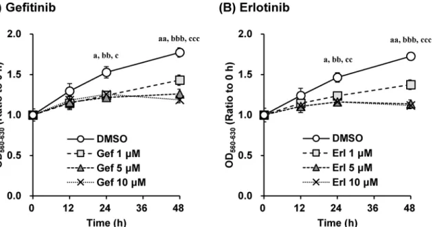
Based on the effect of gefitinib and erlotinib on cell proliferation of A549 cells, the EGFR inhib-itors suppressed proliferation of A549 cells in a concentration-dependent manner (Fig 1).
Fig 1. Gefitinib and erlotinib suppressed growth of A549 cells in a concentration-dependent manner.The effects of (A) gefitinib and (B) erlotinib (1– 10μM) on cell proliferation of A549 cells were investigated. Cell counts were estimated by the MTT assay. Data are expressed as means±S.E.M. of three independent experiments. Each symbol indicates significant differences from DMSO group; a,p<0.05; aa,p<0.01 (DMSO vs. 1μM), bb,p<0.01; bbb,
p<0.001 (DMSO vs. 5μM), c,p<0.05; cc,p<0.01; ccc,p<0.001 (DMSO vs. 10μM), one-way ANOVA with Dunnett’s post hoc tests.
Gefitinib and Erlotinib Induced the Up-Regulation of
PARK2
and
CHOP
mRNA in A549 Cells
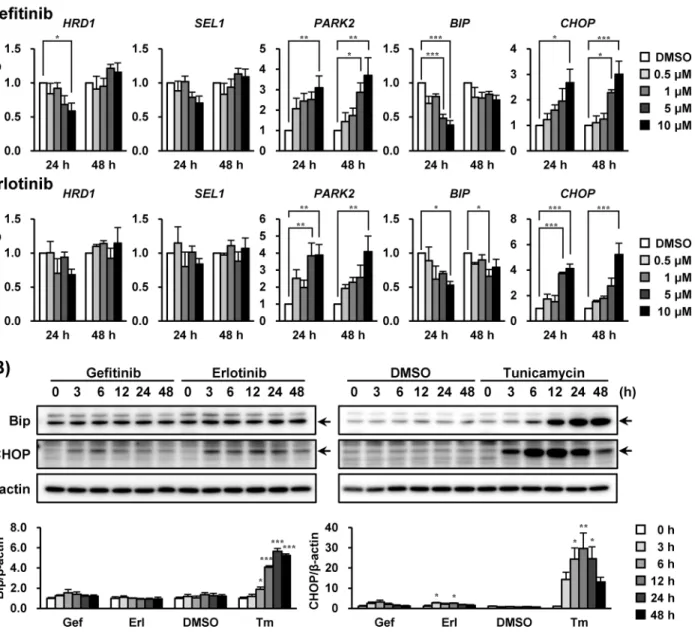
Next, we investigated if unfolded protein response (UPR)-related molecules were influenced by EGFR inhibitors in A549 cells. After incubation with these drugs for 24 and 48 h in increasing concentrations, both gefitinib and erlotinib significantly increased the mRNA levels ofPARK2 (P<0.01, 10μM) andCHOP(P<0.001, 10μM) (Fig 2A), encoding as E3 ubiquitin ligase
[20] and an apoptosis mediator [21,22], respectively. TheBIPmRNA level, where protein is the molecular chaperone induced by ER stress, was transiently decreased by exposure to
Fig 2. Gefitinib and erlotinib induced the up-regulation ofPARK2andCHOPmRNA in A549 cells.The effects of gefitinib and erlotinib on (A) mRNA and (B) protein expression levels of unfolded protein response (UPR)-related genes in A549 cells were investigated. (A) A549 cells were treated with gefitinib or erlotinib (0.5–10μM) for 24 and 48 h accordingly. Each mRNA expression level was normalized to18S rRNAlevel, and plotted relative to the control value (designated as 1.0). Data are expressed as means±S.E.M. of at least three independent experiments. (B) A549 cells were treated with gefitinib (10μM), erlotinib (10μM) or tunicamycin (2.5μg/mL) for indicated times. Representative images of three independent experiments are shown. Asterisks indicate significant differences from (A) DMSO or (B) 0 h group (*;p<0.05,**;p<0.01,***;p<0.001, one-way ANOVA with Dunnett’s post hoc tests).
gefitinib and erlotinib before being reversed to the baseline level without affecting mRNA expressions ofHRD1(an E3 ubiquitin ligase) [23] andSEL1(an HRD1 stabilizer) [24].
At 10μM, gefitinib and erlotinib transiently elicited a slight increase in CHOP levels without altering Bip levels during the assay (up to 48 h). Parkin protein encoded byPARK2gene was not detected (data not shown). Parkin in A549 cells could be under the detection limit because the expression might be low in these cells. We also confirmed that 2.5μg/mL of tunicamycin (an ER stress inducer) substantially elevated CHOP and Bip protein levels in A549 cells (Fig 2B).
Gefitinib and Erlotinib Decreased Cyclin-D1 without Activating
Caspase-3
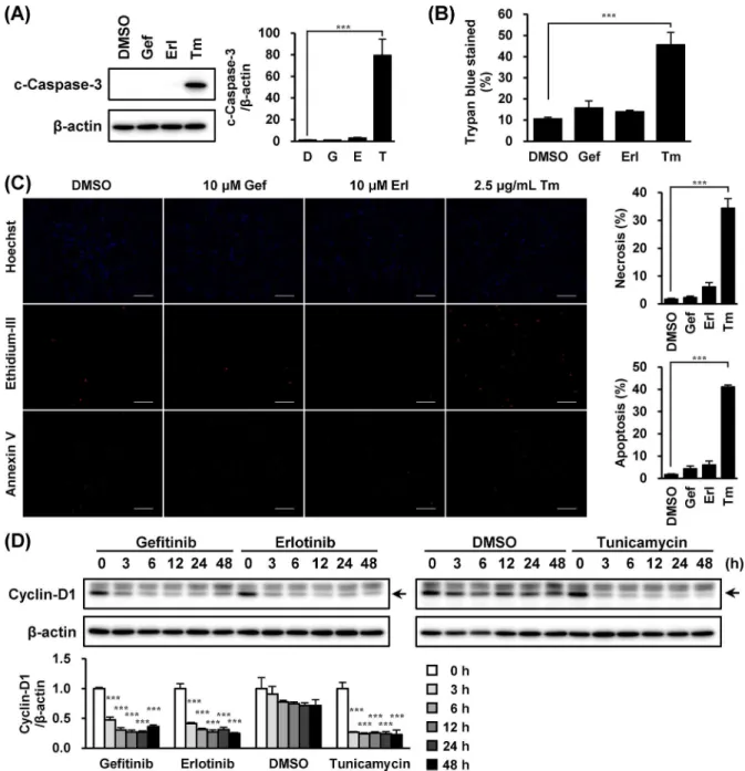
Because proapoptotic-signaling from CHOP leads to caspase-3 activation [25], we examined if EGFR inhibitors would activate caspase-3 in A549 cells. In accordance with CHOP alteration, cleaved caspase-3 was observed in cells treated with tunicamycin; however, such an event was not detected in cells treated with EGFR inhibitors (Fig 3A). Although tunicamycin evoked cell death in A549 cells, gefitinib and erlotinib did not affect the cells (Fig 3B and 3C). Meanwhile, treatment of these EGFR inhibitors reduced cyclin-D1 expression (Fig 3D).
Gefitinib and Erlotinib Activated eIF2
α
/ATF4 Signaling
Because ATF4 analogously targets upstream ofCHOPandPARK2genes [26,27], we next investigated if EGFR inhibitors activated the PERK/eIF2α/ATF4 pathway. As shown inFig 4A and 4B, ATF4 was induced and eIF2αwas phosphorylated in A549 cells treated with EGFR inhibitors. In addition, ATF4 induction was observed to lag behind eIF2αphosphorylation. When PERK is phosphorylated in ER stress conditions, gel mobility of PERK shifts upward [28]. However, this phenomenon was not confirmed when A549 cells were exposed to EGFR inhibitors (Fig 4C).
TUDCA Suppressions of eIF2
α
Phosphorylation and Cyclin-D1
Reduction by EGFR Inhibitors
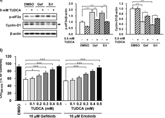
As phosphorylation of eIF2αinduces cyclin-D1 reduction [8,9], the association of eIF2α phos-phorylation with cyclin-D1 reduction caused by EGFR inhibitors was investigated. TUDCA, which is known to inhibit the phosphorylation of eIF2α[29,30], suppressed eIF2α phosphory-lation induced by gefitinib and erlotinib (Fig 5A). The reduction of cyclin-D1 expression by EGFR inhibitors was also alleviated by concomitant treatment with TUDCA (Fig 5A). In accor-dance with cyclin-D1 alteration by EGFR inhibitors, TUDCA suppressed the inhibition of cell growth induced by gefitinib and erlotinib (Fig 5B).
Phosphorylation of eIF2
α
Induced by EGFR Inhibitors Was Independent
of EGFR Inhibition
Finally, we examined if eIF2αphosphorylation and cyclin-D1 reduction by EGFR inhibitors were due to their inhibitory effect against EGFR.EGFRsiRNA strongly silenced EGFR expres-sion without suppressing phosphorylation of eIF2αin A549 cells treated with the EGFR inhibi-tors (Fig 6). The reduction of cyclin-D1 induced by EGFR inhibitors did not fully recover (to baseline) by EGFR-silencing.
Discussion
mRNA expressions ofCHOPandPARK2in this cell line.CHOPgene can be activated via the ER stress response element (ERSE) [33], unfolded protein response element (UPRE) [34], and amino acid response element (AARE). ATF4 targets at AARE [27], and transcriptionally regu-lates thePARK2gene [26]. We therefore investigated the effects of gefitinib and erlotinib on the PERK/eIF2α/ATF4 pathway by EGFR inhibitors in A549 cells. Our results demonstrated that these drugs induced phosphorylation of eIF2αand activation of ATF4, thus confirming that gefitinib and erlotinib (EGFR inhibitors) inducedCHOPandPARK2mRNA expressions via activation of the eIF2α/ATF4 pathway.
Despite eliciting eIF2αphosphorylation, gefitinib and erlotinib did not activate PERK, which is one of the sensor molecules in ER stress [35,36]. Additionally, these two drugs did not change the mRNA expressions ofHRD1andSEL1L. Transcriptions of bothHRD1and SEL1mRNA are regulated by UPRE and/or ERSE, which are the targets influencing the ATF6 and inositol requiring kinase 1α(IRE1α/X-box binding protein 1 pathway [37,38]. From the speculative‘calmness’of these three sensors (PERK, IRE1αand ATF6) responsive to ER stress, our results indicate that gefitinib and erlotinib would not induce ER stress response in A549 cells. Since eIF2αhas been reported to be phosphorylated not only by PERK, but also by gen-eral control non-derepressible-2 [39], RNA-activated protein kinase R [40], and heme-regu-lated inhibitor [41], examination of the effects of these EGFR inhibitors on the aforesaid molecules are warranted.
Although CHOP protein was transiently and slightly increased by the EGFR inhibitors tested here, the fact that CHOP induction precedes ATF4 induction suggests that this CHOP protein increase was probably not mediated by ATF4. Moreover, phosphorylation of eIF2α preferentially enhancesCHOPmRNA translation mediated by an upstream open-reading frame through intracellular eIF2-GTP depletion [42]. As such, a mechanism involving Fig 4. Gefitinib and erlotinib activated eIF2α/ATF4 signaling.The effects of gefitinib and erlotinib (10μM) on (A) eIF2α/ATF4 activation and (C) PERK
migration in A549 cells were investigated. A549 cells were treated with gefitinib (10μM) or erlotinib (10μM) for indicated times and tunicamycin (0.005–10μg/mL) for 6 h. Whole-cell lysates were analyzed by immunoblotting using antibodies respectively specific against (A) eIF2α, phosphorylated eIF2α, ATF4,β-actin, and (C) PERK. Representative images of three independent experiments are shown. (B) Asterisks indicate significant differences from 0 h group (*;p<0.05,
**;p<0.01,***;p<0.001, one-way ANOVA with Dunnett’s post hoc tests).
eIF2-GTP depletion to enhanceCHOPmRNA translation after eIF2αphosphorylation induced by these EGFR inhibitors may be plausible.
In this study, gefitinib and erlotinib did not induce caspase-3 activation when an increase in CHOP was observed. An adequate CHOP expression level can actually beget apoptosis in cells [43]. However, the induction of CHOP by gefitinib and erlotinib was much lower than that by tunicamycin (which induced significant cell death). Alternatively, persistent translational sup-pression by phospho-eIF2αmight have prevented downstream mRNA of CHOP being trans-lated into protein production. When exposed to concomitant treatment with TUDCA and EGFR inhibitors, the repression of phospho-eIF2αlevels was observed coincidently with an alleviation of cyclin-D1 decrease, indicating that EGFR inhibitors reduced cyclin-D1 expres-sion via eIF2αphosphorylation. Furthermore, when TUDCA prevented cyclin-D1 reduction induced by the EGFR inhibitors, growth inhibition was not detected. These results suggest that TUDCA might have promoted alveolar repair by preventing EGFR inhibitors from suppressing type-II cell growth. Although both drugs also phosphorylated eIF2αin PC-9 cells, which are human lung cancer cell line and are extremely sensitive to EGFR inhibitors, the protective effect of TUDCA was not found in this cell line (S1andS2Figs), suggesting that TUDCA Fig 5. The eIF2αphosphorylation and cyclin-D1 reduction by EGFR inhibitors were suppressed by tauroursodeoxycholic acid (TUDCA).The
effects of TUDCA on growth inhibition by EGFR inhibitors in A549 cells were investigated. (A) A549 cells were treated with gefitinib or erlotinib (10μM) with/ without TUDCA (0.5 mM) for 3 h (A) and 48 h (B). (A) Whole-cell lysates were analyzed by immunoblotting using antibodies respectively specific against phosphorylated eIF2α, cyclin-D1, andβ-actin. Representative images of five independent experiments are shown. (B) Cell counts were estimated by the MTT assay. Data are expressed as means±S.E.M. of three independent experiments. Asterisks indicate significant differences between two groups (*;p<0.05,**;p<0.01,***;p<0.001, one-way ANOVA with Tukey-Kramer’s tests).
treatment would not disturb the anti-cancer efficacy of EGFR inhibitors in non-small cell lung cancer patients. In this study, gefitinib and erlotinib did not induce apoptosis or necrosis in A549 cells. Since resistance of A549 cells to EGFR inhibitors is due to v-Ki-ras2 Kirsten rat sar-coma viral oncogene homolog (KRAS) mutation, the present data obtained from this cell line do not inform whether these drugs induce cell death in normal lung cells, which express nor-mal KRAS. This is the limitation of our study to clarify the cellular mechanism of pulmonary toxicity related to ILD using A549 cells.
Finally, we demonstrated in the siRNA study that eIF2αphosphorylation induced by gefiti-nib and erlotigefiti-nib was observed not to involve EGFR function. This result strongly supports the view that the eIF2αphosphorylation occurred independent of the EGFR inhibitory action. In contrast, EGFR-silencing partly ameliorated the cyclin-D1 reduction by EGFR inhibitors, implying that the event could occur in a manner both dependent and independent of EGFR, possibly via the eIF2αphosphorylation pathways. Cyclin-D1 repression induced by EGFR inhibitors has been believed to depend on the EGFR inhibitory effect, because EGF-signaling is associated with the activation of signal transducer and activator of transcription 3 and mito-gen-activated protein kinase, as well as direct induction of cyclin-D1 transcription via EGFR Fig 6. Phosphorylation of eIF2αinduced by EGFR inhibitors was independent of EGFR inhibitory
effect.Effects of EGFR knockdown on eIF2αphosphorylation and alteration of cyclin-D1 levels induced by gefitinib and erlotinib were investigated. A549 cells were transfected with 50 pmol of siRNA againstEGFRor control siRNA for 48 h before treated with gefitinib or erlotinib (10μM) for further 3 h. Whole-cell lysates were analyzed by immunoblotting using antibodies respectively specific against EGFR, phosphorylated eIF2α, cyclin-D1, andβ-actin. Representative images of five independent experiments are shown. Asterisks indicate significant differences between two groups (*;p<0.05,**;p<0.01,***;p<0.001, one-way ANOVA with Tukey-Kramer’s tests).
binding to the cyclin-D1 promoter [44–46]. However, our results suggest that the cyclin-D1 reduction caused by EGFR inhibitors also occurred via the non-EGFR pathway.
In conclusion, the EGFR inhibitors used in our study, gefitinib and erlotinib, induced eIF2α phosphorylation independently via EGFR inhibition. Followed by both EGFR inhibition and eIF2αphosphorylation, cyclin-D1 reduction raised growth inhibition of A549 cells (the model of alveolar epithelial cells). Because growth inhibition could be responsible for lung injury [47, 48], phosphorylation of eIF2αinduced by these EGFR inhibitors may potentially contribute to repair inhibition of alveoli. However, further studies to clarify the mechanism of targeting eIF2αphosphorylation by EGFR inhibitors are warranted. The present findings may serve as a potential approach to preventing incidences of ILD and pulmonary fibrosis induced by EGFR tyrosine kinase inhibitors, such as gefitinib and erlotinib.
Supporting Information
S1 Fig. Gefitinib and erlotinib arised cell death of PC-9 cells.PC-9 cells were treated with gefitinib or erlotinib (10μM) for indicated times (A and B) and 24 h (C). Cell counts were esti-mated by the MTT assay. Data are expressed as means ± S.E.M. of three independent experi-ments. Each symbol indicates significant differences from DMSO group; a,p<0.05; aa,
p<0.01; aaa,p<0.001 (DMSO vs. 0.01μM), bbb,p<0.001 (DMSO vs. 0.1μM), ccc,
p<0.001 (DMSO vs. 1μM), ddd,p<0.001 (DMSO vs. 10μM), one-way ANOVA with
Dun-nett’s post hoc tests. (C) Cells were stained by Hoechst 33342, Ethidium-III and Annexin V (Magnification ×100, scale bar; 200μm). Asterisks indicate significant differences from DMSO group. (;p<0.001, one-way ANOVA with Dunnett’s post hoc tests).
(TIFF)
S2 Fig. Gefitinib and Erlotinib phosphorylated eIF2αin PC-9 cells.PC-9 cells were treated
with gefitinib or erlotinib (10μM) with/without TUDCA for indicated times (A), 3 h (B) and 48 h (C). Whole-cell lysates were analyzed by immunoblotting using antibodies respectively specific against phosphorylated eIF2α, cyclin-D1 andβ-actin. Representative images of three independent experiments are shown. (C) Cell counts were estimated by the MTT assay. Data are expressed as means ± S.E.M. of three independent experiments. Asterisks indicate signifi-cant differences between two groups. (;p<0.05,;p<0.01,;p<0.001, one-way ANOVA with Tukey-Kramer’s tests).
(TIFF)
Author Contributions
Conceived and designed the experiments: SK TO. Performed the experiments: SK. Analyzed the data: SK TO AY SI SN TN IY. Contributed reagents/materials/analysis tools: TO KM. Wrote the paper: SK TO KM.
References
1. Inoue A, Saijo Y, Maemondo M, Gomi K, Tokue Y, Kimura Y, et al. (2003) Severe acute interstitial pneu-monia and gefitinib. Lancet 361: 137–139. PMID:12531582
2. Cersosimo RJ (2006) Gefitinib: an adverse effects profile. Expert Opin Drug Saf 5: 469–479. PMID:
16610973
3. Kudoh S, Kato H, Nishiwaki Y, Fukuoka M, Nakata K, Ichinose Y, et al. (2008) Interstitial lung disease in Japanese patients with lung cancer: a cohort and nested case-control study. Am J Respir Crit Care Med 177: 1348–1357. doi:10.1164/rccm.200710-1501OCPMID:18337594
4. Uhal BD (1997) Cell cycle kinetics in the alveolar epithelium. Am J Physiol 272: L1031–L1045. PMID:
5. Petty WJ, Dragnev KH, Memoli VA, Ma Y, Desai NB, Biddle A, et al. (2004) Epidermal growth factor receptor tyrosine kinase inhibition represses cyclin D1 in aerodigestive tract cancers. Clin Cancer Res 10: 7547–7554. PMID:15569985
6. Orzáez M, Guevara T, Sancho M, Pérez-Payá E (2012) Intrinsic caspase-8 activation mediates sensiti-zation of erlotinib-resistant tumor cells to erlotinib/cell-cycle inhibitors combination treatment. Cell Death Dis 3: e415. doi:10.1038/cddis.2012.155PMID:23096116
7. Kalish LH, Kwong RA, Cole IE, Gallagher RM, Sutherland RL, Musgrove EA (2004) Deregulated cyclin D1 expression is associated with decreased efficacy of the selective epidermal growth factor receptor tyrosine kinase inhibitor gefitinib in head and neck squamous cell carcinoma cell lines. Clin Cancer Res 10: 7764–7774. PMID:15570011
8. Raven JF, Baltzis D, Wang S, Mounir Z, Papadakis AI, Gao HQ, et al. (2008) PKR and PKR-like endo-plasmic reticulum kinase induce the proteasome-dependent degradation of cyclin D1 via a mechanism requiring eukaryotic initiation factor 2αphosphorylation. J Biol Chem 283: 3097–3108. PMID:
18063576
9. Hamanaka R, Bennett B, Cullinan S, Diehl J (2005) PERK and GCN2 contribute to eIF2α phosphoryla-tion and cell cycle arrest after activaphosphoryla-tion of the unfolded protein response pathway. Mol Biol Cell 16: 5493–5501. PMID:16176978
10. Rowlands AG, Panniers R, Henshaw EC (1988) The catalytic mechanism of guanine nucleotide exchange factor action and competitive inhibition by phosphorylated eukaryotic initiation factor 2. J Biol Chem 263: 5526–5533. PMID:3356695
11. Gebauer F, Hentze MW (2004) Molecular mechanisms of translational control. Nat Rev Mol Cell Biol 5: 827–835. PMID:15459663
12. Namba T, Tanaka K, Hoshino T, Azuma A, Mizushima T (2011) Suppression of expression of heat shock protein 70 by gefitinib and its contribution to pulmonary fibrosis. PLoS One 6: e27296. doi:10. 1371/journal.pone.0027296PMID:22096546
13. Thomas AQ, Lane K, Phillips J, Prince M, Markin C, Speer M, et al. (2002) Heterozygosity for a surfac-tant protein C gene mutation associated with usual interstitial pneumonitis and cellular nonspecific interstitial pneumonitis in one kindred. Am J Respir Crit Care Med 165: 1322–1328. PMID:11991887
14. Lawson WE, Cheng D-S, Degryse AL, Tanjore H, Polosukhin VV, Xu XC, et al. (2011) Endoplasmic reticulum stress enhances fibrotic remodeling in the lungs. Proc Natl Acad Sci U S A 108: 10562– 10567. doi:10.1073/pnas.1107559108PMID:21670280
15. Jorgensen E, Stinson A, Shan L, Yang J, Gietl D, Albino AP (2008) Cigarette smoke induces endoplas-mic reticulum stress and the unfolded protein response in normal and malignant human lung cells. BMC Cancer 8: 229. doi:10.1186/1471-2407-8-229PMID:18694499
16. Omura T, Asari M, Yamamoto J, Oka K, Hoshina C, Maseda C, et al. (2013) Sodium tauroursodeoxy-cholate prevents paraquat-induced cell death by suppressing endoplasmic reticulum stress responses in human lung epithelial A549 cells. Biochem Biophys Res Commun 432: 689–694. doi:10.1016/j. bbrc.2013.01.131PMID:23416354
17. Lin Y-C, Wu M-H, Wei T-T, Lin Y-C, Huang W-C, Huang L-Y, et al. (2014) Metformin sensitizes antican-cer effect of dasatinib in head and neck squamous cell carcinoma cells through AMPK-dependent ER stress. Oncotarget 5: 298–308. PMID:24457597
18. Kerkelä R, Grazette L, Yacobi R, Iliescu C, Patten R, Beahm C, et al. (2006) Cardiotoxicity of the cancer therapeutic agent imatinib mesylate. Nat Med 12: 908–916. PMID:16862153
19. Rahmani M, Davis EM, Crabtree TR, Habibi JR, Nguyen TK, Dent P, et al. (2007) The kinase inhibitor sorafenib induces cell death through a process involving induction of endoplasmic reticulum stress. Mol Cell Biol 27: 5499–5513. PMID:17548474
20. Imai Y, Soda M, Takahashi R (2000) Parkin suppresses unfolded protein stress-induced cell death through its E3 ubiquitin-protein ligase activity. J Biol Chem 275: 35661–35664. PMID:10973942
21. Zinszner H, Kuroda M, Wang X, Batchvarova N, Lightfoot RT, Remotti H, et al. (1998) CHOP is impli-cated in programmed cell death in response to impaired function of the endoplasmic reticulum. Genes Dev 12: 982–995. PMID:9531536
22. Song B, Scheuner D, Ron D, Pennathur S, Kaufman RJ (2008) Chop deletion reduces oxidative stress, improves beta cell function, and promotes cell survival in multiple mouse models of diabetes. J Clin Invest 118: 3378–3389. doi:10.1172/JCI34587PMID:18776938
23. Kaneko M, Ishiguro M, Niinuma Y, Uesugi M, Nomura Y (2002) Human HRD1 protects against ER stress-induced apoptosis through ER-associated degradation. FEBS Lett 532: 147–152. PMID:
12459480
24. Kaneko M, Nomura Y (2003) ER signaling in unfolded protein response. Life Sci 74: 199–205. PMID:
25. Yamaguchi H, Wang H-G (2004) CHOP is involved in endoplasmic reticulum stress-induced apoptosis by enhancing DR5 expression in human carcinoma cells. J Biol Chem 279: 45495–45502. PMID:
15322075
26. Bouman L, Schlierf A, Lutz AK, Shan J, Deinlein A, Kast J, et al. (2011) Parkin is transcriptionally regu-lated by ATF4: evidence for an interconnection between mitochondrial stress and ER stress. Cell Death Differ 18: 769–782. doi:10.1038/cdd.2010.142PMID:21113145
27. Bruhat A, Jousse C, Carraro V, Reimold AM, Ferrara M, Fafournoux P (2000) Amino acids control mammalian gene transcription: activating transcription factor 2 is essential for the amino acid respon-siveness of the CHOP promoter. Mol Cell Biol 20: 7192–7204. PMID:10982836
28. Cybulsky AV, Takano T, Papillon J, Bijian K (2005) Role of the endoplasmic reticulum unfolded protein response in glomerular epithelial cell injury. J Biol Chem 280: 24396–24403. PMID:15863508
29. Henkel AS, Dewey AM, Anderson KA, Olivares S, Green RM (2012) Reducing endoplasmic reticulum stress does not improve steatohepatitis in mice fed a methionine- and choline-deficient diet. Am J Phy-siol Gastrointest Liver PhyPhy-siol 303: G54–G59. doi:10.1152/ajpgi.00052.2012PMID:22556147
30. Macedo B, Batista AR, Ferreira N, Almeida MR, Saraiva MJ (2008) Anti-apoptotic treatment reduces transthyretin deposition in a transgenic mouse model of Familial Amyloidotic Polyneuropathy. Biochim Biophys Acta 1782: 517–522. doi:10.1016/j.bbadis.2008.05.005PMID:18572024
31. Higenbottam T, Kuwano K, Nemery B, Fujita Y (2004) Understanding the mechanisms of drug-associ-ated interstitial lung disease. Br J Cancer 91: S31–S37. PMID:15340376
32. Ware LB, Matthay MA (2000) The acute respiratory distress syndrome. N Engl J Med 342: 1334–1349. PMID:10793167
33. Yoshida H, Okada T, Haze K, Yanagi H, Yura T, Negishi M, et al. (2000) ATF6 activated by proteolysis binds in the presence of NF-Y (CBF) directly to the cis-acting element responsible for the mammalian unfolded protein response. Mol Cell Biol 20: 6755–6767. PMID:10958673
34. Lee A-H, Iwakoshi NN, Glimcher LH (2003) XBP-1 regulates a subset of endoplasmic reticulum resi-dent chaperone genes in the unfolded protein response. Mol Cell Biol 23: 7448–7459. PMID:
14559994
35. Harding HP, Zhang Y, Ron D (1999) Protein translation and folding are coupled by an endoplasmic-reticulum-resident kinase. Nature 397: 271–274. PMID:9930704
36. Harding HP, Zhang Y, Bertolotti A, Zeng H, Ron D (2000) Perk is essential for translational regulation and cell survival during the unfolded protein response. Mol Cell 5: 897–904. PMID:10882126
37. Kaneko M, Yasui S, Niinuma Y, Arai K, Omura T, Okuma Y, et al. (2007) A different pathway in the endoplasmic reticulum stress-induced expression of human HRD1 and SEL1 genes. FEBS Lett 581: 5355–5360. PMID:17967421
38. Yamamoto K, Suzuki N, Wada T, Okada T, Yoshida H, Kaufman RJ, et al. (2008) Human HRD1 pro-moter carries a functional unfolded protein response element to which XBP1 but not ATF6 directly binds. J Biochem 144: 477–486. doi:10.1093/jb/mvn091PMID:18664523
39. Zhang P, McGrath BC, Reinert J, Olsen DS, Lei L, Gill S, et al. (2002) The GCN2 eIF2alpha kinase is required for adaptation to amino acid deprivation in mice. Mol Cell Biol 22: 6681–6688. PMID:
12215525
40. Barber GN (2005) The dsRNA-dependent protein kinase, PKR and cell death. Cell Death Differ 12: 563–570. PMID:15846372
41. Chen J-J (2007) Regulation of protein synthesis by the heme-regulated eIF2alpha kinase: relevance to anemias. Blood 109: 2693–2699. PMID:17110456
42. Palam LR, Baird TD, Wek RC (2011) Phosphorylation of eIF2 facilitates ribosomal bypass of an inhibi-tory upstream ORF to enhance CHOP translation. J Biol Chem 286: 10939–10949. doi:10.1074/jbc. M110.216093PMID:21285359
43. Gotoh T, Oyadomari S, Mori K, Mori M (2002) Nitric oxide-induced apoptosis in RAW 264.7 macro-phages is mediated by endoplasmic reticulum stress pathway involving ATF6 and CHOP. J Biol Chem 277: 12343–12350. PMID:11805088
44. Lin SY, Makino K, Xia W, Matin A, Wen Y, Kwong KY, et al. (2001) Nuclear localization of EGF receptor and its potential new role as a transcription factor. Nat Cell Biol 3: 802–808. PMID:11533659
45. Park OK, Schaefer TS, Nathans D (1996) In vitro activation of Stat3 by epidermal growth factor receptor kinase. Proc Natl Acad Sci U S A 93: 13704–13708. PMID:8942998
47. McGrath-Morrow S, Lauer T, Yee M, Neptune E, Podowski M, Thimmulappa RK, et al. (2009) Nrf2 increases survival and attenuates alveolar growth inhibition in neonatal mice exposed to hyperoxia. Am J Physiol Lung Cell Mol Physiol 296: L565–L573. doi:10.1152/ajplung.90487.2008PMID:19151108