Molecular cloning and expression analysis of a zebrafish novel zinc finger
protein gene
rnf141
Wenqian Deng, Huaqin Sun, Yunqiang Liu, Dachang Tao, Sizhong Zhang and Yongxin Ma
Department of Medical Genetics, West China Hospital & Division of Morbid Genomics, State Key
Laboratory of Biotherapy, Sichuan University, Chengdu, Sichuan, People’s Republic Of China.
Abstract
ZNF230 is a novel zinc finger gene cloned by our laboratory. In order to understand the potential functions of this gene in vertebrate development, we cloned the zebrafish orthologue of humanZNF230, named rnf141. The cDNA fragment ofrnf141 was obtained by rapid amplification of cDNA ends (RACE). The open reading frame (ORF) en-codes a polypeptide of 222 amino acids which shares 75.65% identity with the humanZNF230. RT-PCR analysis in zebrafish embryo and adult tissues revealed thatrnf141 transcripts are maternally derived and that rnf141 mRNA has a broad distribution. Zygoticrnf141 message is strongly localized in the central nervous system, as shown by whole-mountin situ hybridization. Knockdown and over expression of rnf141 can induce abnormal phenotypes, in-cluding abnormal development of brain, as well as yolk sac and axis extendsion. Marker gene analysis showed that rnf141 may play a role in normal dorsoventral patterning of zebrafish embryos, suggesting that rnf141 may have a broad function during early development of vertebrates.
Key words: rnf141, zebrafish (Danio rerio), development, zinc finger protein.
Received: October 20, 2008; Accepted: May 13, 2009.
Introduction
The zinc finger gene family, one of the largest gene families in mammals, is defined by a conserved cysteine and histidine rich domain essential for the binding of zinc ions (Freemont 1993; Klug and Schwabe, 1995). This gene family can be divided into several subfamilies, including ring finger, C2H2, glucocorticoid receptor, GATA1, GAL4, and LIM (Barlow et al., 1994; Borden and Freemont, 1996; Hammarstromet al., 1996).
In accordance with their diverse structures, zinc fin-ger proteins have been assigned multiple functions, includ-ing DNA recognition, transcriptional activation, RNA packaging, regulation of apoptosis, ubiquitination and many others (Coleman, 1992; Wolfeet al., 2000; Laityet al., 2001; Vazquezet al., 2007). More than 20 different zinc finger genes located on sex chromosomes or autosomes have been proposed to play a regulatory role in mammalian spermatogenesis (Noceet al., 1992; Pieler and Bellefroid, 1994; Yanet al., 2002).
The humanZNF230, which maps to the short arm of chromosome 11 (11p15), encodes a C3HC4-type zinc
fin-ger protein motif (ring finfin-ger motif), and, consistent with a role in premeiotic or postmeiotic sperm development, one of its transcripts has been identified in abundance in the testicular tissue of fertile men, but neither in fetus nor in azoospermic patients. This suggested thatZNF230may be involved in spermatogenesis, and loss of its expression may lead to azoospermia (Zhanget al., 2001). But so far, no clear biological function and mechanism had been eluci-dated.
In order to analyse the function of this novel zinc fin-ger gene during vertebrate development, we decided to identify a potential orthologue in the teleost fish Danio rerio. This animal has become widely used as a genetic model to uncover specific functions of unknown proteins (Dooley and Zon, 2000; Rubinstein, 2003). Being transpar-ent early embryonicl stages, easy to manipulate and highly reproductive makes the zebrafish an ideal animal system for molecular studies (Moroet al., 2007).
We here report the cloning and characterization of an 816 bp cDNA sequence, namedrnf141,which represents a candidate zebrafish orthologue of the humanZNF230gene. By using whole-mountin situhybridization, RT-PCR, gene knockdown and overexpression analysis, we further showed its spatiotemporal expression pattern during early developmental stages.
Send correspondence to Yongxin Ma. Department of Medical Ge-netics, West China Hospital & Division of Morbid Genomics, State Key Laboratory of Biotherapy, Sichuan University, Renmin Nanlu, Section 3 #17, Chengdu, Sichuan, People’s Republic Of China. E-mail: [email protected].
Materials and Methods
Zebrafish embryo maintenance
Zebrafish AB strain was provided by National Zebrafish Resources of China, and maintained under stan-dard laboratory conditions at 28.5 °C (Westerfield, 1993). Embryonic stages were identified by morphological fea-tures (Kimmelet al., 1995), and embryos in developmental stages of interest were fixed in 4% paraformaldehyde.
RNA extraction and reverse transcription
Total RNAs was isolated from adult zebrafish tissues using RNeasy Mini Kit (Qiagen) according to the manufac-turer’s instructions. SuperScript TM Reverse Transcriptase (Invitrogen) was used for reverse transcription.
Cloning of the zebrafishrnf141gene
Primers for 5’-RACE (366L 5’-ACGAGACGC CTCTACCATTCCATCC-3’) and the other three pairs of primers (70U 5’-TCTCCATTTGGAGCCAAGATGGGC C-3’ and 763L 5’- TAGATTTTTAAGGTCTGTGTGGG TG-3’; 338U 5’-AGGAGGATGGAATGGTAGAGGCGT C-3’ and 619L 5’- GGCTCTGGCCGCTCCACTTGTCA AT-3’; and 432U 5’- TAGTTCAAATGTGGCGGCAG AGGGA-3’ and 816L 5’- ATATATAGGTGGTCTTTT ATGGGGA-3’) were designed to obtain the complete cod-ing sequence, based on the potential orthologue ofZNF230
in zebrafish. This orthologue sequence was acquired by PSI-BLAST alignment. 5’-RACE experiments were per-formed using SMART RACE cDNA Amplification Kit (Clontech) (Frohmanet al., 1988). cDNAs were reverse-transcribed from total RNAs of zebrafish tissue. The PCR products, including 5’- RACE products, were ligated into the pGEM-T Easy Vector (Promega), cloned and se-quenced bidirectionally. Sequences obtained by 5’-RACE and the other three fragments were assembled, and the contig was queried to the zebrafish genome database to de-termine its chromosomal location, and analyse its genomic structure. The deduced amino acid sequence was searched against InterPro Database for possible functional domains.
Multi-tissue RT-PCR
To reveal the tissue distribution and expression of zebrafishrnf141gene, total RNA was extracted from em-bryos of various developmental stages (Kimmel et al., 1995) and several tissues of adult zebrafish. The gene-specific primers 488U 5’-GGATGGGCAGGGTAAAAC AGTTGA- 3’(forward) and 683L 5’-GCATCCGACATG ACCCAGGATTCATTAG-3’ (reverse) were designed to amplify a 196 bp fragment ofrnf141. Amplification was performed in 30 cycles as follows: 30 s denaturation at 94 °C, 30 s primer annealing at 62 °C and 1 min extension at 72 °C. The PCR products were electrophoresed on 1% agarose gel in 1 x TAE buffer and ethidium bromide stained.
Primer sequences used for amplifying 470 bp ß-actin were 5’-TGTGGCCCTTGACTTTGAGCAG-3’ (forward) and 5’-TAGAAGCATTTGCGGTGGACGA-3’(reverse), according to Kaslin et al., (2004),. In negative controls, ddH2O was used instead of cDNA template. The
gene-specific primers were selected from two exons separated by an intronic sequence to identify possible amplicons from contaminating genomic DNA. All synthetic oligonuleo-tides were purchased from Invitrogen Corporation (CA, USA).
rnf141gene knockdown and overexpression experiments
rnf141 morpholino antisense oligonucleotide (rnf141-MO, 5’-CCAGAAAGCTGCTGGCCCATCTTG G-3’) was used to targetrnf141mRNA, and 5’mispaired control morpholino (5’-CCAcAAAcCTcCTGcCCCATgT TGG-3’) served as a control. Both were designed by using Gene-tools (Philomath, OR). The coding region ofrnf141
was ligated into vector pcDNA3 (Invitrogen) and linearized by appropriate restriction enzymes for mRNA was synthe-sis by using mMESSAGE mMACHINE® Kit (Ambion).
rnf141-MO and control-MO were injected into 1-2 cell zebrafish embryos by using a Model PLI-90 Pico-Injector.
Whole-mountin situhybridization
rnf141sense and antisense RNA probes were labeled with digoxigenin-11-UTP and synthesized by using DIG RNA Labeling Kit (SP6/T7) (Roche). The template for the probe was the entire 668 bp long cDNA. Whole-mountin situhybridizations was performed as described by Wester-field (1995). Images were captured using an Olympus digi-tal camera.
Results and Discussion
Zebrafishrnf141has a C3HC4 zinc finger domain
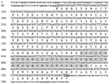
We cloned zebrafishrnf141based on the amino acid sequence of human ZNF230 and mouse znf230 by PSI-BLAST alignment and RT-PCR including RACE. As a re-sult, four fragments were obtained that formed a816 bp cDNA contig. This was submitted to GenBank (accession number: AY621088). Previous studies have shown that hu-manZNF230 has two transcripts (Zhanget al., 2001). In mouse, there are three transcripts of znf230 (Qiu et al.
2003). In contrast, in our analyses on zebrafish we found only one transcript of this gene. Furthermore, our result was confirmed by another mRNA sequence record (GenBank accession numbe BC071534) that encodes the same protein submitted by Strausberget al.. Also, the sequence of our 3’-RACE fragment is identical to this sequence record.
structure generally are nuclear transcriptional factors with the motifs being involved in both protein-DNA and pro-tein-protein interactions. We found that zebrafishrnf141
protein shares 75.65% and 75.22% identity in amino acid sequence with the human and mouse homologues, respec-tively. Furthermore, aligning amino acid sequence of zebrafishrnf141withZNF230of other vertebrate species indicated that this gene domain is highly conserved (date not shown), suggesting functional similarity and conserva-tion.
rnf141is expressed in the CNS, primarily during early embryogenesis
To analyse the spatiotemporal expression ofrnf141
during early embryonic development, whole-mountin situ
hybridizations were performed on two-cell stage to five-day-old embryos using an antisense probe. As a result,
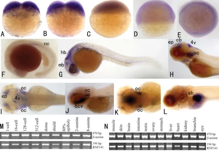
rnf141 transcripts were already detected at the two-cell stage (Figure 2A), thus suggesting a maternal origin of the transcript. From the sphere stage (4 hpf) to the tail bud stage (10 hpf) (Figure 2C-E), thernf141transcripts have a broad distribution. However, at the 5-somite stage (11.6 hpf), a characteristic pattern was displayed with marked staining in the notochord (Figure 2F), and at the Prim-5 stage of the pharyngula period, this pattern was displayed in the mid-brain and hindmid-brain (Figure 2G). Following the long-pec stage (48 hpf), restricted signal localization was evident in the otic capsule, 4th ventricle, epiphysis and cerebellum (Figure 2H-I). When embryos reached the protruding-mouth stage (72 hpf), obvious signals were detected in the oral cavity and otic capsule (Figure 2J-K). In 5 dpf (120 hpf) embryos, an extensivernf141expression was vis-ible in the gut, with a restricted localization in the swim
bladder (Figure 2L). To assess the specificity of the antisense probe, a sense probe was used in a parallel control experiment at all stages. With this sense probe no staining was detected in any embryo (Figure 3).
The consistency of hybridization experiments was confirmed by RT-PCR expression analysis performed on cDNAs from whole zebrafish embryos at various early de-velopmental stages (Figure 2M).
Since the 1 kb transcript of humanZNF230is only ex-pressed in fertile male testis, whereas another 4.4 kb tran-script was detected in many tissues; include heart, brain, skeletal muscle, kidney and pancreas (Zhanget al., 2001), we further addressed the question as to whether zebrafish
rnf141maintains its ubiquitous spatial expression in adult stages. As shown in Figure 2N, RT-PCR based analysis demonstrated that almost all analysed tissues of adult fish do display a high content ofrnf141transcripts.
In conclusion, these results of whole-mountin situ
hybridization and RT-PCR analyses performed both on zebrafish embryos and adult tissues provide evidence that
rnf141may have multiple functions. The detection of its transcripts in the CNS of early embryos, especially re-stricted in the notochord at the 5-somite of the segmenta-tion period, suggests a funcsegmenta-tion for rnf141 in zebrafish development. Further analysis is ongoing in order to im-prove knowledge on the role ofrnf141.
rnf141may play a part in normal dorsoventral patterning of zebrafish embryos
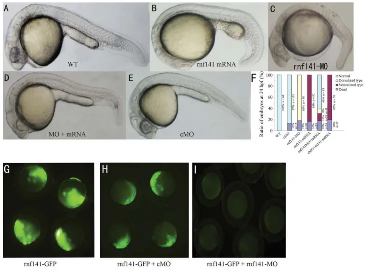
To further study the potential function ofrnf141, we first injected zebrafish embryos with synthetic rnf141
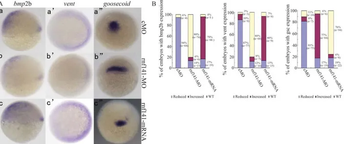
mRNA. Injection of 200 pgrnf141 mRNA caused 84% (n = 92) of the embryos to show phenotypes that are charac-teristic of embryonic ventralization at 24 hpf (Figure 4B). The expression of the shield-specific genegoosecoidwas decreased at the shield stage (Figure 5Ac”). In contrast, the ventral markersbmp2bandventexpanded dorsally during gastrulation (Figure 5Ac-c’). The ratios of embryos with al-tered marker gene expression are summarized in Figure 5B. To investigate the role of endogenous rnf141, a morpholino antisense oligonucleotide (rnf141-MO) was in-jected into one-cell embryos. As a result, 81% (n = 88) of the embryos injected with 12 ng rnf141-MO exhibited dorsalized phenotypes at 24 hpf: complete loss of the yolk sac extension and partial loss of the caudal ventral fin (Fig-ure 4C). The effects ofrnf141knockdown on the expres-sion of the marker genes bmp2b, vent and goosecoid
(Figure 5Ab-b”) tended to be opposite to those ofrnf141
overexpression. In contrast, injection with 15 ng of control morpholino, which differs from rnf141-MO in five mis-matched nucleotides, did not cause developmental defects (Figure 4E).
To test the efficiency of the morpholinos, fertilized eggs were injected with 12 ngrnf141-MO in combination with 100 pg of prnf141-GFP DNA, an expression construct
containing the full coding region ofrnf141 cDNA fused in-frame to a GFP coding sequence. At this dose ofrnf141
-MO, the injected embryos almost lacked green fluores-cence from the GFP fusion protein (Figure 5I), while the
Figure 2- Expression analysis ofrnf141in early embryos and adult zebrafish tissues.rnf141mRNA was initially detected at the 2-cell stage (A) and 4-cell stage (B), the signal become weaker at the sphere stage (C), shield stage (D) and bud stage (E). At the 5-somite stage (11.6 hpf), a characteristic pat-tern was displayed with marked staining in the notochord (F). At the 5-prim stage (24 hpf), a strong signal was detected in the head, particularly in the midbrain, hindbrain and the otic capsule (G). At the long-pec stage (48 hpf) the signal localization became restricted to the otic capsule, the 4thventricle,
as well as the epiphysis and tegmentum (H-lateral view from left; I-dorsal view). An even more restricted expressions was detected in the oral cavity and otic capsule when embryos reached the mouth-protruding stage (72 hpf) (J- lateral view from left; K-dorsal view). An extensive expression in the gut and a restricted localization in swim bladder was found in 5 dpf embryo (L). Expression analysis ofrnf141detected by RT-PCR in different developmental stage embryos (M) and adult zebrafish tissues (N). Abbreviations: mb, midbrain; hb, hindbrain; ep, epiphysis; cb, cerebellum; 4v, 4th ventricle; oc, otic capsule; ocv, oral cavity; sb, swim bladder.
same dose ofrnf141-cMO injected embryos retained visi-ble fluorescence (Figure 5H), suggesting thatrnf141-MO could effectively block translation ofrnf141mRNA.
To test the specificity of rnf141-MO, a 5 mis-pair
rnf141mRNA corresponding to 5 mis-pair control mor-pholino was synthesized for rescuing the phenotype medi-ated with rnf141-MO. These results showed that the
rnf141-MO-induced dorsalization could be neutralized by coinjection with a smaller amount of 5 mis-pair rnf141
(Figure 4D), suggesting thatrnf141-MO specifically tar-getsrnf141.
In conclusion, knockdown ofrnf141by using special morpholino-induced abnormal outcomes, including inordi-nate development of the CNS with an atrophic hindbrain, thin and crooked notochord, as well as disappearance of yolk sac extension, abnormality of axis, and partial loss of the caudal ventral fin. These embryos are characteristic of
weakly dorsalized phenotypes, reminiscent of mini fin (mfn) and lost-a-fin (laf) mutant embryos, which were first described by Mullinset al(1996) and were subsequently found to be caused by inefficient BMP signaling (Baueret al., 2001; Connors et al., 1999; Mintzer et al., 2001). Overexpressing this gene by injection of rnf141 mRNA may caused embryos to show ventralized phenotypes. We also noted thatrnf141-MO-induced dorsalization could be neutralized by coinjection of a smaller amount ofrnf141
mRNA, suggesting that rnf141-MO specifically targets
rnf141mRNA. The expression of ventral markers (bmp2b
andvent) and of a dorsal marker (goosecoid) were impacted by altered expression of rnf141, thus suggesting that zebrafish rnf141 may participate in normal dorsoventral embryonic patterning. Further research is needed to better understand the respective biological pathway(s) and im-prove the knowledge on the function ofrnf141.
Figure 4- Knockdown and overexpression analysis ofrnf141. All images are lateral views of live embryos at 24 hpf, anterior is to the left. (A) Wild-type embryo. (B) Injection with 200 pgrnf141mRNA resulted in enlargement of the yolk sac, in addition an extension and broadening of caudal ventral fin. (C) Injection with 12 ngrnf141-MO led to caudal ventral fin loss of and yolk sac extension. (D) Coinjection with 100 pgrnf141mRNA and 12 ng
Acknowledgments
This research is supported by the National Natural Science Foundation of China (30770812, 90408025 and 30500186) and National High-Tech Research and Devel-opment Program of China (2008AA02Z102).
References
Barlow PN, Luisi B, Milner A, Elliott M and Everett R (1994) Structure of the C3HC4 domain by 1H-nuclear magnetic resonance spectroscopy. A new structural class of zinc-finger. J Mol Biol 237:201-211.
Bauer H, Lele Z, Rauch GJ, Geisler R and Hammerschmidt M (2001) The type I serine/threonine kinase receptor Alk8/Lost-a-fin is required for Bmp2b/7 signal transduction during dorsoventral patterning of the zebrafish embryo. De-velopment 128:849-858.
Borden KL and Freemont PS (1996) The RING finger domain: A recent example of a sequence-structure family. Curr Opin Struct Biol 6:395-401.
Coleman JE (1992) Zinc proteins: Enzymes, storage proteins, transcription factors, and replication proteins. Annu Rev Biochem 61:897-946.
Connors SA, Trout J, Ekker M and Mullins MC (1999) The role of tolloid/mini fin in dorsoventral pattern formation of the zebrafish embryo. Development 126:3119-3130.
Dooley K and Zon LI (2000) Zebrafish: A model system for the study of human disease. Curr Opin Genet Dev 10:252-256. Freemont PS (1993) The RING finger. A novel protein sequence
motif related to the zinc finger. Ann NY Acad Sci 684:174-192.
Frohman MA, Dush MK and Martin GR (1988) Rapid production of full-length cDNAs from rare transcripts: Amplification using a single gene-specific oligonucleotide primer. Proc Natl Acad Sci USA 85:8998-9002.
Hammarstrom A, Berndt KD, Sillard R, Adermann K and Otting G (1996) Solution structure of a naturally-occurring zinc-peptide complex demonstrates that the N-terminal zinc-bin-ding module of the Lasp-1 LIM domain is an independent folding unit. Biochemistry 35:12723-12732.
Kaslin J, Nystedt JM, Ostergard M, Peitsaro N and Panula P (2004) The orexin/hypocretin system in zebrafish is con-nected to the aminergic and cholinergic systems. J Neurosci 24:2678-2689.
Kimmel CB, Ballard WW, Kimmel SR, Ullmann B and Schilling TF (1995) Stages of embryonic development of the zebrafish. Dev Dyn 203:253-310.
Klug A and Schwabe JW (1995) Protein motifs 5. Zinc fingers. FASEB J 9:597-604.
Laity JH, Lee BM and Wright PE (2001) Zinc finger proteins: New insights into structural and functional diversity. Curr Opin Struct Biol 11:39-46.
Mintzer KA, Lee MA, Runke G, Trout J, Whitman M and Mullins MC (2001) Lost-a-fin encodes a type I BMP receptor, Alk8, acting maternally and zygotically in dorsoventral pattern formation. Development 128:859-869.
Moro E, Maran C, Slongo ML, Argenton F, Toppo S and Onisto M (2007) Zebrafish spata2 is expressed at early develop-mental stages. Int J Dev Biol 51:241-246.
Mullins MC, Hammerschmidt M, Kane DA, Odenthal J, Brand M, van Eeden FJ, Furutani-Seiki M, Granato M, Haffter P, Heisenberg CP, et al. (1996) Genes establishing dorso-ventral pattern formation in the zebrafish embryo: The ven-tral specifying genes. Development 123:81-93.
Noce T, Fujiwara Y, Sezaki M, Fujimoto H and Higashinakagawa T (1992) Expression of a mouse zinc finger protein gene in both spermatocytes and oocytes during meiosis. Dev Biol 153:356-367.
Pieler T and Bellefroid E (1994) Perspectives on zinc finger pro-tein function and evolution - An update. Mol Biol Rep 20:1-8.
Figure 5- Expression patterns of marker genes in injected embryos at shield stage. (A) Expression patterns ofbmp2b,vent,goosecoidin embryos injected with 15 ngrnf141-5mis-MO, 12 ngrnf141-MO, or 200 pgrnf141mRNA, respectively. Forbmp2bexpression, the embryo is shown in lateral view with dorsal pointed towards right; the embryo showingventexpression is depicted in animal pole view with dorsal oriented towards left; and the embryo with
Qiu W, Zhang S, Xiao C, Xu W, Ma Y, Liu Y and Wu Q (2003) Molecular cloning and characterization of a mouse sperma-togenesis-related ring finger gene znf230. Biochem Biophys Res Commun 306:347-353.
Rubinstein AL (2003) Zebrafish: From disease modeling to drug discovery. Curr Opin Drug Discov Dev 6:218-223.
Strausberg RL, Feingold EA, Grouse LH, Derge JG, Klausner RD, Collins FS, Wagner L, Shenmen CM, Schuler GD, Altschul SF,et al.(2002) Generation and initial analysis of more than 15,000 full-length human and mouse cDNA se-quences. Proc Natl Acad Sci USA 99:16899-16903.
Vazquez O, Vazquez ME, Blanco JB, Castedo L and Mascarenas JL (2007) Specific DNA recognition by a synthetic, mono-meric Cys2His2 zinc-finger peptide conjugated to a mi-nor-groove binder. Angew Chem Int Ed Engl 46:6886-6890.
Westerfield M (1993) The Zebrafish Book: A Guide for the Labo-ratory Use of Zebrafish. University of Oregon Press, Ore-gon, pp 8-48.
Westerfield M (1995) The Zebrafish Book: A Guide for the Labo-ratory Use of Zebrafish. Eugene Press, Oregon, pp 200-204.
Wolfe SA, Nekludova L and Pabo CO (2000) DNA recognition by Cys2His2 zinc finger proteins. Annu Rev Biophys Biomol Struct 29:183-212.
Yan W, Burns KH, Ma L and Matzuk MM (2002) Identification of Zfp393, a germ cell-specific gene encoding a novel zinc fin-ger protein. Mech Dev 118:233-239.
Zhang S, Qiu W, Wu H, Zhang G, Huang M, Xiao C, Yang J, Kamp C, Huang X, Huellen K,et al.(2001) The shorter zinc finger protein ZNF230 gene message is transcribed in fertile male testes and may be related to human spermatogenesis. Biochem J 359:721-727.
Internet Resources
Zebrafish Genome Resources, http://www.ncbi.nlm.nih.gov/pro-jects/genome/guide/zebrafish/ (U.S. National Library of Medicine, Maryland MD, 2005).
InterPro Database, http://www.ebi.ac.uk/interpro/ (European Bioinformatics Institute, 2006).
Associate Editor: André Luiz Paranhos Perondini