Cloning and Characterization of SmZF1, a Gene Encoding
a
Schistosoma mansoni
Zinc Finger Protein
Paulo R Eleutério de Souza*, Analina F Valadão*, Carlos E Calzavara-Silva*,
Glória R Franco*, Marcos A de Morais Júnior, Frederico GC Abath**/
+Laboratório de Imunopatologia Keizo Asami and Departamento de Genética, Universidade Federal de Pernambuco, Recife, PE, Brasil *Laboratório de Genética-Bioquímica, Departamento de Bioquímica e Imunologia,
ICB, Universidade Federal de Minas Gerais, Belo Horizonte, MG, Brasil **Departamento de Imunologia, Centro de Pesquisas Aggeu Magalhaes-Fiocruz, Av. Moraes Rego s/no, Cidade Universitária, 50670-420
Recife, PE, Brasil
The zinc finger motifs (Cys2His2) are found in several proteins playing a role in the regulation of transcripton. SmZF1, a Schistosoma mansoni gene encoding a zinc finger protein was initially isolated from an adult worm cDNA library, as a partial cDNA. The full sequence of the gene was obtained by subcloning and sequencing cDNA and genomic fragments. The collated gene sequence is 2181 nt and the complete cDNA sequence is 705 bp containing the full open reading frame of the gene. Analysis of the genome sequence revealed the presence of three introns interrupting the coding region. The open reading frame theoretically encodes a protein of 164 amino acids, with a calculated molecular mass of 18,667Da. The predicted protein contains three zinc finger motifs, usually present in transcription regulatory proteins. PCR amplification with specific primers for the gene allowed for the detection of the target in egg, cercariae, schistosomulum and adult worm cDNA libraries indicating the expression of the mRNA in these life cycle stages of S. mansoni. This pattern of expression suggests the gene plays a role in vital functions of different life cycle stages of the parasite. Future research will be directed to elucidate the functional role of SmZF1.
Key words: DNA binding protein - gene cloning - Schistosoma mansoni - zinc finger protein
Schistosomiasis is a human disease caused by several trematodes of the genus Schistosoma, with approximately 200 milion people currently infected and a further 500-600 milion worldwide at risk of infection throughout tropical and subtropical ar-eas of the world (Savioli et al. 1997). In South America, S.mansoni is the only causative agent of schistosomiasis. Schistosome worms have a com-plex life-cycle involving molluscus and vertebrate hosts as well as short periods of larvae swimming freely in water. The evolution from one develop-mental stage to another involves modifications in the morphology, physiology and biochemistry of the parasite, being associated with the activation/ inactivation of stage-specific genes. Indeed, sev-eral stage-specific genes with different patterns of
expression during the schistosome life-cycle have been described (Simpson et al. 1984, Davis et al. 1985, Grossman et al. 1990, Chen et al. 1992, Abath et al. 1994, Mei & Lo Verde 1997).
The recognition of specific sequences of DNA by proteins is a central mechanism for a number of important biological processes, including gene ex-pression, recombination, and DNA repair (Mitchell & Tjian 1989). Many of those proteins, including transcription factors, present zinc finger motifs in their structure, that are responsible for the binding to DNA sequences (Bernstein et al. 1994, Yokono et al. 1998, Clarke & Berg 1998). The classical C2H2 zinc finger motif consists of about 30 amino acids, four of which (two cysteines and two histidines -Cys2His2) coordinate tetrahedrically a single zinc atom, forming a loop of twelve amino acids between the second cysteine residue and the first histidine residue. The cysteine residues are separated by two or four amino acids and the two histidine resi-dues are separated by three to five amino acids (Berg 1990, Bernstein et al. 1994). The sequence, number and organization of the zinc finger motifs, are important for the biological function of the pro-tein.
The study of gene expression regulation in S. mansoni is still incipient. Thus, the molecular
char-This work was supported by grants from Brazilian Re-search Council, Fundação de Amparo à Ciência e Tecnologia do Estado de Pernambuco and Fundação de Amparo à Pesquisa do Estado de Minas Gerais.
+Corresponding author. Fax: +55-81-3453.2449. E-mail:
acterization of these regulatory proteins in Schis-tosoma may contribute to a better understanding of the biology of the parasite as well as the evalua-tion of these proteins as targets for immunotherapy or drug therapy. The present communication re-port on the cloning and structural characterization of SmZF1, a gene encoding a putative transcrip-tion regulatory protein of S. mansoni containing three zinc finger motifs. In addition, evidence is provided indicating that SmZF1 is expressed in dif-ferent life-cycle stages of the parasite.
MATERIALS AND METHODS
DNA purification and cDNA libraries - Ge-nomic DNA was purified from S. mansoni LE strain adult worms as described previously (Simpson et al. 1982). The plasmids and polymerase chain reac-tion (PCR) fragments were purified with the Wizard DNA Purification Systems (Promega). S. mansoni adult worm cDNA libraries were con-structed in λgt11 (Abath et al. 1993). In addition, egg, cercariae, 3 h schistosomulum, and adult worm cDNA libraries UUEE and SmZU were constructed in λZAP as part of the Schistosoma genome project (Franco et al. 2000).
PCR - A number of primers, targeting specific regions of the gene, were used to amplify DNA fragments for cloning into the vectors pBlueScript KS+ (Stratagene) and pUC18 (Amersham Pharmacia Biotech), and further sequencing (Table, Fig. 1).
For the amplification of the SmZF1 gene from the λgt11 library, 100 µl reaction mixture was used containing approximately 4 µl of the cDNA library, 10 mM Tris HCl pH 8.4, 50 mM KCl, 1.5 mM MgCl2, 200 µM each deoxynucleotide triphosphate, 250 nM each primer and 2.5 U of Taq polymerase. The con-ditions used for amplification were 94oC for 4 min, followed by a step cycle program set to denature at 94oC for 1 min, anneal at 55oC for 1 min, and extend at 72oC for 2 min for a total of 30 cycles. Amplifica-tions of the other cDNA libraries were performed in a 30 µl volume containing 1 µl of the cDNA library, 10 mM Tris HCl pH 8.3, 75 mM KCl, 3.5 mM MgCl2, 200 µM each deoxynucleotide triphosphate, 200 nM each primer and 2 U of Taq DNA polymerase. PCR of genomic DNA was performed in a 30 µl volume containing 20 ng of genomic DNA, 10 mM Tris HCl pH 8.8, 75 mM KCl2, 3.5 mM MgCl2, 200 µM each deoxynucleotide triphosphate, 400 nM each primer and 2U of Taq DNA polymerase. The conditions used for the amplifications were 94oC for 4 min, followed by a step cycle program set to denature at 94oC for 1 min, anneal at 52oC for 1 min, and extend at 72oC for 1 min for a total of 30 cycles. The amplicons were analyzed in 1% agarose gel stained by ethidum bromide or in 6% polyacrylamide gels silver stained (Santos et al. 1993).
DNA cloning and sequencing - An adult worm
λgt11 cDNA library was screened by hybridization with the cDNA A157 (GenBank U67153) as a probe, following the instructions of the Digoxigenine-DNA labelling/Detection kit (Boehringer Mannheim). The PCR product of the cDNA clone isolated was cloned into the EcoRI digested pBlueScript KS+. Other clones were inserted into the SmaI site of pUC18 using the Surclone ligation kit (Amersham Pharmacia Biotech).
The sequencing reactions were performed us-ing the Thermo Sequenase fluorescent labelled primer cycle sequencing kit with 7-deaza-dGTP (Amersham Pharmacia Biotech). Fluorescent prim-ers targeting the margins of the cloning sites were used for DNA sequencing of both strands, using the A.L.F. DNA Automated Sequencer (Amersham Pharmacia Biotech).
Sequence analysis - Search for homologous sequences was undertaken using the BLAST pro-gram (Altschul et al. 1997, http://www. ncbi.nlm.nih.gov). Open reading frame (OFR) search and DNA translation were performed using the DNAsis program. The PredictProtein server (http:/ /cubic.bioc.columbia.edu/predictprotein/) was used for prediction of secondary structure and post-translational modification sites, whereas the “SDSC1” - SDSC Protein Structure Homology Mod-eling Server (http://cl.sdsc.edu/hm.html) was used for prediction of the three-dimensional structure of the SmZF1 gene product, using the Zif268 mouse protein (PDB accession 1mey_C) as a model, due to its high similarity with SmZF1. The hydropho-bicity plot was calculated according to Kyte and Doolittle (1982).
RESULTS
Cloning and sequencing of SmZF1, a gene encoding a S. mansoni zinc finger protein - The cDNA ZNF-17 was casually isolated from an adult worm λgt11 cDNA library during attempts to ob-tain the complete cDNA for Sm13, a S. mansoni tegumental antigen. This cDNA was amplified and cloned into pBlueScript KS+, and DNA sequenc-ing revealed that the 603 bp cDNA shared no sig-nificant homology to the A157 clone (GenBank U67153) of the Sm13 gene (Abath et al. 2000). In-stead, it encoded a 88 amino acid protein which contained three zinc finger motifs, although lack-ing its N-terminal region.
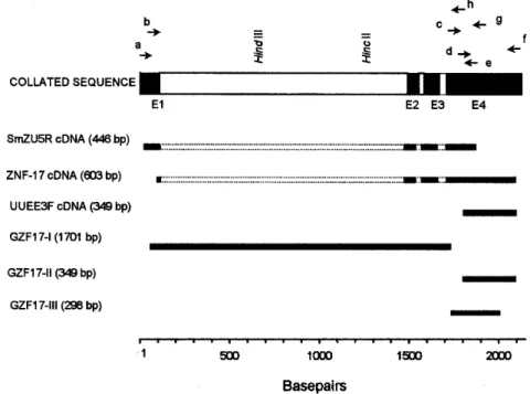
On the basis of the sequence of the ZNF-17 cDNA, several primers were sinthesized to se-quence the complete open reading frame of the gene and detect the existence of possible introns (Fig. 1). The 446 bp fragment SmZU-5R amplified from a
of an additional sequence to the 5' region, while the 349 bp fragment UUEE-3F amplified from another
λZAP adult worm cDNA library with primers ZFF1 and ZnFLower, identified the poly-A tail of the 3’ region of the gene.
Characterization of the intron-exon pattern of SmZF1 - Primers were designed based on the col-lated SmZF1 cDNA sequence to characterize the genomic structure of the SmZF1 gene (Fig. 1, Table). The genomic fragment GZF17-I (obtained by am-plification of genomic DNA with primers ZnFStart and ZnFMR) of approximately 1.7 kb was larger than the corresponding cDNA sequence, indicat-ing the presence of introns in that region. The par-tial sequencing of the terminal regions of GZF17-I allowed the identification of restriction sites (HindIII and HincII) (Fig. 1) used in the subcloning strategies to sequence this DNA fragment. The
sequences of the genomic fragments GZF17-II (ob-tained using primers ZFF1 and ZnFLower) and GZF17-III (amplification with primers ZnFMF and ZnFStop) (Fig. 1) were identical to the cDNA se-quence, indicating absence of introns. After com-plete sequencing of the clones and subclones, se-quence alignment revealed the presence of three introns: the first intron is 1408 bp (nucleotides 109 to 1516), the second 32 bp (nucleotides 1578 to 1609) and the third 37 bp (nucleotides 1697 to 1733) (Figs 1, 2). The canonical donor (GT)/acceptor (AG) splicing sites are present at all exon/intron junc-tions (von Heijne 1987). The mean A + T content of the introns was around 68%, higher than that ob-served for the coding region. The 2181 bp collated sequence may represent the region that corre-sponds to the primary transcript of the SmZF1 gene (Figs 1, 2).
TABLE
Set of specific primers used for cloning and sequencing of the SmZF1 gene
Primer Sequence Nucleotide position
ZFR1 5’-GTTTTACACCTTTATGG-3’ Reverse (1906-1922)
ZFF1 5’-GAGTTCTGAATTGTGCG-3 Forward (1833-1849)
ZnFLower 5’-GAGGAATTCTGAAAGAATAATAAATGTA-3’ Reverse (2164-2181)
ZnFMF 5’-ACGGAAATAGATACTGAA-3’ Forward (1736-1753)
ZnFStart 5’-GAGGAATTCATGGAATTTTACTTCACA-3’ Forward (53-70) ZnFStop 5’-GAGAAGCTTCTGGCATACTTCACAT-3’ Reverse (2018-2033)
ZnFMR 5’-TTCAGTATCTATTTCCGT-3 Reverse (1736-1753)
ZnFUpper 5’-GAGCTGCAGGTTCAGTAAAATGTTCAA-3’ Forward (1-18) Restriction sites inserted at the 5´end of some primers are underlined.
Theoretical analysis and structural predictions - The final SmZF1 cDNA sequence obtained re-vealed a 5’ untranslated region (5’ UTR) of 52 bp, an ORF of 492 bp which encodes a 164 aa protein with a calculated molecular mass of 18,667Da (esti-mated pI of 7.62) and a 3’ UTR of 161 bp without any characteristic polyadenylation site but present-ing a poly-A tail. The putative ATG initiation codon
is in agreement with the Kosak consensus sequence (Kosak 1987) (Fig. 2). The A+T content of the cDNA sequence, about 61% within the coding region and 65% in both 5’ and 3’ untranslated regions, and its codon usage are in accordance to the values de-scribed previously for S. mansoni genes (Milhon & Tracy 1995). The derived amino acid sequence is rich in lysine (9.8%), threonine (7.9%),
phenylala-GTTCAGTAAAATGTTCAATTGTGCGCTGGAATCTATTGTGTAGACTTTAACTATGGAATTTTACTTCACA 70 M E F Y F T 6 TTGACTAAAAAGCTGAGCAAATATACCTGGAGCGTTCAGgtacgtttggggatacatcatatgttgcgtt 140 L T K K L S K Y T W S V Q 19 agtgtaacgcatattggtcgctacatttgatgacacttttgaatattccttaatcggattatctcgaagt 210 agaagcgtcatgggagaagaagcatttaggttttgtatttcaaatttggatataaagtaattagtatgcc 280 accatacatatttacggtgagctattttagtcgtcaaaaatatgagataatggaagcattttgaatgccg 350 attatgctatgtttttcgtcttcagcctcatgtttcactgaccggaaattcttgcttacgatagtcatcg 420 ggaatacagagttgtgttcactcgacacatctttctcaaatgccaggctaatgtataaaatcagtctact 490 aagtgaataaaattactttacattaggatgcttgtgctctttcaataacataatgctgtataacaaaaat 560 ctgtctatttcatgttcctaaagttatcagcaagtctatttgtcgttgttttgtctcgagaaaagcttgt 630 tgtctaggtttcatcgtattctaggtattacaattaagacacaataccagccaacgtaataaaattggtc 700 ttccgtaataacctggtttttcgctgcgcatgaatatggtatatagttatataaaataactgatagtgtc 770 acgagctgctgtttggcctgataggtttgtcactgaatatctgctatctcccattcgaaattccctattt 840 gttaaaaaatccccttcaaacaaactagtaaaatatcgtgagtccaaatggtatgaaagtttggcgttga 910 tcattgtttctaaactgtagtttattgcctagatttaattagccagtaaatgacgatctctactccacat 980 ttattagtaacagacgttgagaattcggcatcatagatgacgtcaattactgaataagggttcagtcaat 1050 agaccaagatgtcatcagcccctactgccataagagcctatctagtagtagtgcgtccggacggtaccgg 1120 ttatcctacaaacatactagtaaagcagcactccaaatctttataaaaaacatccgcaacatacccatgt 1190 caccaaggttactcaactaacttatgtttccgcgtcttgatcctataagagtacatggccagcatatccc 1260 acgccagttaaagaatcagctattcaactctttaagcacgttaacagtaaacttgttgcaatatgatatt 1330 gtggttgccccttcagattaaatactttaggtgaatgcttccgcaaacgcaaatataagttcgtagttcc 1400 catactacttctgtttactactgctactacgagttttttttattcattttcctgtgtattcatccccgga 1470 ttcctagtatcccaatgctgttgatcttgtcgccttatatctgtagACTTTCAAGATGAACGAACCAACT 1540 T F K M N E P T 27 GGTGTCGGGCCAACATTTGCTGATGCATGCGATGATGgtaatctttaactataatcctatcgttttcagG 1610 G V G P T F A D A C D D G 40 CGAACTTATCAGCATTTGTTGTCTTTGTGGTAAAACGTTTTCAAGTCAGAGTCTTCTACACAAACATTTT 1680 E L I S I C C L C G K T F S S Q S L L H K H F 63 GAATTGATGCATGAAGgtaactcgtcaagaggatttcaaacagtttaaattagGTACGGAAATAGATACT 1750 E L M H E G T E I D T 74
GAACAGTATGATCTAAGTGGATTTGCCGCTATGGGGAATGAACAAGGTCGTAAAAGTAATGGTGAAGAAG 1820 E Q Y D L S G F A A M G N E Q G R K S N G E E D 98 ATGCAAATTTCCGAGTTCTGAATTGTGCGTTTTGCAACAAAGTATTTACTAAACACTGTAATTTAAACAC 1890 A N F R V L N C A F C N K V F T K H C N L N T 121
ACATATCAAAGCAGTCCATAAAGGTGTAAAACCGTTTGAATGCACTTATTGTTATAAAGGATTCACTCGA 1960 H I K A V H K G V K P F E C T Y C Y K G F T R 144 AATTCTGATCTTCATAAGCACATCGACGCTGTTCACAAAGGTCTCAAGCCTTTCCGGATGTGAAGTATGC 2030 N S D L H K H I D A V H K G L K P F R M * 164 CAGCGAAACTTCTCTCAGAAATCCAGCCTAAAACGACACATAGAAGCAATTCACGAAGATCCTCGGCATC 2100 GCTGAAGAGAAACCAGATTGTATAATCCTCTCCAATTTTCATATGATTTCATGTTCAAAAATATACATTT 2170 ATTATTCTTTC 2181
nine (7.9%) and glycine (7.3%), however very poor in arginine (2.4%) and tryptophan (0.6%).
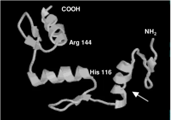
Protein database searches revealed that the derived SmZF1 protein shows about 47% similarity with many eukaryotic zinc finger proteins that are involved in transcriptional regulation (data not shown). When EST database (dbEST) was searched, one S. mansoni cDNA sequence (GenBank accesion number AA559461) with simi-larity to the gene described herein was found, indi-cating the isolation of a homologous cDNA by the Schistosoma Genome Initiative. An attribute of the SmZF1 protein was the presence of four amino acid residues between the two histidines residues in all three zinc finger motifs (Fig. 2), whereas in the other proteins analyzed three residues predominated at that position (not shown). The three Cys2His2 zinc fingers motifs of 22 aa extension fit to the consen-sus sequence Cys-X2-4-Cys-X3-Phe-X5-Leu-X2 -His-X3-5-His (Berg 1990, Bernstein et al. 1994). The putative three-dimensional structure of the protein was obtained by comparative molecular modelling using the Zif268 mouse protein (PDB accession 1mey_C). Computational modelling clearly demon-strated the presence of three zinc fingers motifs for the SmZF1 protein (Fig. 3), where each finger unit is composed by two antiparallel β-strands at the cysteine side and one α-helix at the histidine side. Together, these structures confer a semicircular C-shape arrangment for SmZF1 protein. A 27-amino acid deletion (residues 64 to 90) was generated by the modelling program at the end of the α-helix of finger 1, since there was no correspondence of this
region in the Zif268 protein. Thus, this helix in the model presented here is formed by the junction of SmZF1 residues 56 to 63 and 91 to 95. According to the secondary structure prediction, using the PredictProtein server, another two putative α-helix can be present in the protein: a1 at the N-terminal region residues 11 to 14 and α3 between the first and the second fingers residues 89 to 95 (data not shown). Interestingly, fingers 1 and 2 are distant to each other, separated by the putative α3-helix, while fingers 2 and 3 are very close. A detailed analysis revealed the existence of two residues that can be involved in forming strong hydrogen bonds with nucleotides in the putative target DNA sequence: His116 at finger 2 and Arg144 at finger 3 (Fig. 3). Those residues are located just before the begin-ning of the α-helix structures of the zinc finger mo-tifs and suggest the function of fingers 2 and 3 in binding DNA.
Six putative phosphorylation sites were identi-fied: one for cAMP- and cGMP-dependent protein kinases (residues 9-12), two for protein kinase C (residues 8-10 and 20-22), two for casein kinase II (residues 32-35, 70-73 and 93-96) and one for ty-rosine kinase (residues 131-137), suggesting the protein can be regulated by phosphorylation. No N-terminal signal peptide was recognized and the prediction of cellular location showed that SmZF1 seems to be preferentially cytoplasmatic. The hy-drophobicity plot demonstrated this protein is very hydrophylic, without any apparent transmembrane domains (not shown). However, two regions spaning from Gly40 to Lys51 and Arg102 to Lys111 showed to be more hydrophobic. These regions included the β-sheet structures at the cysteine side of the two first fingers. The antigenic index analy-sis (Jameson & Wolf 1988) showed that this pro-tein seemed to be very antigenic, excepting the two small hydrophobic regions described above (not shown).
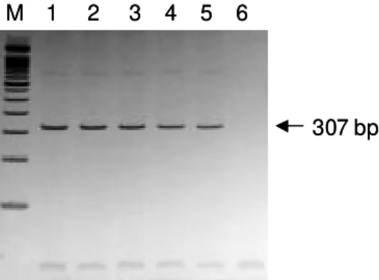
Expression of SmZF1 in S. mansoni life cycle stages - cDNA libraries of different stages of the parasite development (egg, cercariae, 3h schistosomulum and adult worm) were amplified by PCR using the primers ZnFMF and ZnFStop (Table). Amplicons of 307 bp were detected in all the stages of the life cycle evaluated (Fig. 4), indi-cating that this gene is expressed during the whole life cycle of the parasite.
DISCUSSION
There are relatively few papers on the charac-terization of transcription factors in Schistosoma, although the promoter regions of several schisto-some genes have been shown to contain putative recognition sites for a number of trans-acting fac-tors (El-Sherbeini et al. 1991, Abath et al. 1994, NH2
COOH
Arg 144
His 116
Zemzoumi et al. 1995). Analysis of the 5´ flanking region of the gene encoding the 28-kDa glutathione S-transferase of S. mansoni (Sm28GST) have iden-tified several regulatory sites able to bind nuclear factors such as AP-1, NF-Y and NF-AT-like pro-teins (Zemzoumi et al. 1995, Serra et al. 1999). The gene encoding the S. mansoni A subunit of the nuclear factor Y (SMNF-YA) was cloned and shown to be able to bind to the CCAAT boxes present in the promoter region of the Sm28GST gene (Serra et al. 1996, Zemzoumi et al. 1996). The gene encoding the S. mansoni hsp70 protein possesses two se-quences similar to heat shock elements (HSE) and three inverted CCAAT boxes. Levy-Holtzman and Schechter (1994) demonstrated by electrophoretic mobility shift assays that S. mansoni extracts con-tain active transcription factors for hsp70 of S. mansoni. In fact, several cDNAs encoding the heat-shock transcription factor of schistosome have been cloned (Lantner et al. 1998). SMNF-YA is present in all the stages of the life cycle of the para-site, which is compatible with the idea that this tran-scription factor is involved with the regulation of several genes. On the other hand, HSE binding fac-tor was detected only in schistosomula and adult worms, correlating with the pattern of hsp70 mRNA expression. In addition, a transcription factor ho-mologous to the human Y-box binding protein was also cloned and structurally characterized in S. mansoni (Franco et al. 1997).
However, none of those yet characterized DNA binding proteins show zinc finger motifs in their structure. This work reports on the cloning and structural characterization of a gene that encodes a S. mansoni zinc finger protein. This class of pro-teins usually plays a role in the regulation of gene
expression through interaction with nucleic acids, and have not been reported in S. mansoni previ-ously. The derived amino acid sequence showed an ORF of 492 bp encoding a 164 amino acid pro-tein interrupted by three Cys2His2 zinc finger mo-tifs. The A+T content, high inside the ORF, and the codon usage are in agreement with data previously described for S. mansoni (Milhon & Tracy 1995).
The zinc fingers are among the most important structural motifs involved in the interaction between proteins and nucleic acids. Zinc-binding units such as the Cys2 His2 zinc finger domins are present in a large number of gene products, representing some of the largest protein families in the Caenorhabditis elegans genome (Clarke & Berg 1998). Although bacteria and archaea contain some proteins that bind zinc they appear to lack the large families of zinc-binding domains like those families in yeast, worms, and other eukaryotes. Proteins containing zinc finger motifs are involved in many aspects of the gene regulation in eukaryotes, for instance, the proteins that induce differentiation and growth, proto-oncogenes and transcription factors in gen-eral (Wingender 1993). Recently, a subgroup of zinc finger motifs associated to protein-protein interac-tions was described, the ring finger motifs, which are also able to interact with DNA (Tanimura et al. 1999). The three zinc finger motifs of SmZF1 pro-tein fit to the consensus zinc finger sequence (Berg 1990, Bernstein et al. 1994). Computational model-ling based on the crystal structure of the Zif268 mouse protein, that also contain three zinc fingers, demonstrated that the three SmZF1 zinc finger mo-tifs are formed by finger structures with two β -strands to form one β sheet at the cysteine side and one α-helix at the histidine side, separated by a loop composed of 12 amino acid residues. The pres-ence of four amino acids residues between the two histidines in all motifs might indicate that the SmZF1 fingers adopt a non-standard loop structure pre-ceding the α-helix, as described for TFIIIA protein (Kochoyan et al. 1991). Elrod-Erickson et al. (1996) have demonstrated that α-helix structures of Zif268 protein fit to the major groove of B-DNA mainly by arginine-guanine and histidine-guanine interac-tions. Of particular importance are the arginine just preceding the α-helix structures. Our analysis dem-onstrated that SmZF1 residues His116 at finger 2 and Arg144 at finger 3, located just before the begining of the α-helix structures, might form strong hydrogen bonds to citosines in DNA sequences. This evidence suggests the function of fingers 2 and 3 in binding DNA, while the function of the first finger is not clear. Since zinc finger motifs can also mediate protein-protein interaction, one could speculate that the function of SmZF1 finger 1 is binding other co-associated factors.
M 1 2 3 4 5 6
307 bp M 1 2 3 4 5 6
307 bp
Our results show that the SmZF1 mRNA is present in all stages of the parasite cycle analyzed, indicating a constitutive expression of SmZF1 throughout the developmental cycle of S. mansoni. However, it does not mean necessarily that the en-coded protein is functionally active in all the para-site life cycle stages. In addition to transcriptional control, regulation of the protein activity by phos-phorylation/dephosphorylation is the major intra-cellular control mechanism in eukaryotic cells. Many structural or regulatory proteins are targeted by protein kinases that introduce a phosphate group in specific residues of tyrosine, threonine and serine, resulting in inactivation, activation or intermediate forms of the protein (Cohen 1989). The presence of six potential phosphorilation sites in the SmZF1 protein suggests it might be submitted to such type of regulation, although this was not addressed in the present study.
To our knowledge, this is the first report on a gene encoding a S. mansoni zinc finger protein, and certainly the understanding of gene regulation in this human parasite will demand further studies on this category of proteins. In conclusion, this paper describes the structural characterization and expression of a S. mansoni gene encoding a puta-tive zinc finger transcriptional regulatory protein. Some speculations were withdrawn from theoreti-cal predictions, and should be validated by future experimental studies aiming at elucidating the func-tional role of this protein.
ACKNOWLEDGEMENTS
To Neuza Antunes Rodrigues and Kátia Barroso (UFMG, Brazil) for technical support and automated DNA sequencing, Dr H-J Hecht (GBF, Germany) for comments and suggestions, and Dr Mohamed Saber (TBRI, Cairo, Egypt) for supplying the egg, cercariae, 3h schistosomulum, and adult worm cDNA libraries.
REFERENCES
Abath FGC, Hagan P, Jeffs A, Simpson AJG 1994. Par-tial characterization and kinetics of expression of Sm15, a Schistosoma mansoni tegumental antigen.
Parasitol Res80: 64-69.
Abath FGC, Hagan P, Jeffs SA, Schechter I, Meadows HM, Holder AA, Simpson AJG 1993. Structure of the gene encoding a putative Schistosoma mansoni
tegumental antigen precursor. Mol Biochem Parasitol 60: 81-92.
Abath FGC, Xavier EM, Allen R, Gomes YM, Lucena-Silva N, Baliza M, Simpson AJG 2000. Character-ization of Sm13, a tegumental antigen of Schisto-soma mansoni. Parasitol Res86: 745-752. Altschul SF, Thomas L, Madden AA, Schaffer JZ, Zheng
Z, Webb M, David JL 1997. Gapped blast PSI-Blast; a new generation of protein database search pro-grams. Nucl Acids Res25: 3389-3402.
Berg JM 1990. Zinc fingers and other metal-binding
do-mains. Elements for interactions between macromol-ecules. J Biol Chem265: 6513-6516.
Bernstein BE, Hoffman RC, Klevt RE 1994. Sequence-specific DNA recognition by Cys2, His2 zinc fin-gers. Ann N Y Acad Sci726: 95-104.
Chen LL, Rekosh DM, Loverde PT 1992. Schistosoma mansoni p48 egg shell protein gene: characteriza-tion, developmental regulated expression and on parition to the p14 egg shell protein gene. Mol Biochem Parasitol52: 39-52.
Clarke ND, Berg JM 1998. Zinc finger in Caenorhabditis elegans: finding families and probing pathways.
Science282: 2018-2022.
Cohen P 1989. The structure and regulation of protein phosphatases. Rev Biochem58: 453-508.
Davis AH, Blanton R, Klich P 1985. Stage and sex spe-cific differences in actin gene expression in Schisto-soma mansoni. Mol Biochem Parasitol17: 289-298. Elrod-Erickson M, Rould MA, Nekludova L, Pabo CO 1996. Zif268 protein-DNA complex refined at 1,6 Aº: A model system for understanding zinc finger-DNA interactions. Structure4: 1171-1180. El-Sherbeini M, Ramadan N, Bostian KA, Knopf PM
1991. Cloning and sequence analysis of the Schisto-soma mansoni membrane glycoprotein antigen GP22.
Mol Biochem Parasitol49: 83-98.
Franco GR, Garratt RT, Tanaka M, Simpson AJ, Pena SDJ 1997. Characterization of Schistosoma mansoni
gene encoding a homologue of the Y-box binding pro-tein. Gene198: 5-16.
Franco GR, Valadão AF, Azevedo V, Rabelo EML 2000. The Schistosoma gene discovery program; state of the art. Int J Parasitol30: 453-463.
Grossman Z, Ram D, Markovics A, Tarrab-Hazdai R, Lantner F, Ziv E, Schechter I 1990. Schistosoma mansoni: stage specific expression of muscle spe-cific genes. Exp Parasitol70: 62-71.
Jameson BA, Wolf H 1988. The antigenic index: a novel algorithm for predicting antigenic determinants. Comp Appl Biosci4: 181-186.
Kochoyan M, Keutman HT, Weiss MA 1991. Alternat-ing zinc fAlternat-inger in the human male-associated protein ZFY: HX3H and HX4H motifs encodes a local struc-tural switch. Biochemistry30: 9396-9402. Kosak M 1987. An analysis of 5'-coding region sequence
from 699 vertebrate messenger RNAs. Nucl Acids Res15: 8125-8148.
Kyte J, Doolittle RF 1982. A simple method for dis-playing the hydropatic character of a protein. J Mol Biol157: 105-132.
Lantner F, Ziv E, Ram D, Schechter I 1998. Different forms of the mRNA encoding the heat-shock tran-scription factor are expressed during the life cycle of the parasitic helminth Schistosoma mansoni. Eur J Biochem253: 390-398.
Levy-Holtzman R, Schechter I 1994. Schistosome ex-tracts with heat shock factor activity revealed by the gel shift assay. Parasitology108: 35-42.
Mei H, Lo Verde PT 1997. Schistosoma mansoni: the developamental regulation and immunolocalization of antioxidant enzymes. Exp Parasitol86: 69-78. Milhon JL, Tracy JW 1995. Update condon usage in
Mitchell PL, Tjian R 1989. Transcriptional regulation in mammalian cells by sequence-specific DNA binding proteins. Science245: 371-378.
Santos FR, Pena SDJ, Epplen JT 1993. Genetic and population study of a Y-linked tetranucleotide re-peat DNA polymorphism with a simple non-isoto-pic technique. Hum Genet90: 655-656.
Savioli L, Renganathan E, Montresor A, Davis A, Behbehani K 1997. Control of schistosomiasis - A global picture. Parasitol Today 13: 444-448. Serra E, Zemzoumi K, Trolet J, Capron A, Dissous C
1996. Functional analysis of the Schistosoma mansoni 28 kDa glutathione S-transferease gene pro-moter: involvement of SMNF-Y transcription fac-tor in multimeric complex. Mol Biochem Parasitol 83: 69-80.
Serra EC, Lardans V, Dissous C 1999. Identification of NF-AT-like transcription factor in Schistosoma mansoni: its possible involvment in the antiparasite action of cyclosporin A. Mol Biochem Parasitol101: 33-41.
Simpson AJ, Payares G, Walker T, Knight M, Smithers SR 1984. The modulation of expression of polypep-tide surface antigens on developing schistosomula of
Schistosoma mansoni. J Immunol 133: 2725-2730. Simpson AJG, Sher A, McCutchan TF 1982. The
ge-nome of Schistosoma mansoni: isolation of DNA, its size, bases and repetitive sequences. Mol Biochem Parasitol22: 169-176.
Tanimura S, Ohtsuka S, Mitsui K, Shirouzu K, Yoshimura A, Ohtsubo M 1999. MDM2 interacts with MDMX through their ring finger domains. FEBs Letters447: 5-9.
von Heijne G 1987. Sequence Analysis in Molecular Bi-ology: Treasure Trove or Trivial Pursuit, Academic Press Inc, San Diego, 97 pp.
Wingender E 1993. Gene Regulation in Eukaryotes, VCH Verlagsgesellschaft mbH, Weinheim, 430 pp. Yokono M, Saegusa N, Matsushita K, Sugiura Y 1998.
Unique DNA binding mode of the N-terminal zinc finger of transcription factor Sp-1. Biochemistry37: 6824-6832.
Zemzoumi K, Dissous C, Cochu A, Trolet J, Capron A, Mcnair A 1995. Interaction of nuclear extracts with the CCAAT-binding site revealed by gel shift assay.
Exp Parasitol80: 149-154.
Zemzoumi K, Serra E, Montovani R, Trolet J, Capron A, Dissous C 1996. Cloning of Schistosoma mansoni