Separomics applied to the proteomics and peptidomics of low-abundance
proteins: Choice of methods and challenges – A review
Maria Cristina Baracat-Pereira
1, Meire de Oliveira Barbosa
1, Marcos Jorge Magalhães Júnior
1,
Lanna Clicia Carrijo
1, Patrícia Dias Games
1, Hebréia Oliveira Almeida
1, José Fabiano Sena Netto
1,
Matheus Rodrigues Pereira
2and Everaldo Gonçalves de Barros
21
Departamento de Bioquímica e Biologia Molecular, Instituto de Biotecnologia Aplicada à Agropecuária,
Universidade Federal de Viçosa, Viçosa, MG, Brazil.
2Departamento de Biologia Geral, Universidade Federal de Viçosa, Viçosa, MG, Brazil.
Abstract
The enrichment and isolation of proteins are considered limiting steps in proteomic studies. Identification of proteins whose expression is transient, those that are of low-abundance, and of natural peptides not described in databases, is still a great challenge. Plant extracts are in general complex, and contaminants interfere with the identification of proteins involved in important physiological processes, such as plant defense against pathogens. This review dis-cusses the challenges and strategies of separomics applied to the identification of low-abundance proteins and pep-tides in plants, especially in plants challenged by pathogens. Separomics is described as a group of methodological strategies for the separation of protein molecules for proteomics. Several tools have been used to remove highly abundant proteins from samples and also non-protein contaminants. The use of chromatographic techniques, the partition of the proteome into subproteomes, and an effort to isolate proteins in their native form have allowed the iso-lation and identification of rare proteins involved in different processes.
Key words:sample preparation, complex protein extract, subproteomes, low-abundance proteins, cell wall proteins.
Introduction
The separomics challenges in plants
Proteomics tools have been widely used in recent years. Proteomics and peptidomics involve sophisticated methodologies which accurately detect alterations respec-tively in protein and peptide synthesis, under different physiological situations. Two main tools are widely used to isolate proteins, especially so two-dimensional electropho-resis (2-DE) associated with mass spectrometry (MS), and liquid chromatography associated with MS (LC-MS). Yet both present limitations inherent to the techniques (Cho, 2007). Multi-dimensional liquid chromatography has been valued in recent years as a technique to obtain native sam-ples, due to the need to validate biological events observed in proteomic studies. Obtaining native proteins is one of the biggest challenges in proteomics. Difficulties in isolating and identifying protein and peptide groups occur due to the high complexity of proteins present in the samples, the presence of non-protein contaminants that are difficult to
remove and the occurrence of post-translational modifica-tions. The difficulties are even greater for proteins and pep-tides present in low abundance (LAP) in the tissues. These, however, have attracted the attention of researchers since they are in general very effective and/or transiently present as components of finely controlled metabolic pathways. Thus, alternative methods to detect and identify these pro-teins are necessary.
Alternative strategies applied to the extraction, purifi-cation and biochemical and functional analyses of these molecules have been proposed, favoring access to struc-tural and functional information of hard-to-reach proteins and peptides (Kolodziejek and van der Hoorn, 2010). Sepa-romics is described as a group of methodological strategies aimed at separating protein molecules for proteomics, in-cluding fractionation and enrichment of specific molecules (Fang and Zhang, 2008). The use of separomic tools is es-pecially important for peptidomics, which is described as the group of methodologies and procedures applied to the analysis of native peptides by means of proteomics tools, since they are small and non-abundant proteins (Jurgens and Schrader, 2002).
Especially for peptidomics, 2-DE is difficult to apply, due to the low concentration of the peptide molecules, their
www.sbg.org.br
Send correspondence to Maria Cristina Baracat-Pereira. Departa-mento de Bioquímica e Biologia Molecular, Instituto de Biotec-nologia Aplicada à Agropecuária, Universidade Federal de Viçosa, 36.570-000 Viçosa, MG, Brazil. E-mail: [email protected].
small sizes (up to 10 kDa), their partial hydrophobic char-acter, and their ionic characteristics, as many peptides are strongly cationic. For the identification of these molecules, the greatest challenges are the small number of available specific databases and the low number of studied and posted molecules, which makes their identification through limited proteolysis techniques and MALDI-MS difficult. In addition, the partial hydrophobicity characteristics and sur-face charges facilitate peptide molecular associations, mak-ing them unavailable for analysis by any known proteomics tools.
For plant proteomics, the greatest challenges are to reduce sample complexity and remove contaminants which are incompatible with the isolation and identification tools (Kolodziejek and van der Hoorn, 2010). The correct use of separomics is imperative, especially for the identification of proteins and peptides expressed in low concentrations. This said, the current review aimed at discussing difficul-ties and challenges in plant proteomics and peptidomics, and to point out methodologies and strategic tools capable of detecting low-abundance proteins, especially biological peptides, differentially expressed in soybean plants after bi-otic and abibi-otic stresses.
Proteomic analysis applied to the response of soybean plants to pathogens
Proteomic analysis has become the most powerful tool for the functional characterization of plants. Informa-tion on soybean sequencing (Kimet al., 2010; Schmutzet al., 2010) and the soybean genome database (Phytozome v7.0:Glycine max) are available. Soybean was the first le-gume species to have its complete genome sequenced, be-coming therefore a key reference for the more than 20,000 legume species (Schmutzet al., 2010). Several genomes are emerging as model for plants, including that from soy-beans. Komatsu and Ahsan (2009) have discussed the ad-vantages and limitations of different proteomics tools ap-plied to the study of soybean defense. In the last two years, different research groups have also discussed the difficul-ties and alternatives concerning proteomic analyses of plants in general (Sudaric et al., 2010; Yamaguchi and Sharp, 2010; Bindschedler and Cramer, 2011). Low protein concentration, difficulties in protein extraction, genome ploidy, interference of highly abundant proteins in green tissue are some of the main challenges in plant proteomics (Bindschedler and Cramer, 2011). In general, there are many additional challenges for plant proteomics, such as the identification of proteins expressed transiently and in relatively low concentration, or the fact that the natural pep-tide has not yet been deposited in any of the databases.In silicoinformation is essential for proteomic analysis of im-portant physiological processes, such as plant defense. The subcellular proteome analysis of soybean plants submitted to stress conditions aims to identify proteins and peptides that are differentially expressed and potentially involved in
plant defense or pathogen resistance induction. Biotechno-logical methods may then be developed in order to intercept the pathogen action before infection, or to produce defense agents to boost the plant’s defense system. Organelle pro-teins and specific soybean tissues have been studied, such as membrane (Komatsu and Ahsan, 2009; Bindschedler and Cramer, 2011), primary roots (Nouri and Komatsu, 2010) and cell wall (Yamaguchi and Sharp, 2010).
Searching For Native Low-Abundance Proteins
and Peptides By Proteomics and Peptidomics
Techniques with different sensitivities and accuracies should be used in the analysis of the proteome. Due to the physical, chemical and biological diversity of proteins, proteomics tools present limitations that make it unfeasible to analyze the entire proteome with only a single separation strategy, even if it is orthogonal, such as the 2-DE or multi-dimensional liquid chromatography. Furthermore, the cellular protein concentration may vary from mg mL-1 to pg mL-1(Fang and Zhang, 2008; Jorrín-Novoet al., 2009).
Different proteomic analysis platforms were devel-oped in recent decades based on biochemical tools already available for protein isolation and identification. By means of these strategies, thousands of proteins have been identi-fied, especially high-abundance proteins (HAP), whereas the identification of low-abundance proteins (LAP) is still lim-ited. The search for LAP is growing, for they can represent important biological markers (Lescuyeret al., 2007), be re-sponsible for eliciting important cellular responses, and may even correspond to low molecular weight transcription fac-tors. Not only is their synthesis transient but they can also be easily lost (Corthalset al., 2000). Different procedures have been used to reduce HAP levels in extracts and to improve LAP detection, such as: (1) partial removal of RUBISCO by increasing dithiothreitol concentration in extracts of rice leaves (Choet al., 2008), or by fractionating extracts of soy-bean leaves with calcium chloride and sodium phytate (Krishnan and Natarajan, 2009), (2) addition of solvents such as the isopropanol to remove storage proteins from ex-tracts of soybean seeds (Natarajanet al., 2009), (3) division of the proteome into subproteomes, as done for banana leaf membranes (Vertommenet al., 2011), or (4) the use of affin-ity and immunoaffinaffin-ity columns for protein purification (Azarkanet al., 2007; Fang and Zhang, 2008).
Due to the need for improvement and/or development of protein separation methods for proteomic studies, sepa-romics makes its appearance as the science for proteomic separation. The main goal of separomics is to obtain a spe-cific set of proteins in a given biological system for proteo-me composition analysis, for protein-protein interaction studies, or for analysis of alterations in protein synthesis in biological materials submitted to different physiological conditions. The concept ofSeparomics (originally called
technolo-gies, processes, requirements, patterns and applications on proteomic separation, including fractionation and enrich-ment (Huanget al., 2005). The greatest challenge was to re-duce the complexity of a given proteome, aiming to increase the effectiveness of proteomic analysis as well as the identification of new proteins through mass spectrome-try (Fang and Zhang, 2008; Kosováet al., 2011).
Specific structures in plants, such as the cell wall and vacuoles, contain substances responsible for the inferior and non-reproductive outcomes during the separation of proteins due to proteolytic breakdown, streaking and char-ge heterochar-geneity.In most plant tissues, the proteins are part of complex structures, requiring special care for their isola-tion in a soluble form, as a native or non-native protein. The most commonly found interfering molecules during protein isolation are phenolic compounds, proteolytic and oxida-tive enzymes, terpenes, pigments, organic acids, inhibiting ions, and carbohydrates (Carpentieret al., 2005).
A further limiting factor in proteomic studies in plants is the loss of protein solubility caused by either the enrich-ment of protein extracts or by the properties of the solvents. Solubility of proteins is usually dependent on their concen-tration and amino acid composition, type and solvent dielec-tric constant, ionic strength, and on the presence of contami-nants (Jorrín-Novo et al., 2009). The optimum solubility conditions will be empirically defined for each system.
Preparing and Isolating Low-Abundance
Proteins and Peptides From Plants
Plant extracts require specific sample preparation procedures especially adjusted for proteomic and pepti-domic studies. These procedures should take into account the solubility, the physico-chemical characteristics and the cellular localization of the proteins, as well as the presence of interfering molecules (Chen and Harmon, 2006; Matros
et al., 2010), which correspond to the most challenging and critical aspects (Komatsu and Ahsan, 2009).
Subproteomes
Since the preparation protocols do not allow evalua-tion of the complete proteome and peptidome, the protocol definition should take into consideration protein subgroups and source material of interest, whether it is an organelle, a cell or a tissue, in order to reduce sample complexity, en-rich and increase the possibility of identifying LAP of inter-est involved in different cellular mechanisms and different locations. Hence, the information obtained for each subpro-teome of the same source material contributes to achieve greater coverage of the proteome being studied. This strat-egy has attracted the attention of several researchers (Cáno-vaset al., 2004; Zhanget al., 2004; Natarajanet al., 2005, 2009; Oehrleet al., 2008; Jorrin-Novoet al., 2009; Krish-nan and Natarajan, 2009; Agrawalet al., 2010; Matroset al., 2010; Kota and Goshe, 2011).
Plant cell wall proteins
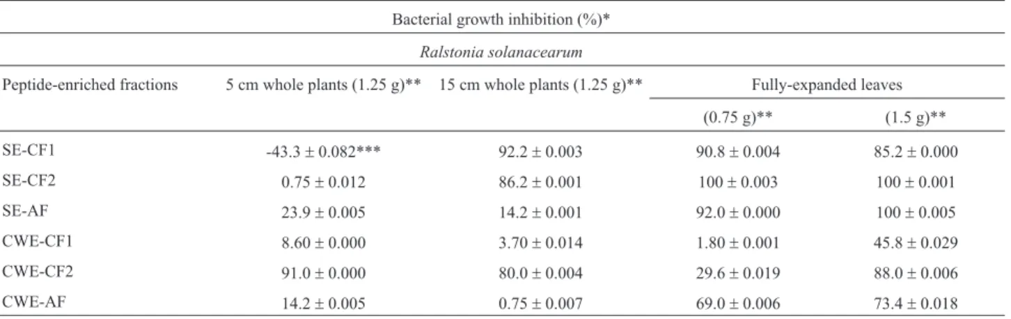
For the study of plant proteomes, the extracts may be successfully fractionated into soluble proteins and cell wall (CW) proteins (Figure 1). CW proteins represent a subpro-teome of great importance, since many of these molecules are involved in the maintenance of the cellular structure and in processes of plant defense, for instance in responses to abiotic and biotic stresses (Jametet al., 2006; Konget al., 2010), or as a constitutive barrier against pathogenic micro-organisms. Studies in our laboratory evidenced high consti-tutive antimicrobial activity against two plant-pathogenic bacteria by the peptide fraction obtained from CW extracts from leaves of bell pepper (Figure 2, Teixeiraet al., 2006) and 60-day-old eggplant (Almeidaet al., 2008). Also for eggplants, the highest inhibition levels were obtained with CW extracts from 5-cm-tall plants, while soluble extracts promoted the highest inhibition rate when fully expanded leaves were analyzed (Table 1). These results suggested that young plants exhibit an innate defense mechanism, very likely to minimize plant microbial invasion, whereas expanded leaves produce soluble defense molecules (Al-meidaet al., 2007).
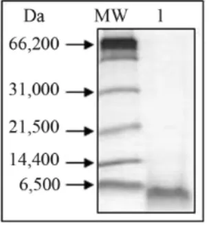
Figure 1- Denaturing electrophoresis (16.5%) of soluble extracts (SE) and cell wall extracts (CWE) of leaves of soybean genotypes ‘PI 561356’ (Embrapa CNPSo) subjected to injury from seven wounds on the leaves. SE and CWE were fractionated by ultrafiltration and molecules contained between 1 and 10 kDa (1-10) and greater than 30 kDa (> 30). MM: 5 uL of the Wide-Range Molecular Weight Markers (Bio-Rad, USA). The peptide bands were stained with Coomassie Brilliant Blue R250. (Courtesy of José Fabiano Sena-Netto, from the final paper for his undergraduate degree in Biochemistry in 2010 at the Federal University of Viçosa, Brazil).
Plant CWs are highly dynamic and contain chemi-cally active compounds secreted by the cells, which are es-sentially polysaccharides and proteins, the latter compris-ing approximately 10% of the CW mass (Jamet et al., 2008). These proteins, which are difficult to isolate from these complex matrixes, require specific extraction meth-ods (Teixeiraet al., 2006; Zhuet al., 2006; Almeidaet al., 2007, 2008; Konget al., 2010), due to their low abundance. Differential extraction enriches the extract, allows access to the CW LAP, and facilitates the comparison of the expres-sion profiles under different stress conditions (Watson and Summer, 2006; Negriet al., 2008).
Protein extraction from CWs can be achieved by methods that may involve or not cell disruption. For each procedure there are advantages and disadvantages, espe-cially concerning contamination and experimental proce-dures. Methods commonly used involve calcium chloride and lithium chloride solutions (Feizet al., 2006; Konget al., 2010). Calcium chloride does not disrupt the cell wall but is capable of releasing ionic molecules externally an-chored to the CW, whereas lithium chloride solution is used to extract proteins intrinsic to the CW, such as glyco-proteins (Feizet al., 2006). When comparatively evaluating the CW subprotreomes of soybean leaves submitted or not to biotic stress using reversed-phase high performance liq-uid chromatography we obtained a group of differential protein peaks in the comparative chromatograms, demon-strating that sample complexity was reduced. This proce-dure allows the identification of the isolated proteins.
Proteins with different ranges of molecular masses
When the aim is to identify proteins of a specific group of molecular mass present in a complex sample, methodologies based on ultrafiltration have an advantage, since they are capable of fractionating, concentrating and exchanging solutions, and thus recovering native samples with molecular masses of interest. Ultrafiltration is a
low-cost process requiring simple equipment only, such as fil-tering membranes of different cut-off values, which allow the fractionation of different protein groups. Soluble (SE) and CW extracts (CWE) of soybean leaves submitted (Fig-ure 1) or not to stress were successfully fractionated in our laboratory through ultrafiltration. Fractions for peptido-mics and proteopeptido-mics, with proteins smaller than 30 kDa and greater than 30 kDa, respectively, were recovered. After separating the fractions by reversed-phase chromatography (RPC) and recovering the proteins and peptides for mass spectrometry analysis, differentially expressed molecules were successfully identified.
Molecular exclusion chromatography (MEC) is also an alternative used by our group for this purpose. This method favors isolating a specific protein group according to the selected resin. In general, MEC presents low accu-racy in the separation of small proteins and peptides, yet it is a fast procedure that generates clean samples with low salt concentration, which is important for later stages of pu-rification, for instance during RPC. MEC has been success-fully used in our proteomic analyses using multi-dimen-sional liquid chromatography. A constitutive antimicrobial peptide was identified in CW extracts from tomato plant af-ter MEC following RPC while an induced defense peptide was detected in plants after abiotic stress (submitted for publication).
Proteins from different cellular compartments
Ultra-centrifugation enables the separation of cellular organelles according to their sizes and densities. Zonal centrifugation or centrifugation by differential velocity may be used for fractionation, the latter being a technique which generates greater sedimentation strength, enabling the separation of organelles of similar characteristics (Ko-matsu, 2006), such as nuclei, mitochondria or plastids,
Table 1- Antimicrobial activity of cationic (CF1, CF2) and anionic (AF) fractions obtained from soluble (SE) or cell-wall (CWE) extracts of young plants or fully-expanded leaves of eggplant (Almeidaet al., 2007, with authorization from the authors).
Bacterial growth inhibition (%)*
Ralstonia solanacearum
Peptide-enriched fractions 5 cm whole plants (1.25 g)** 15 cm whole plants (1.25 g)** Fully-expanded leaves
(0.75 g)** (1.5 g)**
SE-CF1 -43.3±0.082*** 92.2±0.003 90.8±0.004 85.2±0.000
SE-CF2 0.75±0.012 86.2±0.001 100±0.003 100±0.001
SE-AF 23.9±0.005 14.2±0.001 92.0±0.000 100±0.005
CWE-CF1 8.60±0.000 3.70±0.014 1.80±0.001 45.8±0.029
CWE-CF2 91.0±0.000 80.0±0.004 29.6±0.019 88.0±0.006
CWE-AF 14.2±0.005 0.75±0.007 69.0±0.006 73.4±0.018
while maintaining their integrity (Wijk, 2004; Kosováet al., 2011).
Contaminant removal and use of additives
A limiting factor for proteomic studies is the presence of protein and non-protein contaminants in the samples. Contaminants may interfere with the detection, isolation or identification of proteins of interest, acting by aggregation with the proteins, by affecting the signal/noise relationship of the detection equipment, by promoting protein degrada-tion or reducing the activity of enzymes, among other fac-tors. Under native conditions, the removal of these contam-inants may be achieved by dialysis, salt fractionation, or with organic cold solvents, through ultrafiltration or pre-parative chromatography. Under denaturing conditions, steps including selective heating or addition of concen-trated acids may favor sample bleaching and the enrich-ment of the protein group of interest. Also, anionic deter-gents like sodium dodecyl sulfate (SDS) or nonionic detergents such as Triton X-100 and Nonidet P-40 can be added in different concentrations. The concentration to be used must be empirically defined.
Fractionation by ultrafiltration allows the simulta-neous desalting and concentration of the samples in the na-tive form (Konget al., 2010). However, during the ultra-filtration procedure certain caution is required so as to maintain the sample in its native form, as well as to avoid protein agglomeration. Some of the factors to be controlled are hydrophobicity, pH and ionic strength of the buffer or solution being used. The choice of salts and pH of the buffer solution for ultrafiltration depends on the subpro-teome under study. For instance, as pH values around 5.5 are close to the pH of the cell wall, the fractionation of CW proteins should be done at pH values close to 5.5 to avoid precipitation of these proteins (Watson and Summer, 2006). On the other hand, many storage proteins, which are usually contaminants, precipitate between pH 4.5 and 4.8 (Speroniet al., 2010), and extraction at these pH values produces less complex extracts. For ultrafiltration of mem-brane proteins and partially hydrophobic proteins, deter-gents should be used, as these become soluble in the presence of amphipathic compounds, such as 3-[(3-chola-midopropyl) dimethylammonio]-1-propanesulfonate (CHAPS), SDS and the nonionic surfactant Triton X-100, among others.
Pressure is considered an important factor which fre-quently is not controlled during ultrafiltration. There are not sufficient studies reporting protein agglomeration, sol-ubility and protein conformation under pressure (Chalikian and MacGregor, 2009; Speroniet al., 2010). Iwabuchi and Yamauchi (1987), fractionating glycinin (360 kDa) and
b-conglycinin (180 kDa), found low molecular mass teins in the protein fraction containing these storage pro-teins, suggesting the occurrence of agglomeration of low
molecular mass proteins together with high molecular mass proteins.
In our experience, when a low salt concentration buffer (5 mM Tris buffer, pH 7.0) was used after the ul-tra-filtration procedure, the occurrence of agglomeration in extracts from soybean leaves increased. Agglomeration was also increased under cold temperatures, and an insolu-ble precipitate was observed in fractions containing low and high molecular masses. Replacing this buffer with 20 mM Tris-HCl, pH 7.0, containing 20 mM ammonium acetate, agglomeration was no longer observed, suggesting that agglomeration was an ionic behavior. In an attempt to disaggregate the precipitate formed in the high molecular fraction, different procedures were used. Disaggregation was not achieved in the presence of 0.5% (v/v) Triton X-100 or by dilution (up to 10 times) with 100 mM Tris-HCl, pH 7.0, but the sequential addition of 0.4, 1.0 and 2.0% (w/v) SDS partially reduced the agglomeration, thus corroborating the hypothesis of ionic behavior.
Denaturing one-dimensional electrophoresis (16.5% T) done with extracts from soybean leaves enriched for low molecular mass proteins (up to 30 kDa) yielded pro-tein bands higher than 30 kDa, suggesting the occurrence of agglomeration after ultrafiltration. This was confirmed when a 60-kDa band was eluted from the gel and again sep-arated under the same conditions, this time producing 6-kDa and 30-kDa bands. Similarly, the elution and new separation of a 6-kDa band has also produced the same bands, confirming that heating the sample for 10 min in the sample buffer (Teixeiraet al., 2006) was unable to solubi-lize proteins in the sample.
Fractionation with ammonium sulfate has been used by our group since 2000 for bleaching plant samples, to re-move contaminants such as carbohydrates, phenolic com-pounds and pigments, among others. Plant extracts ob-tained in the absence of this compound presented high noise/signal relationship in the spectrometry analyses and protein agglomeration was observed. Recently, Parket al.
(2008) stressed the importance of this procedure to remove interfering molecules. Nevertheless, caution should be taken, as the presence of salt may interfere with electropho-retic properties and the MS procedure.
inhibitors with different specificities (unpublished date). The use of non-interfering additives is an empirical deci-sion, which should be carefully evaluated because these compounds may have to be removed in a subsequent purifi-cation step.
Refined methods for isolation
Separation by two-dimensional electrophoresis
Two-dimensional electrophoresis (2-DE) is the most commonly used separation technique in proteomics for comparative and global protein analyses under differential conditions. A high number of proteins can be identified in a single gel. Considering that in general, plant tissues do not present a high protein concentration, and that the presence of proteases and interfering molecules can drastically affect proteomic analyses, an efficient protein extraction protocol must be used. This protocol should be able to eliminate sec-ondary metabolites, remove additives which are not com-patible with the different stages of purification, and enrich the LAP. Extraction methods using phenol in conjunction with ammonium acetate/methanol precipitation have pro-ved to be highly efficient (Isaacsonet al., 2006). Using this procedure Xuet al.(2006) were able to obtain a large num-ber of intense and well-resolved spots from soybean leaves in 2-D gels. Similar results were obtained in our laboratory. Methods based on precipitation with trichloroacetic acid (TCA) have also been used in the literature (Chen and Harmon, 2006) and in our laboratory. Using a method based on an initial TCA precipitation step followed by ex-traction by the dense phenol/SDS method and a final pre-cipitation with ammonium acetate/methanol, Wanget al.
(2003) obtained a high number of soluble and membrane proteins from soybean leaves. To optimize this method, we introduced some modifications, such as the use of protease inhibitors (1 mM phenylmethylsulfonyl fluoride), reducing agents (different concentrations ofb-mercaptoethanol and dithiothreitol) and 1% polyvinylpolypyrolidone, which eli-minate or prevent the action of proteases and compounds that interfere with the subsequent steps in protein separa-tion and identificasepara-tion.
Liquid chromatography (LC) can be used as an alter-native to 2-DE when the proteins of interest present ex-tremes of molecular mass and pI, when they are highly hydrophobic or LAP (Natarajanet al., 2009; Gilmore and Washburn, 2010; Kolodziejek and van der Hoorn, 2010).
Separation by chromatographic methods
Chromatographic methods using a combination of different separation principles (multi-dimensional) in on-line or off-on-line processes, or the selective separation by af-finity have been used and improved to resolve the complex-ity of the extracts and/or to enrich them. Commercially available on-line chromatographic systems generally use a strong ionic exchange column (IEC) followed by a reverse phase. Molecular exclusion chromatography (MEC)
fol-lowed by reverse phase chromatography (RPC) is also used (Issaq, 2001), though less often. Our group has worked with off-line chromatography with success, especially associat-ing MEC and RPC for prospection of proteins with differ-ent ranges of molecular masses, when two antimicrobial peptides from tomato leaves were purified under native conditions and subsequently identified (data submitted for publication). These procedures are orthogonal, creating a large number of fractions to be analyzed, making proteomic analyses by MDLC difficult and lengthy. However, it al-lows a greater proteome and peptidome prospection, with little restriction for very large or very small and electrically charged membrane proteins. And in particular, it allows the isolation of native purified samples for evaluation of the proteome functionality.
Although these methods are very efficient, comple-mentary techniques are required for obtaining further infor-mation regarding the proteome. Thus, the development and application of technologies and methods for separation by affinity and enrichment have become a high priority for de-fining separomic procotols to be used for different organ-isms.
A great variety of affinity and immuno-affinity col-umns are already available and allow the separation of complex biological extracts into different protein classes, or even to fractionate LAP. These commercially available columns are important for the identification of protein-protein interaction networks (Azarkanet al., 2007) and to remove unwanted HAP proteins, peptides and nucleotides, metals, etc. (Keshishianet al., 2007; Cellaret al., 2008; Fang and Zang, 2008; Huang and Fang, 2008; Miernyk and Hajduch, 2011).
Identifying Soybean Proteins and Peptides By
Proteomics Tools
allowed the identification and characterization of proteins with accuracy, thanks to genomic and protein sequence in-formation deposited in public databases (Schmutz et al., 2010).
Conclusions
Proteomics is an invaluable tool for functional geno-mic analysis of plants. Techniques with different sensitivi-ties and accuracies should be used in the analysis of the re-spective proteome. Highly sensitive and accurate identification equipment is available; however, better pro-tein and peptide extraction procedures and methods for en-richment and isolation of low-abundance proteins with transient expression and of rare peptides are needed. Some alternatives to overcome or minimize these limitations are: (1) the decrease of extract complexity to allow evaluation of subproteomes, (2) use of methodologies and/or additives to remove highly abundant proteins known as contami-nants, (3) use of chromatographic techniques which allow the enrichment of specific protein fractions, and (4) isola-tion of proteins in their native form to aid their identifica-tion and validaidentifica-tion of metabolic pathways in which they are involved. The use of basic biochemistry tools which sepa-rate protein and peptides based on their physico-chemical characteristics can further the enrichment process. Knowl-edge about the chemical and structural characteristics of non-protein contaminants is helpful in the choice of meth-ods and tools for their removal or inactivation. This infor-mation will also aid in defining methods to prevent solu-bilization of these contaminants and their consequent contact with the proteins of interest.
Acknowledgments
The authors wish to express their gratitude to the Bra-zilian agencies CNPq, FAPEMIG, FINEP and CAPES for financial support and fellowships, and to Embrapa Soja and NuBioMol/UFV for partnership and technical support.
References
Almeida HO, Mattos EC, Barbosa MO, Teixeira FR, Magalhaes RDM, Romeiro RS, Fontes EPB and Baracat-Pereira MC (2007) Peptide fraction inhibiting plant pathogen growth predominated in cell wall extracts from young plants or in soluble cell fraction from expanded leaves from eggplants. J Phytopathol 155:735-737.
Almeida HO, Teixeira FR, Romeiro RS, Silva DJH, Pereira PRG, Fontes EPB and Baracat-Pereira MC (2008) Atividade anti-microbiana de extratos peptidicos de folhas de berinjela na inibiçao do crescimento de Ralstonia solanacearum e Clavibacter michiganensis subsp. michiganensis. Summa Phytopathol 34:62-64 (Abstract in English).
Azarkan M, Huet J, Baeyens-Volant D, Looze Y and Vanden-bussche G (2007) Affinity chromatography: A useful tool in proteomics studies. J Chromatogr B Analyt Technol Biomed Life Sci 849:81-90.
Bindschedler LV and Cramer R (2011) Quantitative plant pro-teomics. Proteomics 11:756-775.
Brandão AR, Barbosa HS and Arruda MA (2010) Image analysis of two-dimensional gel electrophoresis for comparative pro-teomics of transgenic and non-transgenic soybean seeds. J Proteomics 73:1433-1440.
Cánovas FM, Dumas-Gaudot E, Recorbet G, Jorrin J, Mock H-P and Rossignol M (2004) Plant proteome analysis. Proteo-mics 4:285-298.
Carpentier SC, Witters E, Laukens K, Deckers P, Swennen R and Panis B (2005) Preparation of protein extracts from recalci-trant plant tissues: An evaluation of different methods for two-dimensional gel electrophoresis analysis. Proteomics 5:2497-2507.
Cellar NA, Kuppannan K, Langhorst ML, Ni W, Xu P and Young SA (2008) Cross species applicability of abundant protein depletion columns for ribulose-1,5-bisphosphate carboxy-lase/oxygenase. J Chromatogr B Analyt Technol Biomed Life Sci 861:29-39.
Chalikian TV and MacGregor Jr RB (2009) Origins of pres-sure-induced protein transitions. J Mol Biol 394:834-842. Chamrad DC, Korting G, Stuhler K, Meyer HE, Klose J and
Bluggel M (2004) Evaluation of algorithms for protein iden-tification from sequence databases using mass spectrometry data. Proteomics 4:619-628.
Chen S and Harmon AC (2006) Advances in plant proteomics. Proteomics 6:5504-5516.
Cho JH, Hwang H, Cho M-H, Kwon Y-K, Jeon J-S, Bhoo SH and Hahn T-R (2008) The effect of DTT in protein preparations for proteomic analysis: Removal of a highly abundant plant enzyme, ribulose bisphosphate carboxylase/oxygenase. J Plant Biol 51:297-301.
Cho WCS (2007) Review – Proteomics technologies and chal-lenges. Genomics Proteomics Bioinform 5:77-85.
Corthals GL, Wasinger VC, Hochstrasser DF and Sanchez JC (2000) The dynamic range of protein expression: A chal-lenge for proteomic research. Electrophoresis 21:1104-1115.
Elias JE, Haas W, Faherty BK and Gygi SP (2005) Comparative evaluation of mass spectrometry platforms used in large-scale proteomics investigations. Nat Meth 2:667-675. Fang X and Zhang W (2008) Affinity separation and enrichment
methods in proteomic analysis. J Proteomics 71:284-303. Feiz L, Irshad M, Pont-Lezica RF, Canut H and Jamet E (2006)
Evaluation of cell wall preparations for proteomics: A new procedure for purifying cell walls fromArabidopsis hypo-cotyls. Plant Meth 2:e10.
Gilmore JM and Washburn MP (2010) Advances in shotgun proteomics and the analysis of membrane proteomes. J Pro-teomics 73:2078-2091.
Huang L, Harvie G, Feitelson JS, Gramatikoff K Herold DA and Allen DL (2005) Immunoaffinity separation of plasma pro-teins by IgY microbeads: Meeting the needs of proteomic sample preparation and analysis. Proteomics 5:3314-3328. Huang L and Fang X (2008) Immunoaffinity fractionation of
plasma proteins by chicken IgY antibodies. Meth Mol Biol 425:41-51.
Issaq HJ (2001) The role of separation science in proteomics re-search. Electrophoresis 22:3629-3638.
Iwabuchi S and Yamauchi F (1987) Electrophoretic analysis of whey proteins present in soybean globulins fraction. J Agric Food Chem 35:205-209.
Jamet E, Canut H, Boudart G and Pont-Lezica RF (2006) Cell wall proteins: A new insight through proteomics. Trends Plant Sci 11:33-39.
Jamet E, Albenne C, Boudart G, Irshad M, Canut H and Pont-Lezica R (2008) Recent advances in plant cell wall proteo-mics. Proteomics 8:893-908.
Jorrín-Novo JV, Maldonado AM, Echevarría-Zomeño S, Valle-dor L, Castillejo MA, Curto M, Valero J, Sghaier B, Donoso G and Redondo I (2009) Plant proteomics update (2007-2008): Second-generation proteomic techniques, an appro-priate experimental design, and data analysis to fulfill MIAPE standards, increase plant proteome coverage and ex-pand biological knowledge. J Proteomics 72:285-314. Jurgens M and Schrader M (2002) Peptidomic approaches in
proteomic research. Curr Opin Mol Ther 4:236-241. Keshishian H, Addona T, Burgess M, Kuhn E and Carr SA (2007)
Quantitative, multiplexed assays for low abundance proteins in plasma by targeted mass spectrometry and stable isotope dilution. Mol Cell Proteomics 6:2212-2229.
Kim MY, Lee S, Van K, Kim TH, Jeong SC, Choi IY, Kim DS, Lee Y S, Park D, Ma J, et al.(2010) Whole-genome se-quencing and intensive analysis of the undomesticated soy-bean (Glycine sojaSieb. and Zucc.) genome. Proc Natl Acad Sci USA 107:22032-22037.
Kolodziejek I and van der Hoorn RAL (2010) Mining the active proteome in plant science and biotechnology. Curr Opin Biotechnol 21:225-233.
Komatsu S (2006) Extraction of nuclear proteins. In: Thiellement H, Zivy M, Damerval C and Méchin V (eds) Methods in Mo-lecular Biology, vol. 335: Plant Proteomics: Methods and Protocols. Humana Press Inc, Totowa, pp 73.
Komatsu S and Ahsan N (2009) Soybean proteomics and its appli-cation to functional analysis. J Proteomics 72:325-336. Kong F-J, Oyanagi A and Komatsu S (2010) Cell wall proteome
of wheat roots under flooding stress using gel-based and LC MS/MS-based proteomics approaches. Biochim Biophys Acta 1804:124-136.
Kosová K, Vítámvás P, Prásil IT and Renaut J (2011) Plant proteome changes under abiotic stress – contribution of proteomics studies to understanding plant stress response. J Proteomics 74:1301-1322.
Kota U and Goshe MB (2011) Advances in qualitative and quanti-tative plant membrane proteomics. Phytochemistry 72:1040-1060.
Krishnan HB and Natarajan SS (2009) A rapid method for deple-tion of Rubisco from soybean (Glycine max) leaf for proteo-mic analysis of lower abundance proteins. Phytochemistry 70:1958-1964.
Lescuyer P, Hochstrasser D and Rabilloud T (2007) How shall we use the proteomics toolbox for biomarker discovery? J Proteome Res 6:3371-3376.
Matros A, Kaspar S, Witzel K and Mock HP (2010) Recent prog-ress in liquid chromatography-based separation and label-free quantitative plant proteomics. Phytochemistry 72:963-974.
Miernyk JA and Hajduch M (2011) Seed proteomics. J Proteo-mics 74:389-400.
Natarajan S, Xu C, Caperna TJ and Garrett WM (2005) Compari-son of protein solubilization methods suitable for proteomic analysis of soybean seed proteins. Anal Biochem 342:214-220.
Natarajan SS, Krishnan HB, Lakshman S and Garrett WM (2009) An efficient extraction method to enhance analysis of low abundant proteins from soybean seed. Anal Biochem 394:259-268.
Negri AS, Prinsi B, Scienza A, Morgutti S, Cocucci M and Spen L (2008) Analysis of grape berry cell wall proteome: A com-parative evaluation of extraction methods. J Plant Physiol 165:1379-1389.
Nielsen ML, Savitski MM and Zubarev RA (2005) Improving protein identification using complementary fragmentation techniques in Fourier transform mass spectrometry. Mol Cell Proteomics 4:835-845.
Nouri MZ and Komatsu S (2010) Comparative analysis of soy-bean plasma membrane proteins under osmotic stress using gel-based and LC MS/MS-based proteomics approaches. Proteomics 10:1930-1945.
Oehrle NW, Sarma AD, Waters JK and Emerich DW (2008) Proteomic analysis of soybean nodule cytosol. Phytoche-mistry 69:2426-2438.
Park J-W, Lee S-G, Song JY, Joo JS, Chung M-J, Kim S-C, Youn H-S, Kang H-L, Baik S-C, Lee W-K,et al.(2008) Proteomic analysis ofHelicobacter pyloricellular proteins fractionated by ammonium sulfate precipitation. Electrophoresis 29:2891-2903.
Roepstorff P and Fohlman J (1984) Proposal for a common no-menclature for sequence ions in mass spectra of peptides. Biomed Mass Spectrom 11:601.
Schmutz J, Cannon SB, Schlueter J, Ma J, Mitros T, Nelson W, Hyten DL, Song Q, Thelen JJ, Cheng J,et al(2010) Genome sequence of the paleopolyploid soybean (Glycine max(L.) Merr.).Nature 463:178-183.
Speroni F, Añón MC and Lamballerie M (2010) Effects of cal-cium and high pressure on soybean proteins: A calorimetric study. Food Res Int 43:1347-1355.
Sudaric A, Vrataric M, Drinic SM and Matosa M (2010) “Bio-technology” in soybean breeding. Genetika-Belgrade 42:91-102.
Teixeira FR, Lima MCO, Almeida HO, Romeiro RS, Silva DJH, Pereira PRG, Fontes EPB and Baracat-Pereira MC (2006) Bioprospection of cationic and anionic antimicrobial pep-tides from bell pepper leaves for inhibition of Ralstonia
solanacearum and Clavibacter michiganensis ssp.
michiganensisgrowth. J Phytopathol 154:418-421. Thiede B, Höhenwarter W, Krah A, Mattow J, Schmid M,
Schmidt F and Jungblut PR (2005) Peptide mass fingerprint-ing. Methods 35:237-247.
Vertommen A, Moller ALB, Cordewenerd JHG, Swennen R, Panis B, Finnie C, America AHP and Carpentiera SC (2011) A workflow for peptide-based proteomics in a poorly se-quenced plant: A case study on the plasma membrane pro-teome of banana. J Proteomics 74:1218-1229.
high levels of interfering compounds. Electrophoresis 24:2369-2375.
Watson BS and Summer LW (2006) Isolation of cell wall proteins from Medicago sativastems. In: Thiellement H, Zivy M, Damerval C and Méchin V (eds) Methods in Molecular Bi-ology, vol. 335: Plant Proteomics: Methods and Protocols. Humana Press Inc, Totowa, pp 79.
Wijk KJ (2004) Plastid proteomics. Plant Physiol Biochem 42:963-977.
Xu C, Garrett WM, Sullivan J, Caperna TJ and Natarajan S (2006) Separation and identification of soybean leaf proteins by two-dimensional gel electrophoresis and mass spectrome-try. Phytochemistry 67:2431-2440.
Yamaguchi M and Sharp RE (2010) Complexity and coordination of root growth at low water potentials: Recent advances from transcriptomic and proteomic analyses. Plant Cell En-viron 33:590-603.
Zhang H, Yan W and Aebersold R (2004) Chemical probes and tandem mass spectrometry: A strategy for the quantitative
analysis of proteomes and subproteomes. Curr Opin Chem Biol 8:66-75.
Zhu J, Chen S, Alvarez S, Asirvatham VS, Schachtman DP, Wu Y and Sharp R (2006) Cell wall proteome in the maize primary root elongation zone. I. Extraction and identification of wa-ter-soluble and lightly ionically bound proteins. Plant Physiol 140:311-325.
Internet Resources
Phytozome ver. 7.0:Glycine max. Center for Integrative Geno-mics (CIG), Joint Genome Institute (JGI), University of Cal-ifornia Regents, http://www.phytozome.net/soybean (Janu-ary 24, 2011).