C
ASER
EPORT|
R
ELATO DEC
ASO473
Granulomatous intersticial nephritis secondary to sarcoidosis
Nefrite intersticial granulomatose secundária a sarcoidose
Authors
Tamires Teixeira Piraciaba 1
Carlos Alberto Balda 1 Luiz Antônio Ribeiro de Moura 1
Carlos Alberto de Castro Pereira 2
Gianna Mastroianni Kirsztajn 1
1 Universidade Federal de São Paulo, Disciplina de Nefrologia, São Paulo - SP, Brasil.
2 Universidade Federal de São Paulo, Disciplina de Pneumologia, São Paulo - SP, Brasil.
Submitted on: 02/14/2017. Approved on: 03/09/2017.
Correspondence to: Tamires Teixeira Piraciaba. E-mail: tamires.nefro@gmail. com
Introdução: a nefrite intersticial granu-lomatosa é uma condição rara, na qual o envolvimento renal é incomum. Sua etiolo-gia é variável e pode ter origem medicinal, infecciosa ou inflamatória. Relato de caso: trata-se de um paciente do sexo masculino, com 65 anos de idade, com lesões renais de etiologia desconhecida, associadas à hipercalcemia. Durante a investigação, ev-idenciaram-se insuficiência cardíaca com disfunção diastólica e envolvimento pul-monar intersticial à tomografia torácica. A função renal (taxa de filtração glomerular) melhorou parcialmente com medidas clíni-cas. Foi realizada biópsia renal, que apre-sentou lesão intersticial moderada com granulomas tuberculoides sem necrose ca-seosa. Conclusão: o objetivo do artigo foi descrever um caso de GIN e alertar para a importância de sua investigação clínica. Neste caso, a biópsia renal, associada a manifestações clínicas sistêmicas, contri-buiu para o diagnóstico de sarcoidose.
R
ESUMOPalavras-chave: hipercalcemia; nefrite in-tersticial; sarcoidose.
Introduction: Granulomatous interstitial nephritis is a rare condition, in which renal involvement is uncommon. Its eti-ology is variable, and may be medicinal, infectious or inflammatory origin. Case report: This is a 65-year-old male patient with renal lesions of unknown etiology, associated with hypercalcaemia. During the investigation, cardiac insufficiency with diastolic dysfunction and interstitial lung involvement on chest tomography were evidenced. Renal function (glomeru-lar filtration rate) has partially improved with clinical measures. Renal biopsy was performed, which showed moderate inter-stitial lesion with tuberculoid granulomas without caseous necrosis. Conclusion:
The objective of the article was to describe a case of NIG and to alert to the impor-tance of its clinical investigation. In this case, renal biopsy, associated with system-ic clinsystem-ical manifestations, contributed to the diagnosis of sarcoidosis.
A
BSTRACTKeywords: hypercalcemia; interstitial; ne-phritis; sarcoidosis.
I
NTRODUCTIONGranulomatous interstitial nephritis (GIN) is a rare condition. Medications, infectious agents, vasculitis and sarcoid-osis are some of its possible etiologies.1
Sarcoidosis is a disease of unknown origin, characterized by the formation of granulomas in the tissues, especially at the lower respiratory tract. Renal damage is uncommon, and it is described in 0.7 to 4.3% of the cases. Major forms of renal impairment are related to alterations in calcium metabolism, manifesting as neph-rocalcinosis and nephrolithiasis.1
DOI: 10.5935/0101-2800.20170084
This paper aims to report a case of GIN in a patient with renal dysfunction of unknown cause, in which the renal biopsy, associated with the clinical mani-festations, contributes to the diagnosis of sarcoidosis.
C
ASEREPORTBraz. J. Nephrol. (J. Bras. Nefrol.) 2017;39(4):473-476
Nefrite granulomatosa intersticial secundária à sarcoidose
474
improvement. He developed fever and weight loss. He reported hyperuricemia, gastritis and dyslipidemia. He was using allopurinol, ranitidine and sinvastatin to treat each of the conditions, respectively. Upon physical examination, on admission, he had bilateral enlarged parotids, which were painless to palpation.
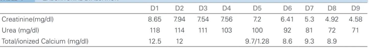
Initial laboratory tests: hemoglobin 9.8g/dl, hema-tocrit 28.7%, 8,140 leukocytes/mm³ (7.6% eosino-phils) and 400.000 platelets; serum creatinine 8.65 mg/dl and urea 118 mg/dl; electrolytes were within the limits of normality, except for total calcium of 12.5 mg/dl. Urinalysis: 11.000 leukocytes/ml, 1000 erythrocytes/ml and proteinuria of 0.5 g/l.
In view of hypercalcemia and renal dysfunction, vigorous venous hydration was prescribed, with a good response (Table 1), that is, there was a progres-sive decrease in serum urea and creatinine, in addi-tion to serum calcium normalizaaddi-tion without the need for other measures for hypercalcemia. Other comple-mentary tests: phosphate 5.3 mg/dl, PTH 7.7 pg/ml, 25-OH-vitamin D 4ng/ml, serum albumin 3.9 g/dl.
Kidney and urinary tract ultrasonography showed right kidney with 10.6 cm and left with 10.8 cm, re-nal parenchyma with preserved thickness and echo-genicity. There was a 0.5 cm stone in the right cali-cinal group, without signs of dilatation of the pelvic system.
Cervical ultrasonography showed enlarged parotid glands, with heterogeneous ecotexture, multiple cys-tic lesions at the border, associated with some promi-nent intraparotid lymph nodes. The submandibular glands were preserved and there was no lymph node enlargement.
The parotid glands returned to their usual size progressively during hospitalization.
A chest X-ray revealed discrete bilateral interstitial infiltrate. Following this, a chest computed tomogra-phy showed an increase of mediastinal lymph nodes, infiltrated diffuse ground-glass, predominant in the bases, and small granulomas (Figure 1).
Transthoracic echocardiography showed a 47% ejection fraction, moderate left ventricular diastolic dysfunction and a PSAP of 38 mmHg, in addition to minimal pericardial effusion.
Stabilization of renal function, with a decrease in serum creatinine at levels around 3.5 mg/dL, was ob-served throughout the evolution. In order to elucidate an etiology of the renal lesion, it was performed a re-nal biopsy (Figures 1 to 4).
In light microscopy showed 22 glomeruli, 3 global-ly sclerosed, the others with conserved cellularity and peripheral capillaries with regular contours were iden-tified. The tubules presented regenerative epithelia, signs of atrophy, surrounded by enlarged interstitium due to edema and inflammatory infiltrate of lymphoid cells. There were clusters of histiocytes, with epithe-lioid characteristics and multinucleated giant cells, in addition to moderate fibrosis. Immunofluorescence microscopy was negative for immunoglobulins, com-plement and fibrinogen. Additional tests in the biopsy material were negative for fungus and BAAR.
Due to the presence of granulomatous non-case-ous lesions, and radiological features suggestive of systemic clinical manifestations, after excluding a role for drugs and infections, prednisone was initiated (40 mg/day orally).
Serum angiotensin I converting enzyme was deter-mined, and its level was elevated: 87.72 mmol/ml/min (reference value: 25-30).
The patient underwent ambulatory follow-up at the Nephrology and Pneumology Services of UNIFESP, and after five weeks of treatment, he pre-sented serum creatinine levels of 2.5 mg/d and ionic calcium of 1.38mg/dl.
D
ISCUSSIONGIN is a rare cause of acute kidney injury. It is diag-nosed in 0.5 to 0.9% of the native kidney biopsies and has several possible causes, among which the most notable are drugs, infections and autoimmune2 diseases (Table 2). Joss et al.2 analyzed a series of 18 cases over 15 years and found that nine cases were id-iopathic, five related to sarcoidosis and two to the use of medication. Patients had varying degrees of renal failure and proteinuria.
Among the drugs that can cause GIN, antibiotics represent one of the main classes. Shah et al.3 reported
a case of NIG related to doxycycline exposure in a 69-year-old patient who, in addition to renal failure, had skin rash and mental impairment.
Other drugs related to NIG are analgesics, diuret-ics and allopurinol.3
The main infectious etiology is tuberculosis. Kidney involvement is insidious and is often diag-nosed post-mortem.3 Other important agents are
fun-gi and atypical bacteria.3
Braz. J. Nephrol. (J. Bras. Nefrol.) 2017;39(4):473-476
Nefrite granulomatosa intersticial secundária à sarcoidose
475
Figure 1. Chest CT scan: diffuse frosted glass infiltrate, mainly in pulmonary bases, and increased septal thickness.
Figure 2. Kidney biopsy fragment (stained by HE), showing interstitial enlargement by inflammatory infiltrate (H & E - 40x).
Figure 3. Renal biopsy (silver impregnation) showing interstitial enlargement with nodular areas. Negative staining, corresponding to granulomas (Jones Silver - 40x).
Figure 4. Histological section of kidney with interstitial agglomerate of epithelioid histiocytes and multinucleated giant cell (PAS - 100x).
D1 D2 D3 D4 D5 D6 D7 D8 D9
Creatinine(mg/dl) 8.65 7.94 7.54 7.56 7.2 6.41 5.3 4.92 4.58
Urea (mg/dl) 118 114 111 103 100 92 81 72 71
Total/ionized Calcium (mg/dl) 12.5 12 9.7/1.28 8.6 9.3 8.9
TABLE 1 LABORITORIALEVALUATION
condition (Blau’s syndrome) or sporadic (sarcoidosis of early onset), should be considered. Unlike sarcoid-osis, PGA has autosomal dominant inheritance and is caused by mutations in the NOD2 gene, also known as CARD15, located on chromosome 16.10 In this
en-tity there is non-caseous granulomatous infiltration in the affected tissues, skin rash, arthritis and uveitis.11
The diagnosis of GIN in sarcoidosis is rare. Granulomatous inflammation in the parenchyma is most commonly found in post-mortem series in up to 40% of cases. This is because this type of involve-ment usually does not lead to renal dysfunction. Most patients with this type of lesion also have extrarenal
manifestations (such as ocular, pulmonary and cuta-neous), but it may also occur in isolation.2
Renal involvement in sarcoidosis is most com-monly due to disorders in calcium metabolism, re-sulting from the deregulated production of 1,25-di-hydroxyvitamin D3 by activated macrophages in the granuloma. Hypercalcemia is detected in 10% of pa-tients, while hypercalciuria is more common, seen in up to 60%.1
Braz. J. Nephrol. (J. Bras. Nefrol.) 2017;39(4):473-476
Nefrite granulomatosa intersticial secundária à sarcoidose
476 Drugs Allopurinol, omeprazole, furosemide, captopril, analgésicos (paracetamol, NSAIS), antibioticos
(penicillin, quinolone, acyclovir, vancomycin, rifamycin)
Infectious Tuberculosis, leprosy, toxoplasmosis, candidiasis, cryptococcosis
Inflammatory Granulomatosis and polyangiitis, eosinophilic granulomatosis and polyangiitis, sarcoidosis Miscellaneous Uric acid cristals, heroin, tubulointerstitial nephritis and uveitis (TINU)
TABLE 2 CAUSESOFGRANULOMATOUSINTERSTITIALNEPHRITIS
be elevated in 60% of cases.7 Their levels may still be
influenced by genetic polymorphisms.7 In the study
by Mahévas et al.,8 55% of the patients studied
pre-sented such disorder.
The enzyme was increased in the presented case, however, it is emphasized that its dosage is not spe-cific, and can be found in diseases such as tubercu-losis, leprosy, lymphoma, diabetes, hyperthyroidism, among others.9 Its follow-up throughout the
evolu-tion of sarcoidosis does not have a well established role.4
Glomerular disorders can also be found, such as minimal change disease, membranous glomerulopa-thy, and focal and segmental glomerulosclerosis.3 In the reported case, renal histopathological evaluation revealed normal glomeruli, and important tubulo-in-terstitial involvement.
Corticosteroid therapy is the mainstay of the treat-ment, with 20 to 40 mg/day of prednisone being used for 6 to 12 weeks, with subsequent dose reduction.4 In cases with neurological, renal, cardiac or ophthal-mologic impairment, it is recommended to administer 1mg/kg/day of prednisone orally. In cases of failure to respond to corticosteroids or if there are contra-indications, immunosuppressive agents may be used in treatment such as azathioprine and mycophenolate mofetil.4 In recent years, the TNF-alpha infliximab antagonist has been used with good response in re-fractory cases.4
In this case, the clinical, laboratorial, radiologi-cal and pathologiradiologi-cal data together contributed to establish the diagnosis of sarcoidosis, an entity that can be treated with corticosteroid with a chance of
adequate response. The reported patient partially re-covered renal function and presented systemic clinical improvement.
R
EFERENCES1. Sharmeen S, Kalkan E, Yi C, Smith SD. Granulomatous In-terstitial Nephritis Presenting as Hypercalcemia and Nephro-lithiasis. Case Rep Nephrol 2016;2016:4186086. DOI: http:// dx.doi.org/10.1155/2016/4186086
2. Joss N, Morris S, Young B, Geddes C. Granulomatous inters-titial nephritis. Clin J Am Soc Nephrol 2007;2:222-30. DOI: http://dx.doi.org/10.2215/CJN.01790506
3. Shah S, Carter-Monroe N, Atta MG. Granulomatous intersti-tial nephritis. Clin Kidney J 2015;8:516-23. DOI: http://dx.doi. org/10.1093/ckj/sfv053
4. Valeyre D, Prasse A, Nunes H, Uzunhan Y, Brillet PY, Mül-ler-Quernheim J. Sarcoidosis. Lancet 2014;383:1155-67. PMID: 24090799 DOI: http://dx.doi.org/10.1016/S0140-6736(13)60680-7
5. Ikeda A, Nagai S, Kitaichi M, Hayashi M, Hamada K, Shi-gematsu M, et al. Sarcoidosis with granulomatous interstitial nephritis: report of three cases. Intern Med 2001;40:241-5. 6. Robson MG, Banerjee D, Hopster D, Cairns HS. Seven cases of
granulomatous interstitial nephritis in the absence of extrarenal sarcoid. Nephrol Dial Transplant 2003;18:280-4. DOI: http:// dx.doi.org/10.1093/ndt/18.2.280
7. Iannuzzi MC, Rybicki BA, Teirstein AS. Sarcoidosis. N Engl J Med 2007;357:2153-65. PMID: 18032765 DOI: http://dx.doi. org/10.1056/NEJMra071714
8. Mahévas M, Lescure FX, Boffa JJ, Delastour V, Belenfant X, Chapelon C, et al. Renal sarcoidosis: clinical, laboratory, and histologic presentation and outcome in 47 patients. Medicine (Baltimore) 2009;88:98-106. DOI: http://dx.doi.org/10.1097/ MD.0b013e31819de50f
9. Studdy PR, Bird R. Serum angiotensin converting enzyme in sarcoidosis-its value in present clinical practice. Ann Clin Biochem 1989;26:13-8. PMID: 2544134 DOI: http://dx.doi. org/10.1177/000456328902600102
10. Jesus A, Oliveira JB, Hilário MOE, Terreri MTRA, Fujihira E, Watase M, et al. Síndromes autoinflamatórias hereditárias na faixa etária pediátrica. J Pediatr 2010;86:353-66