affect the lower respiratory tract, causing bron-chiolitis, bronchitis and pneumonia.(2)
Viral pneumonia is defined as a disease in which there are gas exchange abnormalities at the alveolar level accompanied by inflammation of the lung parenchyma. The pulmonary inflam-matory phenomenon commonly translates to imaging abnormalities that are detectable by X-ray or CT. In viral pneumonia, the clin-ical profiles are quite varied, depending on the infectious agent as well as on the age and immune state of the host. In recent years, severe acute respiratory syndrome (SARS) coronavi-ruses, avian H5N1 influenza A viruses and North American hantaviruses have gained prominence as causative agents of severe pneumonia, which In humans, the most common types of
infection are respiratory tract infections, among which viral infections predominate. Currently, 1,200 viruses that infect the respiratory tract are known to exist, although many of them are not likely to cause disease.(1) Among the diseases that result from viral respiratory tract infections, two that are very common, but difficult to distin-guish, are of note: the common cold, which has low severity and short duration, as well as mani-festing as headache, sneezing, chills and sore throat and evolving to rhinorrhea, nasal obstruc-tion, cough and malaise; and the flu, which is more severe and has an abrupt onset, with fever, headache, sore throat, myalgia, sneezing, weak-ness and appetite loss. However, viruses can also
Viral pneumonia: epidemiological, clinical,
pathophysiological and therapeutic aspects*
Pneumonias virais: aspectos epidemiológicos, clínicos, fisiopatológicos e tratamento
Luiz Tadeu Moraes Figueiredo
Abstract
In humans, the most common types of infection are respiratory tract infections, among which viral infections predominate. Viruses can also infect the low respiratory tract, causing bronchiolitis, bronchitis and pneumonia. The objective of this review article was to show epidemiological, pathophysiological, clinical and therapeutic aspects of viral community-acquired pneumonia. These types of pneumonia are commonly caused by influenza A and B; parainfluenza 1, 2 and 3; respiratory syncytial virus; or adenovirus. We also address the types of pneumonia caused by hantaviruses, metapneumoviruses and rhinoviruses.
Keywords: Pneumonia, viral; Influenza, human; Respiratory syncytial virus infections; Hantavirus.
Resumo
As infecções do trato respiratório são as formas de infecção mais comuns que afetam o homem e, dentre essas, predominam as de causa viral. Os vírus também podem acometer o trato respiratório baixo, causando bronquiolite, bronquite e pneumonia. Neste artigo de revisão, objetivamos mostrar aspectos epidemiológicos, fisiopatológicos, clínicos e do tratamento das pneumonias comunitárias por vírus. Essas pneumonias costumam ser causadas por vírus influenza A e B; parainfluenza 1, 2 e 3; vírus respiratório sincicial; e adenovírus. Também são apresentados aqui os hantavírus, metapneumovírus e rinovírus causando pneumonia.
Descritores: Pneumonia viral; Influenza humana; Infecções por vírus respiratório sincicial; Hantavírus.
* Study carried out at the University of São Paulo at Ribeirão Preto School of Medicine, Ribeirão Preto, Brazil.
Correspondence to: Luiz Tadeu Moraes Figueiredo. Centro de Pesquisa em Virologia, Faculdade de Medicina de Ribeirão Preto - USP, Av. Bandeirantes, 3900, CEP 14049-900, Ribeirão Preto, SP, Brasil.
Tel 55 16 3602-3271. E-mail: [email protected] Financial support: None.
can actively inhibit or delay such responses. In addition, hantaviruses have an inhibiting effect on the cell receptors responsible for the mainte-nance of vascular integrity. More severe clinical profiles have also been associated with high viral loads. Inversely, neutralizing antibodies seem to have a protective effect against the severe forms.(6) Furthermore, in severe RSV pneumonia, high local viral replication, an exacerbated pro-inflammatory response and a high level of T cell activation have been observed.(7)
In patients with pneumonia, there are no clinical criteria that suggest, with proven safety, the viral etiology. In addition, there are difficul-ties in establishing the general etiologic diagnosis of pneumonia, especially of viral pneumonia, which limits the knowledge about this disease and its causative agents. The classical descrip-tion of severe viral pneumonia is based on that of the influenza A virus. Patients present with cough that is initially dry but can evolve to the production of pinkish mucous sputum, and respiratory failure, characterized by cyanosis and hypoxia. At examination, patients are found to be acutely ill, showing increased respiratory rate and crackles disseminated through the lung parenchyma projection area, as well as even-tually presenting rhinitis and conjunctivitis. In RSV pneumonia, bronchitis and bronchiolitis associated with necrotic mucosal lesion and mucus plugging take on added importance.(3) In adults, high fever, leukocytosis with neutrophilia and radiological findings of lobar pneumonia do not predict the viral etiology. Similarly, in children, clinical alterations, such as an axillary temperature > 39°C, and altered complemen-tary test results, such as total neutrophil counts > 8,000/mm3 (hematological test) and well-de-fined, lobar or segmental pulmonary infiltrates (radiological test) , were analyzed. Those clin-ical alterations and altered complementary test results were organized so as to, when present, generate grades whose sum, when high, would indicate a high probability of bacterial pneu-monia. However, a low grade (lower than 4) would be predictive of the viral etiology in cases of childhood pneumonia.(8)
Traditionally, in the diagnosis of respiratory infections, virology laboratories use blood and nasopharyngeal/oropharyngeal swabs or aspi-rates, which are processed for viral isolation in cell culture, for detection of IgM antibodies, leads to respiratory failure and high mortality.(1)
However, in addition to causing primary pneu-monia, viruses impair the local respiratory tract defense mechanisms by damaging the respira-tory tract mucosa, thereby favoring the onset of secondary bacterial pneumonia. In addition, some chronic diseases, such as COPD and heart failure, and even pregnancy have been described as being associated with a greater risk of viral pneumonia.(3)
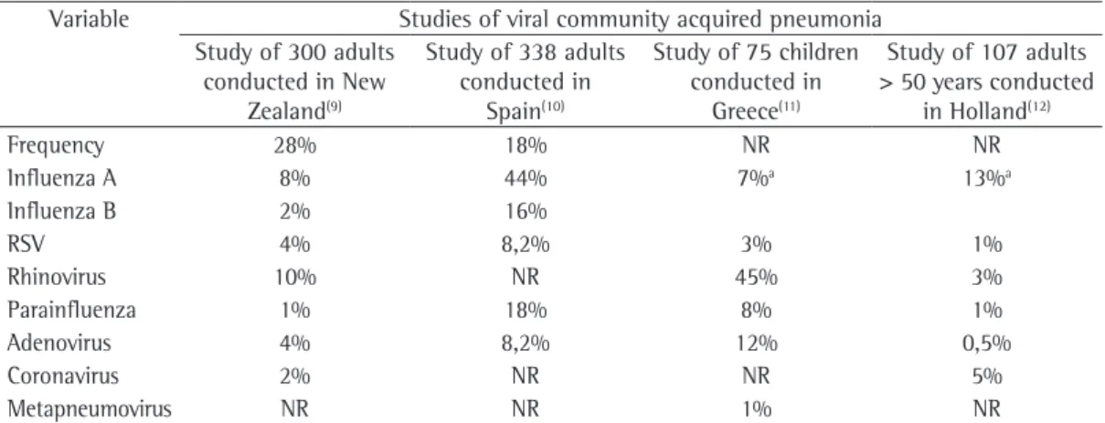
mentioned above. Some of these studies are summarized in Table 1. In a study of 300 adult individuals with community-acquired pneu-monia conducted in New Zealand, 28% of the cases were of viral etiology. The viruses detected were rhinovirus (in 10%), influenza A (in 8%), influenza B (in 2%), RSV (in 4%), adenovirus (in 4%), coronavirus (in 2%) and parainfluenza (in 1%). In addition, more than one type of virus was found in some patients. Those authors observed that the patients with viral pneu-monia presented more myalgia than did those with bacterial infection. They also observed that simultaneous rhinovirus and pneumococcal infection was associated with more severe pneu-monia.(9) In a study of 338 adult individuals with community-acquired pneumonia conducted in Spain, it was demonstrated that 18% presented detectable virus and that the agents found were influenza A (in 44%), parainfluenza (in 18%), influenza B (in 16%), RSV (in 8.2%) and adeno-virus (in 8.2%). Those authors observed that, in 66% of the cases of viral pneumonia, the radio-logical examinations showed alveolar infiltrates, that complaints of cough with expectoration were less frequent in the viral cases and even that most of those patients presented tachypnea (more than 30 breaths/min).(10) Community-acquired pneumonia in other age brackets has also been studied in terms of viral etiology. In a study of 75 school-age children with commu-nity-acquired pneumonia conducted in Greece, the viruses detected were rhinovirus (in 45%), adenovirus (12%), parainfluenza (8%), influenza (7%), RSV (in 3%) and metapneumovirus (1%). Mixed viral-bacterial infections were found in suggestive of recent infection, using
serolog-ical methods and for detection of IgG using immunoenzymatic methods. However, in the last 15 years, highly sensitive techniques for the genomic detection of pathogens in clinical samples, such as PCR and RT-PCR—with their various real-time multiplex variants—as well as microarrays, have become popular. Using variants of those methods, there are various commer-cial diagnostic kits for detection of respiratory viruses. Therefore, the etiologic diagnosis of viral pneumonia on a large scale is currently possible.
Various viruses are recognized as causa-tive agents of pneumonia. Such viruses can be classified as those causing disease in immuno-competent individuals, which are the object of this review article, and as those acting opportun-istically, in the presence of immunodeficiency. In addition, the etiologies of viral pneumonia among immunocompetent individuals depend on the time of the year, since several of these viral infections are seasonal, occurring predomi-nantly in the winter, or even epidemic, as well as also depending on the use of sensitive and reliable diagnostic approaches to viral detection, and here are included molecular methods for genomic detection. Influenza A and B; parain-fluenza 1, 2 and 3; RSV; and adenovirus are traditionally known to be causative agents of pneumonia.
There have been few studies of viral infec-tions causing community-acquired pneumonia. However, in recent years, there have been studies of this subject using multiple diagnostic approaches and including the modern techniques
Table 1 - Studies of viral community-acquired pneumonia.
Variable Studies of viral community acquired pneumonia Study of 300 adults
conducted in New Zealand(9)
Study of 338 adults conducted in
Spain(10)
Study of 75 children conducted in
Greece(11)
Study of 107 adults > 50 years conducted
in Holland(12)
Frequency 28% 18% NR NR
Influenza A 8% 44% 7%a 13%a
Influenza B 2% 16%
RSV 4% 8,2% 3% 1%
Rhinovirus 10% NR 45% 3%
Parainfluenza 1% 18% 8% 1%
Adenovirus 4% 8,2% 12% 0,5%
Coronavirus 2% NR NR 5%
Metapneumovirus NR NR 1% NR
response of the host with the production of neutralizing antibodies. The viral infection is initiated by the action of viral hemagglutinin, which binds, in the cell membrane, to sialic acid residue. At the same time, neuraminidase, which is another viral protein, cleaves the sialic acid, allowing the release and spread of viruses recently produced by the infected cell. Currently, at least 16 antigenically specific hemaggluti-nins (H1-H16) and 9 neuraminidases (N1-N9) are known. Influenza A viruses with H1-H3 are the causative agents of most of the cases in humans. The other hemagglutinins are associ-ated with viruses of aquatic birds or of other mammals. The antigenic variations in H and N contribute to the epidemic nature of the influenza viruses by two mechanisms: minor antigenic variations (drift) that alter epitopes targeted by neutralizing antibodies; and major antigenic variations (shift) that occur by shuf-fling of viral genomic segments when there is simultaneous infection with two types of influ-enza viruses, resulting in the production of a mutant. This was the case of the viruses that caused pandemics in 1918 (H1N1), 1957 (H2N2) and 1969 (H3N2). Influenza A epidemics usually occur annually, during the winter, lasting for 6 to 8 weeks in the area and producing cases of varying severity. Viral transmission occurs via aerosolized droplets from cough or sneeze of infected individuals, and the onset of the disease occurs after a period of incubation of 2-3 days. This efficient mechanism of transmission, asso-ciated with a short period of incubation, results in major and explosive epidemics of this viral infection.(15) Avian influenza H5N1 appeared, in the last decade, as a threat that could cause a severe pandemic. The virus, originating from Asia, spread through migrating aquatic birds and caused hundreds of infections in humans, resulting in severe and highly lethal pneumonia, in different regions of the world. However, to date, the avian H5N1 virus has not been found to be adapted for human-to-human transmis-sion via respiratory secretions.(16)
Belonging to the Paramyxoviridae family, RSVs are enveloped, have single-stranded nonsegmented RNA, are classified into two anti-genic groups, A and B, both of which can cause outbreaks, and are associated with bronchiolitis in childhood. The genome of RSVs is more stable and does not have high mutagenic activity, 35% of the cases.(11) Regarding individuals over
50 years of age with community-acquired pneu-monia, a study of 107 patients conducted in Holland revealed, using RT-PCR and real-time PCR as methods for rapid diagnosis, the presence of influenza virus (in 13%), coronavirus (in 5%), rhinovirus (in 3%), parainfluenza (in 1%), RSV (in 1%) and adenovirus (in 0.5%).(12) It has been reported that influenza A and RSV are the most common viruses in elderly patients with viral pneumonia in the United States.(13)
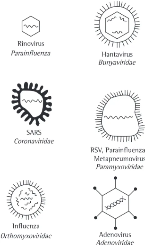
Therefore, influenza A and B, RSV, parainflu-enza, metapneumovirus, coronavirus, rhinovirus, hantavirus and adenovirus are among the viruses that cause community-acquired pneumonia. A chart with the structure of these viruses is shown in Figure 1.(14)
Influenza viruses, which belong to the Orthomyxoviridae family, are enveloped, have 8 single-stranded RNA segments and are classi-fied as types A, B and C. The two glycoproteins of the viral envelope are the target of the immune
SARS Coronaviridae
RSV, Parainfluenza, Metapneumovirus
Paramyxoviridae
Influenza
Orthomyxoviridae Adenovirus Adenoviridae Rinovirus
Parainfluenza Hantavirus Bunyaviridae
Figure 1 - Viruses that cause pneumonia and
families to which they belong. With the exception of adenoviruses, all of them are single-stranded RNA viruses. Adapted from Flores.(14) SARS: severe acute
Hantaviruses belong to the Bunyaviridae family. Hantavirus infection in humans leads to HCPS, an emerging disease in the Americas, caused by inhalation of aerosolized excreta from wild rodents (Sigmodontinae) contaminated with these viruses. In Brazil, HCPS has occurred since 1993, and, as of 2008, approximately 1,100 cases had been reported. The Araraquara, the Juquitiba, the Castelo dos Sonhos, the Rio Mearim and the Laguna Negra-símile hanta-viruses are known to be causative agents of the human form of the disease in Brazil. The analysis, between 1998 and 2007, of a sample of 70 patients with HCPD in the region of Ribeirão Preto, a city in the state of São Paulo, Brazil, revealed a higher incidence of the disease between April and September, the dry season. Of those patients, 75.7% were male, and the mean age was 35.8 ± 11.7 years. After an incubation period of 2 to 30 days, those patients presented the following: dyspnea (in 87%); fever (in 81%); cough and headache (in 44% and 34%, respec-tively), for 3-6 days (mean, 4 days); symptoms accompanied by tachycardia (in 81%); arterial hypotension (in 56%); a decrease in SaO2 (in when compared with that of influenza viruses.
In addition, RSV infections induce incomplete immunity, and RSV reinfection usually causes mild respiratory disease. The rate of occurrence of pneumonia and more severe profiles increases with age. Among elderly individuals residing in nursing homes, 10% are expected to be infected with RSV each year, and, of those, 10% are expected to develop pneumonia.(17)
Parainfluenza viruses, similarly to RSVs, belong to the Paramyxoviridae family and are associated with bronchitis and pneumonia in suckling infants. Three viral types, numbered from 1 to 3, are recognized, causing 4-14% of respiratory infections. These viruses produce recurring infections, including in adult life, causing pneumonia in young individuals and bronchopneumonia in elderly individuals.(18)
Human metapneumoviruses, similarly to parainfluenza viruses and RSVs, belong to the Paramyxoviridae family. They were described recently (in 2001), are globally distributed and have been found to cause bronchiolitis and pneumonia in suckling infants.(19) In young adults, they cause colds, flu and exacerbation of asthma attacks. Metapneumovirus respiratory infections also occur in elderly individuals with cardiopulmonary diseases.(13)
Coronaviruses, which belong to the Coronaviridae family, are commonly associ-ated with colds, and their laboratory isolation is difficult. The OC43 and 229E strains are the most common, having been described in all age brackets. In 2002, a new coronavirus appeared in China, causing a new disease, which was designated SARS. This disease spread rapidly around the world through travelers coming from China, causing pneumonia presenting as respira-tory failure and high mortality, and was initially considered a major public health threat world-wide. However, probably due to the fact that the virus was not easily transmitted, the epidemic decreased in a few months.(20)
Rhinoviruses, which belong to the Picornaviridae family, are the most common causative agents of colds, which occur among individuals of all ages and throughout the year. The role of rhinoviruses as primary causative agents of pneumonia is controversial. However, rhinoviruses have been recovered from the lower airways of neonates and immunocompromised patients with pneumonia.(21)
CB 06/20/99 06/22/99
06/27/99 06/30/99
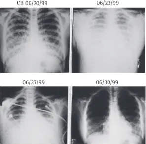
Figure 2 - Radiological evolution of hantavirus
toconjunctivitis, gastroenteritis, hemorrhagic cystitis, meningoencephalitis, hepatitis, myocar-ditis and severe disseminated disease. Severe pneumonia caused by serotype 14 has been described in adults and children with mild or moderate COPD.(23)
The treatment of viral pneumonia depends on the severity of the profile and on the infecting agent. General support measures, especially those with ventilation, for the treatment of hypoxia, can be critical for patient survival. The high frequency at which bacterial infections are associated with viral infections makes it possible that, after microbiological examination, antibi-otics are indicated in such cases. Antiviral therapy is indicated in severe cases and in immunocom-promised individuals, based on diagnostic tests for viruses. This therapy is usually more efficient when it is initiated early, at the onset of symp-toms.(3)
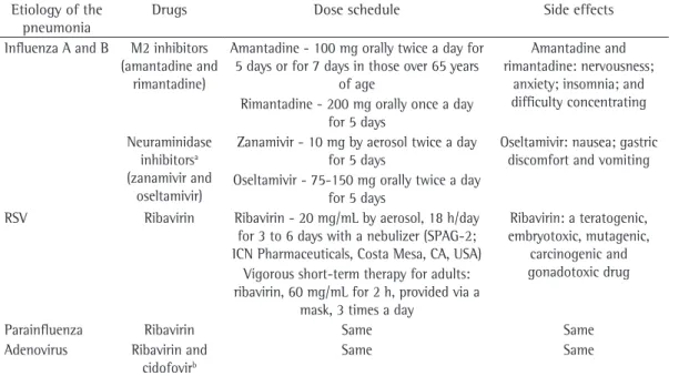
Four antiviral drugs (amantadine, rimanta-dine, zanamivir and oseltamivir) can be used in the treatment of influenza virus infections. Amantadine and rimantadine are active against influenza A virus. Zanamivir and oseltamivir are active against influenza A and B. These drugs, when administered in the first 48 h after disease onset, reduce the severity of the disease and the symptoms. Zanamivir and oseltamivir can 49%); metabolic acidosis (in 57%);
lymphocy-topenia (in 51%); hematocrit > 45% (in 70%); leukocytosis with a left shift (in 67%); increased serum creatinine (in 51%); and increased serum urea (in 42%). In addition, they presented radio-logical alteration with bilateral diffuse interstitial opacification between the first and the fourth day of disease and subsequent alveolarization, as shown in Figure 2. The progression to respi-ratory failure, arterial hypotension and shock occurred 24-48 h after the onset of symptoms, and the highest frequency of deaths occurred on the fourth day of disease. Increased hemat-ocrit and thrombocytopenia were signs strongly suggestive of the disease. The working diagnosis of atypical pneumonia was associated with a good prognosis (p = 0.0136), whereas parenteral fluid resuscitation of more than 2,000 mL and arterial hypotension were associated with a bad prognosis (p = 0.0286 and p = 0.0453, respec-tively). Contact with infected rodents proved to be a risk factor for transmission and morbidity (p = 0.0198). In the sample of individuals with HCPS caused by the Araraquara virus, the mortality rate was 54.3%.(22)
Adenoviruses, DNA viruses that belong to the Adenoviridae family, occur worldwide. Adenoviruses of 52 serotypes can be the cause of asymptomatic infections, pharyngitis,
kera-Table 2 - Antiviral drugs used in the treatment of pneumonia.(3,13)
Etiology of the pneumonia
Drugs Dose schedule Side effects
Influenza A and B M2 inhibitors (amantadine and
rimantadine)
Amantadine - 100 mg orally twice a day for 5 days or for 7 days in those over 65 years
of age
Amantadine and rimantadine: nervousness;
anxiety; insomnia; and difficulty concentrating Rimantadine - 200 mg orally once a day
for 5 days Neuraminidase
inhibitorsa
(zanamivir and oseltamivir)
Zanamivir - 10 mg by aerosol twice a day for 5 days
Oseltamivir: nausea; gastric discomfort and vomiting Oseltamivir - 75-150 mg orally twice a day
for 5 days
RSV Ribavirin Ribavirin - 20 mg/mL by aerosol, 18 h/day for 3 to 6 days with a nebulizer (SPAG-2; ICN Pharmaceuticals, Costa Mesa, CA, USA)
Ribavirin: a teratogenic, embryotoxic, mutagenic,
carcinogenic and gonadotoxic drug Vigorous short-term therapy for adults:
ribavirin, 60 mg/mL for 2 h, provided via a mask, 3 times a day
Parainfluenza Ribavirin Same Same
Adenovirus Ribavirin and cidofovirb
Same Same
RSV: respiratory syncytial virus. aIn the case of influenza B, neuraminidase inhibitors act exclusively. bNo other
trauma. Although ribavirin is effective in vitro against hantaviruses, its activity in vivo has yet to be scientifically proven.(25)
Regarding the treatment of SARS, in addition to ventilatory support measures, it is recom-mended that ribavirin, or even a combination of ribavirin, lopinavir/ritonavir and corticoster-oids—a combination recommended, based on the reduction in the viral load, as the most effective therapy—be used, although its effectiveness has yet to be confirmed in controlled studies.
The prevention of viral infections that cause community-acquired pneumonia includes, depending on the virus, vaccines and passive immunization. For the prevention of influenza A infections, there are inactivated virus vaccines that are produced in embryonated eggs and have a panel of viruses that circulate in the region.(26) In order to prevent RSV infections, especially in high-risk children, the following immunoglobulins are used: RSV-IGIV (RespiGam™; Massachusetts Public Health Biologic Laboratories, Boston, MA, USA) and palivizumab (Synagis®, Abbott, São Paulo, Brazil).(3)
References
1. Nolte FS. Molecular diagnostics for detection of bacterial and viral pathogens in community-acquired pneumonia. Clin Infect Dis. 2008;47 Suppl 3:S123-6.
2. Eccles R. Understanding the symptoms of the common cold and influenza. Lancet Infect Dis. 2005;5(11):718-25.
3. Treanor JJ. Respiratory infections. In: Richman DD, Whitley RJ, Hayden FG. Clinical Virology. Washington: ASM Press; 2002. p.7-26.
4. Ng WF, To KF, Lam WW, Ng TK, Lee KC. The comparative pathology of severe acute respiratory syndrome and avian influenza A subtype H5N1--a review. Hum Pathol. 2006;37(4):381-90.
5. Thomas PG, Keating R, Hulse-Post DJ, Doherty PC. Cell-mediated protection in influenza infection. Emerg Infect Dis. 2006;12(1):48-54.
6. Borges AA, Figueiredo LT. Atualização de conhecimentos sobre a patogênese da síndrome pulmonar e cardiovascular por hantavírus. Rev Patol Trop. 2007;36(3):191-204. 7. Buchholz UJ, Ward JM, Lamirande EW, Heinze B, Krempl
CD, Collins PL. Deletion of nonstructural proteins NS1 and NS2 from pneumonia virus of mice attenuates viral replication and reduces pulmonary cytokine expression and disease. J Virol. 2009;83(4):1969-80.
8. Moreno L, Krishnan JA, Duran P, Ferrero F. Development and validation of a clinical prediction rule to distinguish bacterial from viral pneumonia in children. Pediatr Pulmonol. 2006;41(4):331-7. Erratum in: Pediatr Pulmonol. 2006;41(5):494.
9. Jennings LC, Anderson TP, Beynon KA, Chua A, Laing RT, Werno AM, et al. Incidence and characteristics of
induce bronchospasm in patients with obstruc-tive pulmonary disease. However, although its efficacy has yet to be definitely established by scientific studies, antiviral therapy is indicated in patients with influenza pneumonia.(13)
In order to prevent the introduction of respiratory viruses that are epidemic and cause severe disease, such as avian H5N1 influenza, it is recommended that the patient be imme-diately placed in respiratory isolation in an infirmary with negative pressure and air filtra-tion. A minimal number of professionals, always wearing N95 masks and being attentive to handwashing, should deal with the patient. In addition, all of the objects used by the patient should be decontaminated.(24)
The treatment of RSV pneumonia includes ventilatory support measures and the use of ribavirin aerosol, as shown in Table 2.
18. Parainfluenza infections in the elderly 1976-82. Br Med J (Clin Res Ed). 1983;287(6405):1619.
19. van den Hoogen BG, de Jong JC, Groen J, Kuiken T, de Groot R, Fouchier RA, et al. A newly discovered human pneumovirus isolated from young children with respiratory tract disease. Nat Med. 2001;7(6):719-24. 20. McIntosh K, Anderson LJ. Coronaviruses. In: Mandell GL,
Bennett JE, Dolin R, editors. Principles and practices of infectious diseases. Philadelphia: Churchill Livingston; 2005. p. 1990-8.
21. Papadopoulos NG, Bates PJ, Bardin PG, Papi A, Leir SH, Fraenkel DJ, et al. Rhinoviruses infect the lower airways. J Infect Dis. 2000;181(6):1875-84.
22. Campos GM. Síndrome Pulmonar e Cardiovascular por Hantavirus: estudos sobre uma doença emergente [thesis]. Ribeirão Preto: Universidade de São Paulo; 2008.
23. Louie JK, Kajon AE, Holodniy M, Guardia-LaBar L, Lee B, Petru AM, et al. Severe pneumonia due to adenovirus serotype 14: a new respiratory threat? Clin Infect Dis. 2008;46(3):421-5.
24. Bell DM; World Health Organization Writing Group. Non-pharmaceutical interventions for pandemic influenza, national and community measures. Emerg Infect Dis. 2006;12(1):88-94.
25. Figueiredo LT. Hantavirose. In: Cimerman S, Cimerman B, editors. Condutas em infectologia. São Paulo: Atheneu; 2004. p.123-32.
26. Luke CJ, Subbarao K. Vaccines for pandemic influenza. Emerg Infect Dis. 2006;12(1):66-72.
viral community-acquired pneumonia in adults. Thorax. 2008;63(1):42-8.
10. de Roux A, Marcos MA, Garcia E, Mensa J, Ewig S, Lode H, et al. Viral community-acquired pneumonia in nonimmunocompromised adults. Chest. 2004;125(4):1343-51.
11. Tsolia MN, Psarras S, Bossios A, Audi H, Paldanius M, Gourgiotis D, et al. Etiology of community-acquired pneumonia in hospitalized school-age children: evidence for high prevalence of viral infections. Clin Infect Dis. 2004;39(5):681-6.
12. Oosterheert JJ, van Loon AM, Schuurman R, Hoepelman AI, Hak E, Thijsen S, et al. Impact of rapid detection of viral and atypical bacterial pathogens by real-time polymerase chain reaction for patients with lower respiratory tract infection. Clin Infect Dis. 2005;41(10):1438-44. 13. Falsey AR, Walsh EE. Viral pneumonia in older adults.
Clin Infect Dis. 2006;42(4):518-24.
14. Flores EF. Estrutura das partículas víricas. In: Flores EF, organizer. Virologia Veterinária. Santa Maria: UFSM; 2007. p.21-9.
15. Centers for Disease Control and Prevention (CDC). Pneumonia and influenza death rates--United States, 1979-1994. MMWR Morb Mortal Wkly Rep. 1995;44(28):535-7. Erratum in: MMWR Morb Mortal Wkly Rep 1995;44(41):782.
16. Enserink M. Avian influenza. Infection study worries farmers, bird lovers. Science. 2009;323(5912):324. 17. Thompson WW, Shay DK, Weintraub E, Brammer L,
Cox N, Anderson LJ, et al. Mortality associated with influenza and respiratory syncytial virus in the United States. JAMA. 2003;289(2):179-86.
About the authors
Luiz Tadeu Moraes Figueiredo