Characteristics associated with complete surgical
resection of primary malignant mediastinal tumors*
,**
Características associadas à ressecção cirúrgica completa de tumores malignos primários do mediastino
Jefferson Luiz Gross, Ulisses Augusto Correia Rosalino, Riad Naim Younes, Fábio José Haddad, Rodrigo Afonso da Silva, Antonio Bomfim Marçal Avertano Rocha
Abstract
Objective: To identify preoperative characteristics associated with complete surgical resection of primary malignant mediastinal tumors. Methods: Between 1996 and 2006, 42 patients with primary malignant mediastinal tumors were submitted to surgery with curative intent at a single facility. Patient charts were reviewed in order to collect data related to demographics, clinical manifestation, characteristics of mediastinal tumors and imaging aspects of invasiveness. Results: The surgical resection was considered complete in 69.1% of the patients. Cases of incomplete resection were attributed to invasion of the following structures: large blood vessels (4 cases); the superior vena cava (3 cases); the heart (2 cases); the lung and chest wall (3cases); and the trachea (1 case). Overall survival was significantly better among the patients submitted to complete surgical resection than among those submitted to incomplete resection. The frequency of incomplete resection was significantly higher in cases in which the tumor had invaded organs other than the lung (as identified through imaging studies) than in those in which it was restricted to the lung (47.6% vs. 14.3%; p = 0.04). None of the other preoperative characteristics analyzed were found to be associated with complete resection. Conclusions: Preoperative radiological evidence of invasion of organs other than the lung is associated with the incomplete surgical resection of primary malignant mediastinal tumors.
Keywords: Thoracic surgery; Survival rate; Mediastinal neoplasms; Diagnostic imaging.
Resumo
Objetivo: Identificar características pré-operatórias associadas à ressecção cirúrgica completa de tumores malignos primários do mediastino. Métodos: Entre os anos de 1996 e 2006, 42 pacientes com tumores malignos primários do mediastino foram submetidos a tratamento cirúrgico com intenção curativa em uma única instituição. Dados demográficos, manifestações clínicas, características do tumor mediastinal e aspectos de invasão por métodos de imagem foram identificados através da análise de prontuários. Resultados: A ressecção cirúrgica foi considerada completa em 69,1% dos pacientes. As causas de ressecção incompleta foram atribuídas à invasão das seguintes estruturas: grandes vasos (4 casos); veia cava superior (3 casos); coração (2 casos); pulmão e parede torácica (3 casos); e traqueia (1 caso). Os pacientes que foram submetidos à ressecção cirúrgica completa tiveram sobrevida global significativamente melhor que os pacientes submetidos à ressecção incompleta. A frequência de ressecção incompleta foi significativamente maior nos casos nos quais foram identificadas características radiológicas de invasão de outros órgãos além do pulmão do que nos casos cujas características eram restritas ao pulmão (47,6% vs. 14,3%; p = 0,04). Nenhuma das outras características pré-operatórias analisadas foi associada com a ressecção cirúrgica completa. Conclusões: Evidências radiológicas de invasão de órgãos além do pulmão no pré-operatório estão associadas à ressecção cirúrgica incompleta de tumores primários malignos do mediastino.
Descritores: Cirurgia torácica; Taxa de sobrevida; Neoplasias do mediastino; Diagnóstico por imagem.
* Study carried out in the Department of Thoracic Surgery. A. C. Camargo Hospital, São Paulo, Brazil.
Correspondence to: Jefferson Luiz Gross. Departamento de Cirurgia Torácica, Hospital A. C. Camargo, Rua Professor Antonio Prudente, 211, Liberdade, CEP 01509-010, São Paulo, SP, Brasil.
Tel 55 11 3277-7720. E-mail: [email protected] Financial support: None.
Submitted: 6 February 2009. Accepted, after review: 15 May 2009.
facility between March of 1986 and August of 2006. Inclusion criteria were histological diag-nosis of malignant primary mediastinal tumor and surgical treatment with the objective of radical resection. Patients with mediastinal lymphomas were excluded, as were those with tumors arising from the trachea, esophagus or lung and those with mediastinal goiters.
Data related to demographic characteristics, clinical manifestation, mediastinal tumor loca-tion, invasion of adjacent organs, preoperative (neoadjuvant) treatment, type of thoracic inci-sion and resectability were collected through a review of individual patient charts. In order to classify tumor location, the mediastinum was divided into three compartments (anterior, middle and posterior) based on a review of radi-ology reports. All patients were submitted to preoperative imaging studies—either chest CT or the combination of chest CT and magnetic reso-nance imaging (MRI), at the discretion of the surgeon—and data regarding the infiltration of adjacent organs were classified according to the radiology reports. Complete surgical resection was defined as complete removal of all macro-scopic tumor; and incomplete surgical resection was defined as no resection or partial resection.
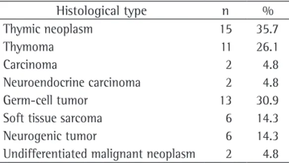
Median age was 39.5 years (range, 1.0-77.4 years), and 30 (71.5%) of the 42 patients were male. Five patients were asymptomatic. The most common initial clinical manifestation was chest pain, which was reported by 18 patients (42.8%). The median duration of symptoms was 4 months (range, 1-36 months). The majority (73.8%) of the tumors were located in the ante-rior mediastinal compartment, whereas 19.1% were located in the posterior compartment, and 7.1% were located in the middle compartment. Thymic malignant neoplasms and germ-cell tumors were the two most common histologic
Introduction
Mediastinal tumors are rare and include a range of different diseases.(1,2) Malignant
medi-astinal tumors can be primary or metastatic from other sites. Surgery, ranging from biopsy to radical resection, plays an essential role in the management of most primary malignant medi-astinal tumors. In addition, surgery can be part of multimodal therapeutic approaches, including chemotherapy or radiotherapy. Surgical resection is rarely used as the sole treatment for primary malignant mediastinal tumors.(3,4)
Since primary malignant mediastinal tumors are rare and heterogeneous, there have been few studies reporting the role of surgical resection in the management of such tumors. Most studies evaluating the management of thoracic tumors have evaluated various benign and malig-nant diseases arising in different mediastinal compartments. Other studies evaluating surgical resection as the primary treatment for tumors arising in the mediastinum were published more than 20 years ago.(5-7) Since then, surgical and
anesthetic techniques have improved consid-erably. Patient characteristics, indications for resection and overall outcomes of surgery have changed significantly. Thymomas and nonsemi-nomatous germ-cell tumors are the primary malignant mediastinal tumors for which surgical treatment is most often applied.(8)
Complete resection has been shown to be a significant determinant of prognosis among patients submitted to surgery for the treatment of invasive primary mediastinal tumors.(9) Age,
together with the characteristics of preopera-tive imaging studies determining the location and consistency of mediastinal tumors, can suggest the diagnosis and the likelihood of malignancy.(1,10-12) However, the factors predicting
the resectability of such tumors have not been reported. The aim of the present study was to identify preoperative characteristics associated with the ability to achieve complete surgical resection in a group of patients with primary malignant mediastinal tumors.
Methods
This was a retrospective study of 42 patients with primary malignant mediastinal tumors submitted to surgery with curative intent. All patients underwent surgical treatment at our
Table 1 - Histologic type of malignant primary mediastinal tumors.
Histological type n %
Thymic neoplasm 15 35.7
Thymoma 11 26.1
Carcinoma 2 4.8
Neuroendocrine carcinoma 2 4.8
Germ-cell tumor 13 30.9
Soft tissue sarcoma 6 14.3
Neurogenic tumor 6 14.3
Results
Among the 42 patients evaluated, surgical resection was considered complete in 29 (69.1%) and incomplete in the remaining 13 (30.9%). Incomplete resection was attributed to tumor invasion of the following structures: large blood vessels (in 4 cases); the superior vena cava (in 3 cases); the heart (in 2 cases); the lung and chest wall (in 3 cases); and the trachea (in 1 case). Among the 13 patients in whom the resection was incomplete, radiotherapy was administered in 6, chemotherapy was adminis-tered in 3, and the chemotherapy-radiotherapy combination was administered in 3. One patient with malignant mesenchymoma received no postoperative treatment and died two months after the surgical procedure due to the progres-sion of malignant disease.
The median follow-up time was 21.5 months (range, 0.2-183 months). Overall five-year survival was 71.3%. Patients submitted to complete surgical resection had significantly better overall survival than did those submitted to incomplete resection (84.4% vs. 42.9%, types, as shown in Table 1. According to the
radiology reports, the tumor was restricted to the mediastinum, without invasion of adja-cent structures, in 14 patients (33.3%). Among the remaining 28 patients (66.6%) in whom there was radiologic evidence of a tumor infil-trating adjacent organs, there was invasion of the lung parenchyma in 18, invasion of large blood vessels in 5, invasion of the pericardium in 6 and invasion of the chest wall in 7. All 28 of those patients were submitted to surgery as the primary treatment. Preoperative chemo-therapy was administered to 13 (31.0%) of the 42 patients. One patient (2.4%) was treated with chemotherapy and radiation therapy prior to surgical resection. The type of thoracic surgical access was selected by the attending surgeon on a case-by-case basis. The most common surgical approach was median sternotomy, employed in 19 patients (45.3%); followed by left lateral thoracotomy, employed in 14 (25.0%); and right lateral thoracotomy, employed in 9 (21.4%). Resection of adjacent structures was performed in 24 patients (57.2%). The lung parenchyma was the structure most frequently excised: wedge/ segmentectomy was performed in 12 patients (28.6%); lobectomy was performed in 2 (4.8%); and pneumonectomy was performed in 1 (2.4%). The following were also resected: the adjacent pericardium in 5 patients (11.9%); the parietal pleura in 5 (11.9%); the chest wall in 4 (9.6%); the phrenic nerve in 2 (4.8%); and the superior vena cava (partial resection) in 1 (2.4%).
Statistical analysis
Overall survival, from the date of surgery to the date of death or the last clinical evaluation, was estimated by the Kaplan-Meier method. The influence that the type of resection (complete or incomplete) had on overall survival was calcu-lated, and comparisons were performed using the log-rank test. The chi-square and Fisher’s exact tests were used, as appropriate, to assess associations between the type of surgical resec-tion and the following variables: demographic data; histologic type; tumor location; inva-sion of adjacent structures; surgical approach; preoperative treatment; and status at admission. Values of p < 0.05 were considered statistically significant. Statistical analyses were performed using the program Statistical Package for the Social Sciences, version 11.5 (SPSS Inc, Chicago, IL, USA).
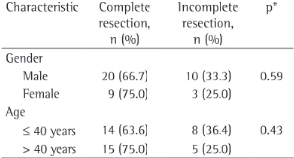
Table 2 - Association between characteristics of the patients and resectability of mediastinal tumors.
Characteristic Complete resection,
n (%)
Incomplete resection,
n (%)
p*
Gender
Male 20 (66.7) 10 (33.3) 0.59 Female 9 (75.0) 3 (25.0)
Age
≤ 40 years 14 (63.6) 8 (36.4) 0.43 > 40 years 15 (75.0) 5 (25.0)
*chi-square test or Fisher’s exact test.
Table 3 - Chest pain and symptom duration (all symptoms): association with resectability of mediastinal tumors.
Variable Complete resection,
n (%)
Incomplete resection,
n (%)
p*
Chest pain
Yes 9 (52.9) 8 (47.1) 0.08
No 17 (81.0) 4 (19.0)
Duration of symptoms
≤ 4 months 13 (72.2) 5 (27.8) 0.26 > 4 months 8 (53.3) 7 (48.7)
mediastinal compartment) was not associated with the ability to achieve complete resection; nor was the type of surgical approach (ster-notomy or lateral thoracotomy). As can be seen in Table 4, the same was true for preop-erative treatment (neoadjuvant treatment or no treatment) and histologic type of the primary mediastinal malignancy (thymic or non-thymic).
In all cases, chest CT or chest CT plus MRI was performed in order to identify invasion of anatomic structures adjacent to the mediastinal tumor.
Resectability was not associated with invasion of mediastinal structures, defined in preoperative imaging evaluation. Refinement of this analysis revealed that tumor invasion of the lung paren-chyma was not associated with resectability. However, invasion of organs other than the lung was strongly associated with resectability. Among the 14 patients in whom there was no invasion of these anatomical structures, complete resec-tion was achieved in 12 (85.7%), compared with 17 (60.7%) of the 28 in whom there was such invasion (Table 5). Involvement of anatomic structures other than the lung was as follows: the heart and large blood vessels (in 8 cases); the chest wall (in 7 cases); the pericardium (in 5 cases); and the esophagus (in 1 case).
Discussion
There have been few studies dedicated to analyzing patients with primary mediastinal tumors. Most studies evaluating the manage-ment of thoracic tumors have included patients with various benign and malignant tumors, treated over long periods.(1,13-16)
The present study evaluated a group of patients with primary malignant mediastinal neoplasms of various histologic types. However, all of the patients were submitted to surgical procedures with curative intent. We selected this specific group of patients in order to identify preoperative factors associated with the ability to achieve complete surgical resection.
Although the present study involved at least six different histologic types of primary medias-tinal tumors, it is not as heterogeneous as some other studies, which have included lymphomas and various types of benign tumors.(16-18)
Lymphomas have been included even in studies that selected patients submitted to surgical resection of malignant mediastinal tumors, p = 0.04). The number of censored cases at 20,
40 and 60 months, respectively, was as follows: complete resection: 16, 8 and 4 patients; and incomplete resection: 5, 4 and 3 patients.
Gender and age were not associated with the resectability of mediastinal tumors (Table 2). Patients with chest pain as an initial clinical mani-festation tended to be unresectable at surgery. Table 3 shows that resection was incomplete in 47.1% of such patients. However, this difference did not reach statistical significance (p = 0.08).
The anatomical location of the primary malignant tumor (anterior, middle or posterior
Table 4 - Tumor location, type of surgical approach, preoperative treatment and histologic type: association with resectability of mediastinal tumors.
Variable Complete resection,
n (%)
Incomplete resection,
n (%) p*
Tumor location Anterior mediastinum
23 (74.2) 8 (25.8) 0.23
Other 6 (54.5) 5 (45.5) Type of surgical approach
Sternotomy 15 (78.9) 4 (21.1) 0.31 Lateral
thoracotomy
14 (60.9) 9 (39.1)
Preoperative treatment
Yes 21 (75.0) 7 (25.0) 0.24
No 8 (57.1) 6 (42.9)
Histological type
Thymic 11 (73.3) 4 (26.7) 0.74 Non-thymic 18 (66.7) 9 (33.3) *chi-square test or Fisher’s exact test.
Table 5 - Association between preoperative radiological evidence of tumor invasion and resectability of mediastinal tumors.
Radiological evidence
Complete resection,
n (%)
Incomplete resection,
n (%)
p*
Invasion of adjacent organs
Yes 17 (60.7) 11 (39.3) 0.16
No 12 (85.7) 2 (14.3)
Invasion of the lung
Yes 11 (61.1) 7 (38.9) 0.51
No 18 (75.0) 6 (25.0)
Invasion of organs other than the lung
Yes 11 (52.4) 10 (47.6) 0.04
No 18 (85.7) 3 (14.3)
One of the most common clinical manifes-tations, chest pain, was observed in 66.7% of the patients submitted to incomplete resection, compared with only 34.6% of those submitted to complete resection, although this difference did not reach statistical significance (p = 0.08). This finding suggests that chest pain is asso-ciated with locally advanced tumors and with lower probability of complete surgical resec-tion. Some authors have described chest pain as a predictor of survival among patients with chest wall tumors.(22) However, there is no data
regarding this characteristic in patients with primary malignant mediastinal neoplasms. Chest pain might be related to chest wall invasion by mediastinal tumors or even to invasion of other adjacent anatomical structures. Invasion of the chest wall (sternum or ribs) should not present technical obstacles to complete surgical resec-tion, since sternum and ribs can be resected with adequate margins. Therefore, we believe that any association between chest pain and resectability is not related to the extent of local invasion, although no definitive conclusion can be made based on the data currently available.
Although imaging of mediastinal tumors is typically performed using chest CT, MRI can be used in order to define great vessel invasion or to identify invasion of the vertebral body. Both radiological methods are employed in order to determine tumor invasion of adjacent anatom-ical structures.(23) One group of authors assessed
the value of CT and MRI in evaluating medias-tinal tumors and concluded that both provide essentially the same information.(24) All of the
patients evaluated in the present study were submitted to chest CT. However, MRI was also performed in certain cases, which were few in number, since MRI has only become available at our institution in the last ten years and was indicated only in specific situations.
To our knowledge, this is the first study correlating radiological findings with the proba-bility of achieving complete surgical resection of mediastinal tumors. We found that radiological invasion of lung parenchyma did not correlate with tumor resectability, probably because resec-tion of the lung parenchyma (wedge resecresec-tion or even lobectomy) can be easily accomplished en bloc with the mediastinal mass. However, inva-sion of structures other than the lung are directly correlated with inability to achieve complete despite the fact that surgery plays a limited role
in the management of mediastinal lymphomas. Accordingly, we decided to exclude patients with lymphomas. In surgical studies of malignant mediastinal tumors,(9,19-21) thymic neoplasms,
mainly thymomas, and primary malignant germ-cell tumors are the histologic types most frequently described, as was also observed in the present study. Another common critique of previous studies of mediastinal tumors is that patients were treated over a long period, during which the therapeutic strategies changed. Because of the low incidence of primary malig-nant mediastinal tumors, the present study described patients treated during a 20-year period, similar to other published studies.(9,16)
Other studies(7) have reported the surgical
treatment of mediastinal neoplasms invading adjacent anatomical structures and requiring extensive surgical resection, followed by recon-structive techniques.(9,16) The authors of those
studies concluded that complete resection is a significant determinant of long-term survival. Our results confirm that patients submitted to complete surgical resection had better overall survival than did those submitted to incom-plete resection. One group of authors reported an overall five-year survival rate of 70% among patients undergoing complete surgical resection,(9) similar to the 85% observed in our
group of patients. Despite the evident impact of complete surgical resection on overall survival, it was not clear whether certain preoperative characteristics can predict the resectability of primary malignant mediastinal tumors.
Complete surgical resection was obtained in 69.1% of the patients evaluated in the present study, similar to the 79% reported in another study, involving 89 patients.(9) These results
8. Whooley BP, Urschel JD, Antkowiak JG, Takita H. Primary tumors of the mediastinum. J Surg Oncol. 1999;70(2):95-9.
9. Bacha EA, Chapelier AR, Macchiarini P, Fadel E, Dartevelle PG. Surgery for invasive primary mediastinal tumors. Ann Thorac Surg. 1998;66(1):234-9.
10. Takeda S, Miyoshi S, Akashi A, Ohta M, Minami M, Okumura M, et al. Clinical spectrum of primary mediastinal tumors: a comparison of adult and pediatric populations at a single Japanese institution. J Surg Oncol. 2003;83(1):24-30.
11. Azarow KS, Pearl RH, Zurcher R, Edwards FH, Cohen AJ. Primary mediastinal masses. A comparison of adult and pediatric populations. J Thorac Cardiovasc Surg. 1993;106(1):67-72.
12. Blegvad S, Lippert H, Simper LB, Dybdahl H. Mediastinal tumours. A report of 129 cases. Scand J Thorac Cardiovasc Surg. 1990;24(1):39-42.
13. Ovrum E, Birkeland S. Mediastinal tumours and cysts. A review of 91 cases. Scand J Thorac Cardiovasc Surg. 1979;13(2):161-8.
14. Dubail D, Nyaruhirira I, Bosschaerts T, Locufier JL, Barthel J, Barroy JP. Primary mediastinal tumors. Acta Chir Belg. 1994;94(4):215-21.
15. Silva R, Gross J, Haddad F, Younes R. Intrathoracic goiter and invasive thymoma: rare concomitant presentation. J Bras Pneumol. 2006;32(4):371-4.
16. Luosto R, Koikkalainen K, Jyrälä A, Franssila K. Mediastinal tumours. A follow-up study of 208 patients. Scand J Thorac Cardiovasc Surg. 1978;12(3):253-9. 17. Davis RD Jr, Oldham HN Jr, Sabiston DC Jr. Primary cysts
and neoplasms of the mediastinum: recent changes in clinical presentation, methods of diagnosis, management, and results. Ann Thorac Surg. 1987;44(3):229-37. 18. Benjamin SP, McCormack LJ, Effler DB, Groves
LK. Primary tumors of the mediastinum. Chest. 1972;62(3):297-303.
19. Couto WJ, Gross JL, Deheinzelin D, Younes RN. Primary mediastinal germ cell tumors [Article in Portuguese]. Rev Assoc Med Bras. 2006;52(3):182-6.
20. Sakurai H, Asamura H, Suzuki K, Watanabe S, Tsuchiya R. Management of primary malignant germ cell tumor of the mediastinum. Jpn J Clin Oncol. 2004;34(7):386-92. 21. Wright CD. Management of thymomas. Crit Rev Oncol
Hematol. 2008;65(2):109-20.
22. Greager JA, Patel MK, Briele HA, Walker MJ, Wood DK, Gupta TK. Soft tissue sarcomas of the adult thoracic wall. Cancer. 1987;59(2):370-3.
23. Tomiyama N, Müller NL, Ellis SJ, Cleverley JR, Okumura M, Miyoshi S, et al. Invasive and noninvasive thymoma: distinctive CT features. J Comput Assist Tomogr. 2001;25(3):388-93.
24. Ikezoe J, Takeuchi N, Johkoh T, Kohno N, Takashima S, Tomiyama N, et al. MRI of anterior mediastinal tumors. Radiat Med. 1992;10(5):176-83.
resection of mediastinal tumors. Invasion of large blood vessels was the most common radi-ological characteristic leading to incomplete surgical resection of mediastinal tumors.
Although the location of mediastinal mass is critical to differential diagnosis of primary malig-nant mediastinal tumors,(1) our results show that
this characteristic did not affect the ability to achieve complete surgical resection. Similarly, the type of surgical approach (sternotomy or thoracotomy) was not found to be associated with the completeness of resection. The type of surgical access should be considered in order to achieve adequate viewing and complete resec-tion with tumor-free margins.
We concluded that patients with primary malignant mediastinal tumors who are surgical candidates should be evaluated preoperatively in order to achieve complete surgical resec-tion, which is associated with better survival. Radiological evidence of invasion of organs other than the lung is associated with incom-plete surgical resection.
References
1. Duwe BV, Sterman DH, Musani AI. Tumors of the mediastinum. Chest. 2005;128(4):2893-909.
2. Cohen AJ, Thompson L, Edwards FH, Bellamy RF. Primary cysts and tumors of the mediastinum. Ann Thorac Surg. 1991;51(3):378-84; discussion 385-6. 3. Temes R, Chavez T, Mapel D, Ketai L, Crowell R, Key C,
et al. Primary mediastinal malignancies: findings in 219 patients. West J Med. 1999;170(3):161-6.
4. Wongsangiem M, Tangthangtham A. Primary tumors of the mediastinum: 190 cases analysis (1975-1995). J Med Assoc Thai. 1996;79(11):689-97.
5. Adkins RB Jr, Maples MD, Hainsworth JD. Primary malignant mediastinal tumors. Ann Thorac Surg. 1984;38(6):648-59.
6. Davis RD Jr, Oldham HN Jr, Sabiston DC Jr. Primary cysts and neoplasms of the mediastinum: recent changes in clinical presentation, methods of diagnosis, management, and results. Ann Thorac Surg. 1987;44(3):229-37. 7. Wychulis AR, Payne WS, Clagett OT, Woolner LB. Surgical
About the authors
Jefferson Luiz Gross
Director. Department of Thoracic Surgery, A. C. Camargo Hospital, São Paulo, Brazil.
Ulisses Augusto Correia Rosalino
Student. Young Investigator Course, A. C. Camargo Hospital, São Paulo, Brazil.
Riad Naim Younes
Tenured Professor. University of São Paulo School of Medicine, São Paulo, Brazil.
Fábio José Haddad
Attending Physician. Department of Thoracic Surgery, A. C. Camargo Hospital, São Paulo, Brazil.
Rodrigo Afonso da Silva
Attending Physician. Department of Thoracic Surgery, A. C. Camargo Hospital, São Paulo, Brazil.
Antonio Bomfim Marçal Avertano Rocha