269
Jornal Brasileiro de Pneumologia 31(3) - Mai/Jun de 2005
Treatment of bronchial stenosis after lung transplantation
using a self-expanding metal endobronchial stent*
MARCOS NAOYUKI SAMANO**, MARLOVA LUZZI CARAMORI,
RICARDO HENRIQUE DE OLIVEIRA BRAGA TEIXEIRA***, HELIO MINAMOTO***,
PAULO MANUEL PÊGO FERNANDES***, FABIO BISCEGLI JATENE***, SÉRGIO ALMEIDA DE OLIVEIRA
*Study carried out at the Instituto do Coração (InCor) of the Hospital das Clínicas da Faculdade de Medicina da Universidade de São Paulo FMUSP, São Paulo, SP
**Specialist, as designated by the Sociedade Brasileira de Pneumologia e Tisiologia ***Specialist, as designated by the Sociedade Brasileira de Cirurgia Torácica
Correspondence to: Fabio Biscegli Jatene. Av. Dr. Enéas Carvalho Aguiar, 44 - 2o andar bloco II sala 9. CEP 05403-000, São Paulo, SP. Phone: 55 11 3069-5248. E-mail: [email protected]
Submitted: 25 May 2004. Accepted, after review: 1 September 2004 J Bras Pneumol 2005; 31(3): 269-72.
Key words: Lung transplantation. Tracheal stenosis. Prosthesis and implants. Stents. Postoperative complications.
Although the incidence of complications resulting from the bronchial anastomosis employed in lung transplantation has decreased in recent years, it remains a significant cause of morbidity and mortality in these patients. Treatment options include balloon dilatation, laser photocoagulation, placement of a stent (silicone or metal), and performing a second operation. We report the case of a patient who presented bronchial stenosis after left lung transplantation and was treated with a self-expanding metal alloy (nitinol) stent (Ultraflex). Despite the fact that this was the first case of stenosis treated in this fashion in Brazil, the positive clinical response, in agreement with results reported in the literature, indicates that this treatment is a viable alternative in such cases.
Complications resulting from bronchial anastomosis still constitute one of the principal factors of increased morbidity and mortality among lung transplant patients. It is estimated that such complications occur in 27% of patients submitted to lung transplant, and that 1 3 % o f t h e s e p a t i e n t s r e q u i r e i n v a s i v e bronc hosc opy for the treatm ent of these
complications(1,2). Although some centers have
experience in the utilization of self-expanding stents, there is no data regarding their use in Brazil. Herein, we report the case of a patient who developed bronchial stenosis after lung transplantation and was treated with a self-expanding metal endobronchial stent.
Case Report
270
Samano, MN, et al.
Treatment of bronchial stenosis after lung transplantation using a self-expanding metal endobronchial stent
CASE REPORT
A 57-year-old male ex-smoker was diagnosed with severe pulmonary emphysema. He presented progressive worsening for two years, to the point of having required hospitalization and mechanical ventilation. Since then, he made continuous use of supplementary oxygen, presenting dyspnea upon exertion and productive cough, mainly in the morning. He had long been making use of prednisone (30 mg/day) and had lost weight, with body mass index of 16.7. Spirometry presented mean forced expiratory volume in one second of 0.59 L (18% of predicted) and forced vital capacity of 1.88 L (46%). A blood gas test was performed with a
catheter of O2 at 2 L/min with PaO2 of 138 mmHg
and PaCO2 of 85 mmHg. A quantitative perfusion
lung scintigraphy presented 35% perfusion in the left lung.
After being submitted to the routine evaluation protocol, the patient was put on a waiting list and was later submitted to left lung transplantation.
The donor was a 38-year-old type B blood male patient, victim of a hemorrhagic cerebrovascular accident. The transplant was performed through a left posterior lateral thoracotomy with bronchial anastomosis using telescope technique, utilizing nonabsorbent propylene 4-0 sutures. Total ischemia time was three hours.
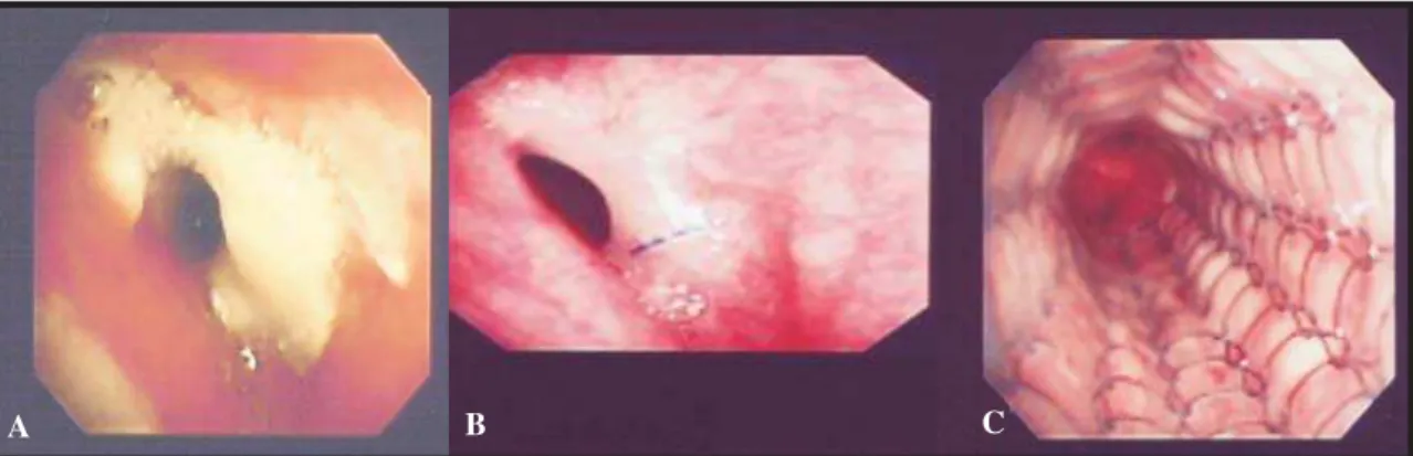
The patient presented uneventful postoperative evolution, with an episode of acute grade II rejection treated with methylprednisolone. On postoperative day 15, bronchial anastomosis evidenced enanthematous reaction and deposition of fibrin (Figure 1) without clinical repercussion, and the
patient was discharged on postoperative day 30. Outpatient fiberoptic bronchoscopy showed anastomosis retraction with decrease of its lumen. Three months after surgery the patient developed cough, dyspnea upon exertion and wheezing in the left hemithorax. A second fiberoptic bronchoscopy showed concentric stenosis of approximately 4 mm in diameter (Figure 1), and spirometry revealed forced expiratory volume in one second of 0.9 L (32% of predicted) and forced vital capacity of 2.73 L (79% of predicted), presenting a flow-volume curve consistent with upper-airway obstruction.
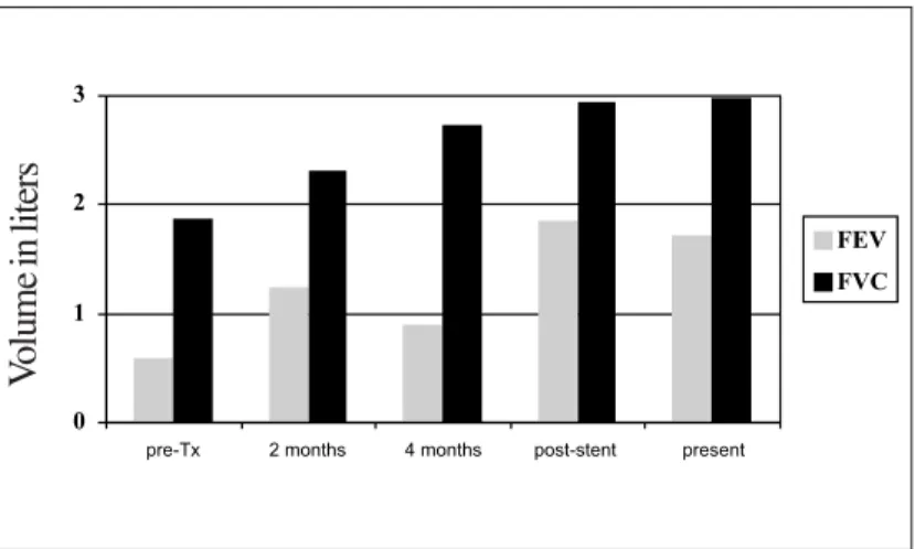
Dilatation of stenosis with a CRE 12-mm balloon (Boston Scientific, San Jose, CA, USA) was initially chosen, with significant improvement of symptoms. However, there was recurrence of cough and dyspnea after three weeks. At that point, it was decided that rigid bronchoscopy should be used. This was performed with the aid of fluoroscopy, dilatation with a CRE 15-mm balloon and treatment with a self-expanding, 14 x 40 mm metallic alloy (nitinol) endobronchial stent (Ultraflex Boston Scientific) (Figure 2). There was immediate improvement of symptoms and a 95% increase in the forced expiratory volume in one second (Figure 3). Four months after this procedure, the patient was breathing normally, and the bronchoscopic findings stabilized, without local complications.
DISCUSSION
During the twenty years between the first lung transplantation performed by James Hardy in 1963 and the first success by the Toronto Lung Transplant Group in 1983, approximately 40 unsuccessful
Figure 1. A: Bronchial anastomosis with intense inflammatory reaction and deposition of fibrin two weeks after transplantation. B: Progression to fibrous cicatricial stenosis after 4 months. C: Final aspect after placement of stent
271
Jornal Brasileiro de Pneumologia 31(3) - Mai/Jun de 2005
transplantations were performed. The unfavorable outcomes were mainly related to poor healing of
the bronchial anastomosis(3). This resulted from
bronchial ischemia since bronchial circulation is not re-established during the transplantation. Although anastomosis of the bronchial arteries have been defended by some authors, it has been found
technically difficult and inefficacious(4). Some
techniques, such as the use of the short bronchial stump from the donor, use of the omentum or pedicle of intercostal muscle and invagination were found effective in reducing problems related to bronchial anastomosis.
A
B
C
Figure 2. A: Multislice computed tomography revealing stenosis from anastomosis (arrow). B: Tomographic reconstruction after placement of stent (arrow). C: Aspect of sheathed stent (Ultraflex®)
Figure 3. Forced expiratory volume in one second (FEV1) and forced vital capacity (FVC), revealing a slight increase after transplantation (Tx) and a sharp increase after placement of the Ultraflex®
Despite all precautions taken, airways complications are still common and are characterized either by obstruction due to fibrosis (stenosis) or dynamic obstruction (bronchomalacia). Therapeutic options for correcting these complications include endoscopic balloon dilatation, laser photocoagulation, placement of a silicone or self-expanding metal stent
and performing a second operation. Burns et al.(5)
consider balloon dilatation a merely palliative treatment, with immediate and transitory improvement of symptoms. In their study, all the patients submitted to dilatation required stent placement. However,
Chhajed et al.(6) observed that 26% of the patients
with stenosis after transplantation required no treatment other than dilatation, considering this to be always the first option since it allows assessment of the extension of the lesion, grade of inflammation and analysis of the bronchial tree in addition to stenosis. Although not efficient in resolving obstructions resulting from bronchomalacia, laser treatment can be used in the thermal ablation of granulation tissue that might be causing bronchial obstruction,. In addition, recurrence appears to be quite common, and we have no experience with the use of laser surgery in Brazil.
Initially, the Hood and Dumon type silicone stents were quite widely used. However, such stents had numerous drawbacks. They were difficult to place and maintain in the appropriate position. They also frequently became impregnated with secretion and their lumens were narrow in comparison with their external caliber, resulting in their eventual replacement by metal stents(7).
0 1 2 3
pre-Tx 2 months 4 months post-stent present
FEV FVC
V
272
Samano, MN, et al.
Treatment of bronchial stenosis after lung transplantation using a self-expanding metal endobronchial stent
There are four types of metal stents currently in use: the Palmaz Gianturco , Wallstent and Ultraflex . The first, a balloon-expandable stent, has no centrifugal radial force, allowing compression of its
sleeve. In fact, using this stent, Lonchyna et al.(7)
were forced to perform more interventions than when using the Wallstent stent (5.22 vs. 1.28
interventions). Burns et al.(5) observed complications
in 36.7% of patients fitted with the Palmaz stent, compared with 10% of patients receiving the Wallstent .
The Gianturco stent, despite its small hooks designed to provide a better fit, may migrate, as was
observed by Chhajed et al.(6) Due to its open mesh
sleeve, it allows the growth of the respiratory epithelium, not interfering in the ciliary beat. It has no longitudinal elasticity, it is difficult to remove, and there are reports of fatal complications such as hemoptysis caused by vascular perforation.
Nevertheless, Herrera et al.(8), using only the Gianturco
stent, reported no complications and obtained good results, with an immediate improvement in mean forced expiratory volume in one second of 87% (range, 50% to 290%).
The Wallstent and Ultraflex metal stents resist compression, feature uniform centrifugal radial force, and do not need hooks for their p l a c e m e n t . T h e y e a s i l y c o n f o r m t o t h e tortuosity of the airways, effectively maintaining a lumen. The complications related to these types of stents include difficulty of removal, formation of granulation tissue and retention of secretions. In the only existing comparative
study, Chhajed et al.(2) analyzed the use of the
Gianturco , Wallstent and Ultraflex stents retrospectively and obtained better results with the last, which presented a lower rate of restenosis (60%, 27% and 0%, respectively), less retention of secretions (0%, 27% and 0%, respectively) and a lower migration rate. They concluded that Ultraflex presents fewer long-term complications than do the other two stent models analyzed.
Since the number of lung transplantations performed in Brazil has increased, complications related to bronchial anastomosis, especially stenosis, have tended to become more common. Stent placement, albeit a palliative treatment, is the most often used due to the difficulties encountered in the second operations on these patients. The positive result obtained with this patient, together with the absence of complications, is in accordance with the few studies reported to date. Although this is the first reported case of the use of an Ultraflex stent in stenosis from bronchial anastomosis in Brazil, the favorable evolution of this patient indicates that its use may constitute a viable option for the treatment of this complication.
REFERENCES
1. Saad CP, Ghamande AS, Minai AO, Murthy S, Petterson G, DeCamp M, et al. The role of self-expandable m e t a l l i c s t e n t s f o r t h e t r e a t m e n t o f a i r w a y c o m p l i c a t i o n s a f t e r l u n g t r a n s p l a n t a t i o n . Transplantation 2003; 75: 1532-8.
2. Chhajed PN, Malouf MA, Tamm M, Glanville AR. U l t r a f l e x s t e n t s f o r t h e m a n a g e m e n t o f a i r w a y c o m p l i c a t i o n s i n l u n g t r a n s p l a n t r e c i p i e n t s . Respirology 2003; 8:59-64.
3. Meyers BF, Patterson GA. Lung Transplantation. In: Pearson FG, Cooper JD, Deslauriers J, Ginsberg RJ, Hiebert CA, Patterson GA, editors. Thoracic Surgery. New York: Churchill Livingstone; 2002: 1085-114. 4. Kshettry VR, Kroshus TJ, Hertz MI, Hunter DW, Shumway
SJ, Bolman III RM. Early and late airway complications after lung transplantation: Incidence and management. Ann Thorac Surg 1997; 63: 1576-83.
5. Burns KEA, Orons PD, Dauber JH, Grgurich WF, Stitt LW, Raghu S, et al. Endobronchial metallic stent placement for airway complications after lung transplantation: Longitudinal results. Ann Thorac Surg 2002; 74: 1934-41. 6. Chhajed PN, Malouf MA, Tamm M, Spratt P, Glanville AR. Interventional bronchoscopy for the Management of airway complications following lung transplantation. Chest 2001; 120: 1894-9.
7. Lonchyna VA, Arcidi Jr. JM, Garrity Jr. ER, Simpson K, Alex C, Yeldandi V, et al. Refractory post-transplant airway strictures: successful management with Wire Stents. Eur J Cardiothorac Surg 1999; 15: 842-50. 8. Herrera JM, McNeil KD, Higgins RSD, Coulden RA,