Radiol Bras. 2017 Mar/Abr;50(2):97–102 97
Percutaneous stent placement for the treatment of malignant
biliary obstruction: nitinol versus elgiloy stents
Tratamento percutâneo de obstrução biliar maligna: comparação entre os stents nitinol e elgiloy
Zurstrassen CE, Bitencourt AGV, Guimaraes MD, Cavalcante ACBS, Tyng CJ, Amoedo MK, Matsushita Junior JPK, Szklaruk J, Marchiori E, Chojniak R. Percutaneous stent placement for the treatment of malignant biliary obstruction: nitinol versus elgiloy stents. Radiol Bras. 2017 Mar/Abr;50(2):97–102.
Abstract
R e s u m o
Objective: This study aimed to compare two self-expanding stents, a nitinol stent and an elgiloy stent, both placed percutaneously, in terms of their efficacy in palliating inoperable malignant biliary obstruction.
Materials and Methods: We retrospectively investigated 99 patients with unresectable malignant biliary obstruction treated with percu-taneous placement of a self-expanding metallic stent at our institution between May 2007 and January 2010. Serum bilirubin and liver enzyme levels were measured before and 30 days after stenting. For all procedures using elgiloy or nitinol stents, stent occlusion and patient survival rates were calculated using Kaplan-Meyer analysis.
Results: All of the patients showed clinical improvement after stent placement, with no difference between the two groups. In both groups, the occlusion-free survival rate was 67% at 30 days, 37% at 90 days, 25% at 180 days, and 10% at 360 days, with no significant difference in relation to the type of stent.
Conclusion: The two stents evaluated showed comparable efficacy for the percutaneous treatment of unresectable biliary malignancy, with good clinical results.
Keywords: Radiology, interventional; Drainage; Stents; Biliary tract/pathology; Oncology.
Objetivo: Este estudo procurou comparar a eficácia do implante percutâneo do stent autoexpansível de nitinol versus o stent de elgiloy para paliação da obstrução biliar maligna irressecável.
Materiais e Métodos: Nós investigamos, retrospectivamente, 99 pacientes com obstrução maligna irressecável tratada com implante percutâneo de stent metálico autoexpansível em nossa instituição, de março de 2007 até janeiro de 2010. Os níveis séricos de bilirru-bina e enzimas hepáticas foram medidos antes e 30 dias após o implante do stent. Para os procedimentos em que foi utilizado o stent
de elgiloy ou o stent de nitinol, as taxas de oclusão dos stents e as taxas de sobrevida dos pacientes foram calculadas pela análise de Kaplan-Meyer.
Resultados: Os pacientes mostraram melhora clínica após o implante dos stents, sem diferença entre os dois grupos. As taxas de sobrevida livre de oclusão foram 67% aos 30 dias, 37% aos 90 dias, 25% aos 180 dias e 10% aos 360 dias para ambos os grupos, sem diferença significativa em relação ao tipo de stent.
Conclusão: Os dois stents mostraram eficácias comparáveis no tratamento da doença biliar maligna irressecável, com bons resultados clínicos.
Unitermos: Radiologia intervencionista; Drenagem; Stents; Trato biliar; Oncologia.
Study conducted at the A.C.Camargo Cancer Center, São Paulo, SP, Brazil.
1. Head of the Department of Interventional Radiology, A.C.Camargo Cancer Center, São Paulo, SP, Brazil.
2. Staff Physician in the Department of Imaging, A.C.Camargo Cancer Center, São Paulo, SP, Brazil.
3. Staff Physician in the Department of Imaging, A.C.Camargo Cancer Center, São Paulo, SP, Assistant Professor of Radiology, Universidade Federal do Vale do São Francisco (UNIVASF), Petrolina, PE, Brazil.
4. Staff Physician in the Department of Interventional Radiology, A.C.Camargo Cancer Center, São Paulo, SP, Brazil.
5. Professor of Radiology, Department of Diagnostic Radiology, Division of Diagnos-tic Imaging, The University of Texas MD Anderson Cancer Center, Houston, TX, USA. 6. Full Professor of Radiology, Universidade Federal do Rio de Janeiro (UFRJ), Rio de Janeiro, RJ, Brazil.
7. Head of the Department of Imaging, A.C.Camargo Cancer Center, São Paulo, SP, Brazil.
INTRODUCTION
Biliary tract obstruction is an important adverse factor for the treatment of patients with unresectable tumors involv-ing the intrahepatic or extrahepatic bile ducts(1).
Percutane-ous or endoscopic biliary drainage and stenting has been the standard palliative treatment in these cases(2), and improved
therapeutic regimens have extended the life expectancy of patients. Therefore, considerable efforts have been made in order to reduce the number of complications associated with
Charles Edouard Zurstrassen1, Almir Galvão Vieira Bitencourt2, Marcos Duarte Guimaraes3, Aline Cristine Barbosa Santos Cavalcante4, Chiang Jeng Tyng2, Mauricio Kauark Amoedo2, João Paulo Kawaoka Matsushita Junior4, Janio Szklaruk5, Edson Marchiori6, Rubens Chojniak7
Mailing Address: Dr. Marcos Duarte Guimaraes. A.C.Camargo Cancer Center – Departamento de Imagem. Rua Paulo Orozimbo, 726, Aclimação. São Paulo, SP, Brazil, 01535-001. E-mail: [email protected].
stent implantation, especially stent occlusion, a major com-plication that can be difficult to manage(1–3).
Self-expanding metallic stents have provided better re-sults than have plastic stents in relation to the occlusion rate and medium- to long-term cost-effectiveness(4,5). However,
even metallic stents may become occluded, mainly because of tumor ingrowth through the stent mesh(6). Therefore,
various types of metallic stents, made of a wide variety of materials and with different mesh characteristics, have been developed in attempt to prevent stent occlusion(7–11).
This study aimed to compare the efficacy of two self-expanding stents—a bare nitinol stent (S.M.A.R.T. CON-TROL; Cordis Corp. Miami, FL, USA) and an elgiloy stent (Wallstent; Boston Scientific, Watertown, MA, USA), both placed percutaneously, in the palliative treatment of inoper-able malignant biliary obstruction.
MATERIALS AND METHODS Study design and patients
The study was a retrospective clinical investigation. All patients undergoing percutaneous treatment of malignant biliary obstruction at our institution between May 2007 and January 2010 were reviewed for eligibility. The study was approved by the research ethics committee of the institution and conducted in accordance with the provisions of the Dec-laration of Helsinki. At the time of the procedure, all patients gave written informed consent for future use of their data.
The inclusion criteria for this study were as follows: diagnosis of malignant biliary obstruction confirmed by percutaneous or surgical biopsy; unresectable disease; and percutaneous placement of a dedicated self-expanding me-tallic stent for the treatment of biliary disease. Unresectability was defined as the presence of vascular invasion, extrahepatic disease, or medical comorbidities that prevented surgical liver resection. The site of biliary obstruction was divided into periampullary and hilar regions, and hilar strictures were subdivided using the Bismuth classification(12).
The following exclusion criteria were applied: resect-able disease; ascites; uncontrollresect-able coagulopathy (interna-tional normalized ratio > 3.0); history of a serious allergic reaction to iodinated contrast media; and previous biliodi-gestive anastomosis.
Stent placement
The procedures, all of which were performed when the patients were under local anesthesia and sedation, were guided by percutaneous transhepatic cholangiography. Antibiotic prophylaxis was given in all cases. Patients with a previous diagnosis of cholangitis were being treated with antibiotics based on bile culture and sensitivity.
Cholangiography was performed using fluoroscopy (Axiom Artis; Siemens, Erlangen, Germany), and a percu-taneous transhepatic puncture was performed with a 22-gauge Chiba needle. When a puncture to the left hepatic lobe was necessary, ultrasound guidance was used.
The bile ducts were punctured on either the right or left side when the stricture was located in the periampullary re-gion or in patients with Bismuth type I hilar strictures. The bile ducts were punctured on both the right and left sides in patients with Bismuth type II hilar strictures. When biliary obstruction involved right or left segments of secondary bile ducts (Bismuth III and IV strictures), more than one punc-ture of the liver lobes involved was performed.
In the presence of cholangitis or when it was not possible to advance the hydrophilic guide wire across the stricture, a temporary external drainage catheter was placed (Ultrathane®
; Cook Medical, Inc., Bloomington, IN, USA). After an ob-servation period of 3–5 days, patients with drainage catheters returned to the operating room for treatment completion (secondary stenting technique). In patients without evidence of cholangitis and in whom the guide wire could be advanced through the stricture to the duodenal loop, a primary stenting technique was performed with direct placement of the stent. Self-expanding metallic stents were chosen according to their availability in the operating room. When a residual stenosis that prevented the contrast medium from flowing into the duodenal loop was observed, the stricture was di-lated with a semi-compliant balloon catheter (Powerflex; Cordis Corp., Miami, FL, USA).
Only one stent was used in patients with periampullary lesions or Bismuth type I hilar strictures. In patients with Bismuth type II hilar strictures, two stents were often em-ployed. In patients with Bismuth type III or IV hilar stric-tures, up to three stents were used in an attempt to drain the largest possible number of obstructed liver segments.
After the success of the procedure was confirmed by proper clearance of the contrast medium injected into the intrahepatic bile ducts, the introducer sheath was removed and the patient was transferred to the recovery room.
Follow-up
Clinical evaluation and laboratory assessment of liver enzymes (aspartate aminotransferase and alanine aminotrans-ferase), canalicular enzymes (alkaline phosphatase and gamma-glutamyl transpeptidase), and serum (total and di-rect) bilirubin levels were performed before and 30 days after the procedure. Patients were followed up for assessment of survival and presence of stent occlusion at 30, 90, 180, and 360 days. Stent occlusion was defined as recurrence of bil-iary obstruction confirmed by imaging studies and labora-tory tests (serum bilirubin levels > 3 mg/dL).
complications as events requiring nominal therapy or obser-vation without sequelae and major complications as events requiring patient hospitalization. Any complication or death occurring within 30 days of stent insertion was considered procedure-related.
Statistical analysis
Patients and procedures were characterized using descrip-tive statistics. Continuous variables were expressed as mean ± standard deviation, and categorical variables were ex-pressed as absolute and relative frequencies. In the compara-tive analysis, we included only those procedures in which self-expanding nitinol S.M.A.R.T. CONTROL stents or elgiloy Wallstents were used. In comparisons between the S.M.A.R.T. CONTROL and Wallstent groups, the Student’s t-test was used in order to compare mean patient ages, and the chi-square test was used in order to compare the levels of biliary ob-struction. Biochemical values obtained before and after stent placement were compared, and the differences were analyzed by nonparametric repeated-measures analysis of variance.
For the S.M.A.R.T. CONTROL and Wallstent groups, stent occlusion and patient survival rates were calculated using Kaplan-Meyer survival (life-table) analysis. The log-rank test was used to assess the differences in occlusion-free progression between the two groups. Survival curves were compared between the two groups using a Cox proportional hazards model adjusted for age and level of biliary obstruc-tion. Data were analyzed using the IBM SPSS Statistics, version 20.0 (IBM Corporation, Armonk, NY, USA). Val-ues of p < 0.05 were considered statistically significant.
RESULTS
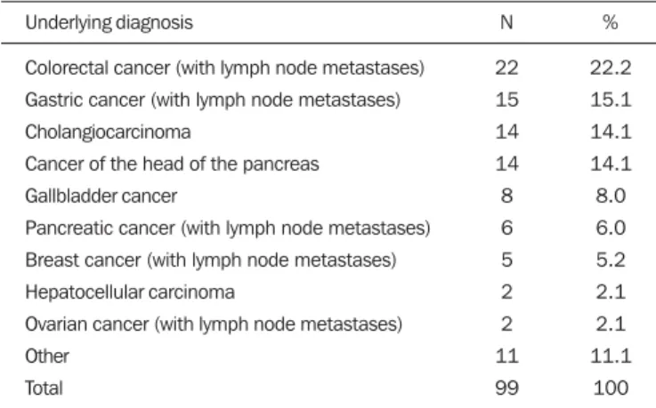
Between May 2007 and January 2010, 99 patients with unresectable malignant biliary obstruction underwent per-cutaneous placement of a self-expanding metallic stent at our institution. Of those 99 patients, 1 (0.9%) underwent three procedures, 8 (7.0%) underwent two procedures, and 90 (92.1%) underwent only one procedure. Therefore, a total of 109 procedures were performed. The mean age of patients was 60.4 ± 12.1 years (range, 29–84 years), and 51% were men. Table 1 shows the causes of biliary tract obstruction among the patients in the sample.
Of the 109 procedures evaluated, 92 (84.4%) were per-formed in order to treat hilar strictures—classified as Bis-muth type I in 33 cases (30.3%), as BisBis-muth type II in 19 (17.4%), as Bismuth type IIIA in 24 (22.0%), and as Bis-muth type IIIB in 16 (14.7%)—the remaining 17 (15.6%) being performed in order to treat periampullary lesions. Percutaneous biliary drainage was performed before stent placement in 37 (33.9%) of the procedures. In 35 proce-dures (32.1%), bilateral drainage was performed. In 80 (73.4%) of the procedures, the biliary stricture was dilated with a balloon catheter after stent insertion.
A single stent was used in 66 (60.4%) of the procedures (all to treat periampullary or Bismuth I strictures), two stents
were used in 36 (33.0%) of the procedures (all to treat Bis-muth II strictures), and three stents were used in 7 (5.6%) of the procedures (all to treat Bismuth III and IV strictures). Wallstents were used in 61 procedures (56%), and S.M.A.R.T. CONTROL stents were used in 48 (44%).
The most common complication was acute pancreati-tis, occurring in 31 cases (28.4%), followed by cholangipancreati-tis, in 12 (11.0%), bronchopneumonia, in 5 (4.6%) thromboem-bolism, in 4 (3.7%), and transient hemobilia, in 3 (2.7%). Twenty-eight patients died within the first 30 days after the procedure, the overall 30-day mortality rate being 28.3%.
Comparative analysis: Wallstents vs. S.M.A.R.T. CONTROL stents
The Wallstent group consisted of 58 patients, with a mean age of 59.4 ± 12.9 years (range, 29–84 years), who collectively underwent 61 procedures, and the S.M.A.R.T. CONTROL group consisted of 46 patients, with a mean age of 62.8 ± 10.7 years (range, 36–85 years), who collectively underwent 48 procedures. There was no statistically signifi-cant difference between the two groups regarding age (p = 0.136) or level of biliary obstruction (p = 0.685).
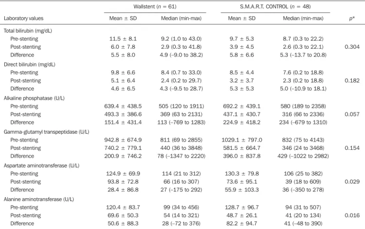
Table 2 shows the laboratory test results before and 30 days after stent placement in the S.M.A.R.T. CONTROL and Wallstent groups. Overall, patients showed post-treatment reductions in the mean values of biochemical variables. There was no statistically significant difference between the two groups in terms of the post-treatment reductions in mean (total and direct) bilirubin levels and mean levels of canali-cular enzymes (alkaline phosphatase and gamma-glutamyl transpeptidase). However, the reductions in aspartate ami-notransferase and alanine amiami-notransferase levels after stent placement were greater in the S.M.A.R.T. CONTROL group than in the Wallstent group (Table 2).
Stent occlusion was identified in 13 patients, 7 in the S.M.A.R.T. CONTROL group and 6 in the Wallstent group (p = 0.989). The number of stent occlusions at 30, 90, 180, and 360 days was 2, 1, 3, and 1, respectively, in the S.M.A.R.T. CONTROL group, compared with 2, 1, 2, and 1, respectively, in the Wallstent group. For the patients in Table 1—Distribution of the etiology of biliary tract obstruction.
Underlying diagnosis
Colorectal cancer (with lymph node metastases) Gastric cancer (with lymph node metastases) Cholangiocarcinoma
Cancer of the head of the pancreas Gallbladder cancer
Pancreatic cancer (with lymph node metastases) Breast cancer (with lymph node metastases) Hepatocellular carcinoma
Ovarian cancer (with lymph node metastases) Other
Total
N
22 15 14 14 8 6 5 2 2 11 99
%
22.2 15.1 14.1 14.1 8.0 6.0 5.2 2.1 2.1 11.1
both groups, occlusion-free survival rates were 67% at 30 days, 37% at 90 days, 25% at 180 days, and 10% at 360 days. When the survival curves were adjusted for age and level of biliary obstruction, there was no difference between the two groups. Occlusion-free survival rates at 30, 90, 180, and 360 days over time according to the type of stent used was 83%, 48%, 31%, and 10%, respectively, in the S.M.A.R.T. CON-TROL group, compared with 57%, 30%, 21%, and 11%, respectively, in the Wallstent group. As can be seen in Fig-ure 1, there was no statistically significant difference in sur-vival curves in relation to the type of stent used (relative risk = 1.52; 95% confidece interval: 0.94–2.44; p = 0.085).
DISCUSSION
Interventional procedures have been adopted at several radiological centers in Brazil(13–24). Placement of self-expand-ing metallic stents in the settself-expand-ing of inoperable biliary malig-nancy is intended to improve the quality of life of affected patients via a minimally invasive procedure. In our series, the performance of the Wallstent was equivalent to that of the S.M.A.R.T. CONTROL stent in the percutaneous treat-ment of unresectable malignant biliary obstruction.
There are a wide variety of metallic stents currently avail-able for the management of patients with biliary obstruction due to inoperable malignancy. The Wallstent is made of elgiloy and was one of the first stents developed for the
treat-Table 2—Biochemical variables before and 30 days after stent placement in patients treated with percutaneous placement of self-expanding elgiloy (Wallstent) or nitinol (S.M.A.R.T. CONTROL) stents.
Laboratory values
Total bilirubin (mg/dL) Pre-stenting Post-stenting Difference Direct bilirubin (mg/dL)
Pre-stenting Post-stenting Difference
Alkaline phosphatase (U/L) Pre-stenting
Post-stenting Difference
Gamma-glutamyl transpeptidase (U/L) Pre-stenting
Post-stenting Difference
Aspartate aminotransferase (U/L) Pre-stenting
Post-stenting Difference
Alanine aminotransferase (U/L) Pre-stenting
Post-stenting Difference
SD, standard deviation. * Nonparametric repeated-measures analysis of variance.
Wallstent (n = 61) S.M.A.R.T. CONTROL (n = 48)
Mean ± SD
11.5 ± 8.1 6.0 ± 7.8 5.5 ± 8.0
9.8 ± 6.6 5.1 ± 6.4 4.6 ± 6.5
639.4 ± 438.5 493.3 ± 386.6 151.4 ± 431.4
942.8 ± 674.9 740.2 ± 779.1 200.9 ± 746.2
124.9 ± 69.9 93.8 ± 72.8 28.4 ± 86.8
120.4 ± 83.7 69.6 ± 50.3 50.6 ± 88.3
Median (min-max)
9.2 (1.0 to 43.0) 2.9 (0.3 to 41.8) 4.9 (–9.0 to 38.2)
8.4 (0.7 to 33.0) 2.4 (0.2 to 29.7) 4.3 (–9.5 to 28.7)
505 (120 to 1911) 369 (63 to 2131) 113 (–769 to 1283)
811 (69 to 2855) 440 (36 to 3848) 78 (–1347 to 2220)
114 (21 to 312) 66 (16 to 307) 27 (–175 to 292)
99 (34 to 456) 54 (14 to 321) 28 (–72 to 376)
Mean ± SD
9.7 ± 5.3 3.9 ± 4.5 5.8 ± 6.6
8.5 ± 4.4 3.2 ± 3.7 5.3 ± 5.3
692.2 ± 439.1 437.1 ± 430.7 224.9 ± 418.2
1029.1 ± 797.0 581.5 ± 664.7 396.0 ± 837.8
130.3 ± 79.8 73.6 ± 95.1 55.9 ± 103.3
128.7 ± 96.7 48.7 ± 26.1 82.2 ± 94.7
Median (min-max)
8.7 (0.3 to 22.2) 2.6 (0.3 to 22.1) 5.3 (–13.7 to 20.8)
7.6 (0.2 to 18.8) 2.3 (0.2 to 18.8) 5.0 (–10.9 to 18.1)
580 (189 to 2358) 316 (66 to 2336) 234 (–679 to 1310)
832 (75 to 4143) 346 (24 to 3468) 429 (–1022 to 2982)
106 (25 to 382) 39 (18 to 609) 36 (–350 to 278)
94 (31 to 507) 41 (20 to 134) 41 (–48 to 390)
p*
0.304
0.182
0.057
0.154
0.029
0.016
Figure 1. Occlusion-free survival curves according to the type of stent used (Kaplan-Meier analysis with log-rank test).
increased radial resistive force, which are associated with a theoretically lower risk of occlusion(26). The S.M.A.R.T. CONTROL stent is made of nitinol, which has thermal shape memory, and has shown greater elasticity and resistance than have Wallstents in experimental studies(27).
Few studies have comparatively analyzed different me-tallic stents to assess whether any one type is superior to the others in terms of long-term stent patency in the manage-ment of malignant biliary obstruction. Gandini et al.(28)
conducted a retrospective, nonrandomized study comparing the use of two different self-expanding metallic stents (Wall-stent and Ultraflex) in 87 patients with unresectable malig-nant biliary obstruction and, as in our study, found that nei-ther was superior to the onei-ther.
Studies evaluating the efficacy of using stents covered with polytetrafluoroethylene, polyethylene terephthalate, or polyurethane for the palliation of inoperable malignancies have reported no significant increase in survival compared with the use of uncovered stents(29–31). However, it has been
reported that patients treated with covered stents require fewer reinterventions and have a better quality of life(30,31).
A retrospective, nonrandomized, comparative study in-volving 101 patients was conducted in order to examine the efficacy of the Niti-D biliary uncovered stent and the uncov-ered Wallstent for the palliative endoscopic management of malignant biliary obstruction(32). The authors of that study found that stent patency tended to be longer in patients re-ceiving the Niti-D stent (n = 41; 153 days) than in those receiving the Wallstent (n = 60; 124 days), although they reported no significant difference between the two stents (p = 0.204). In our study, better results were also observed in the S.M.A.R.T. CONTROL group, mainly for occlusion-free survival, although there was no statistically significant dif-ference in survival curves in relation to the type of stent used (p = 0.085). We believe that the increased radial resistive force of the material employed in the S.M.A.R.T. CONTROL stent (nitinol) may have contributed to the lower rate of stent occlusion(26).
Another issue to be considered is the high (28.3%) 30-day mortality rate observed in our study. That may be asso-ciated with the generally poor clinical condition of the pa-tients, who all suffered from advanced, unresectable malig-nancy and most of whom (50.4%) already had diffuse me-tastases. A study involving 71 patients with obstructive jaun-dice due to solid malignancies was conducted to investigate the overall survival of patients after percutaneous transhepatic biliary drainage and showed that patients with poor perfor-mance status (Eastern Cooperative Oncology Group score > 2) have a dismal prognosis and should not undergo percu-taneous transhepatic biliary drainage(33). Currently, there are few effective treatment options for patients with unresectable malignant biliary obstruction, who have particularly poor survival characteristics. Therefore, although further survival improvement is unlikely to be obtained by stenting alone, we believe that this palliative treatment option meets its primary
goal (i.e., to improve the quality of life of affected patients via a minimally invasive procedure), which appears to be independent of stent type.
The present study has some limitations. The retrospec-tive nature of the study and the heterogeneity of the sample (with lesions of various histological types and at different sites) hinder the extrapolation of our results to other popu-lations. In addition, because of the small number of stent occlusions, it was not possible to detect a statistically sig-nificant difference in occlusion-free survival rates between the two groups, despite the higher rates observed in the S.M.A.R.T. CONTROL group.
In conclusion, our results showed no clinically or sta-tistically significant differences between Wallstents and S.M.A.R.T. CONTROL stents for the percutaneous treatment of unresectable malignant biliary obstruction. Both ap-proaches appear to be safe and effective palliative therapies for affected patients, with good clinical results. Neverthe-less, prospective randomized studies involving samples that are more homogeneous (comprising patients with lesions of a similar histological type, as well as similar obstruction sites and implantation techniques), are warranted in order to clarify this issue.
REFERENCES
1. Chu D, Adler DG. Malignant biliary tract obstruction: evaluation and therapy. J Natl Compr Canc Netw. 2010;8:1033–44. 2. Ho CS, Warkentin AE. Evidence-based decompression in
malig-nant biliary obstruction. Korean J Radiol. 2012;13 Suppl 1:S56– 61.
3. Kida M, Miyazawa S, Iwai T, et al. Recent advances of biliary stent management. Korean J Radiol. 2012;13 Suppl 1:S62–6. 4. Lammer J, Hausegger KA, Fluckiger F, et al. Common bile duct
obstruction due to malignancy: treatment with plastic versus metal stents. Radiology. 1996;201:167–72.
5. Schmassmann A, von Gunten E, Knuchel J, et al. Wallstents versus plastic stents in malignant biliary obstruction: effects of stent pa-tency of the first and second stent on patient compliance and sur-vival. Am J Gastroenterol. 1996;91:654–9.
6. Lee MJ, Dawson SL, Mueller PR, et al. Palliation of malignant bile duct obstruction with metallic biliary endoprostheses: technique, results, and complications. J Vasc Interv Radiol. 1992;3:665–71. 7. Rossi P, Bezzi M, Rossi M, et al. Metallic stents in malignant biliary
obstruction: results of a multicenter European study of 240 patients. J Vasc Interv Radiol. 1994;5:279–85.
8. Hamy A, d’Alincourt A, Paineau J, et al. Percutaneous self-expand-able metallic stents and malignant biliary strictures. Eur J Surg Oncol. 1997;23:403–8.
9. Bezzi M, Orsi F, Salvatori FM, et al. Self-expandable nitinol stent for the management of biliary obstruction: long-term clinical re-sults. J Vasc Interv Radiol. 1994;5:287–93.
10. Adam A, Chetty N, Roddie M, et al. Self-expandable stainless steel endoprostheses for treatment of malignant bile duct obstruction. AJR Am J Roentgenol. 1991;156:321–5.
11. Lee BH, Choe DH, Lee JH, et al. Metallic stents in malignant bil-iary obstruction: prospective long-term clinical results. AJR Am J Roentgenol. 1997;168:741–5.
embolization for unresectable symptomatic giant hepatic heman-giomas: single-center experience using a lipiodol-ethanol mixture. Radiol Bras. 2015;48:154–7.
14. Aguiar-Dias D, Castro-Afonso LH, Abud DG. Femoral artery in-jury during aneurysm coiling. Radiol Bras. 2015;48:335–6. 15. Herrero Lara JA, de Araújo Martins-Romêo D, Caparrós Escudero
C, et al. Hybrid treatment of penetrating aortic ulcer. Radiol Bras. 2015;48:192–4.
16. Ajzen S. Contribution of transrectal ultrasonography-guided biopsy in the diagnosis of prostate cancer: looking back and ahead. Radiol Bras. 2015;48(1):vii.
17. Lopes PM, Sepúlveda L, Ramos R, et al. The role of transrectal ultrasound in the diagnosis of prostate cancer: new contributions. Radiol Bras. 2015;48:7–11.
18. Rocha RD, Girardi AR, Pinto RR, et al. Axillary ultrasound and fine-needle aspiration in preoperative staging of axillary lymph nodes in patients with invasive breast cancer. Radiol Bras. 2015; 48:345–52.
19. Petrilli M, Senerchia AA, Petrilli AS, et al. Computed tomography-guided percutaneous trephine removal of the nidus in osteoid os-teoma patients: experience of a single center in Brazil. Radiol Bras. 2015;48:211–5.
20. Alves MLD, Gabarra MHC. Comparison of power Doppler and thermography for the selection of thyroid nodules in which fine-needle aspiration biopsy is indicated. Radiol Bras. 2016;49:311–5. 21. Cantador AA, Siqueira DED, Jacobsen OB, et al. Duplex ultrasound and computed tomography angiography in the follow-up of endo-vascular abdominal aortic aneurysm repair: a comparative study. Radiol Bras. 2016;49:229–33.
22. Urban LABD. Fine-needle aspiration of axillary lymph nodes: a change of paradigm in the approach to axillary compromise? Radiol Bras. 2016;49(1):ix.
23. Canella EO. Percutaneous biopsy and radiological imaging of the breast. Radiol Bras. 2016;49(2):ix.
24. Matsuoka MW, Rocha SMS, Suzuki L, et al. Ultrasound guided injection of botulinum toxin into the salivary glands of children with neurological disorders. Radiol Bras. 2016;49:131–2.
25. Huibregtse K. The wallstent for malignant biliary obstruction. Gastrointest Endosc Clin N Am. 1999;9:491–501.
26. Barras CD, Myers KA. Nitinol – its use in vascular surgery and other applications. Eur J Vasc Endovasc Surg. 2000;19:564–9. 27. Duda SH, Wiskirchen J, Tepe G, et al. Physical properties of
endovascular stents: an experimental comparison. J Vasc Interv Radiol. 2000;11:645–54.
28. Gandini R, Fabiano S, Pipitone V, et al. Management of biliary neoplastic obstruction with two different metallic stents implanted in one session. Cardiovasc Intervent Radiol. 2005;28:48–52. 29. Rossi P, Bezzi M, Salvatori FM, et al. Clinical experience with
cov-ered wallstents for biliary malignancies: 23-month follow-up. Cardiovasc Intervent Radiol. 1997;20:441–7.
30. Krokidis M, Fanelli F, Orgera G, et al. Percutaneous palliation of pancreatic head cancer: randomized comparison of ePTFE/FEP-covered versus unePTFE/FEP-covered nitinol biliary stents. Cardiovasc Intervent Radiol. 2011;34:352–61.
31. Zurstrassen CE, Santos AC, Tyng CJ, et al. Percutaneous use of ePTFE/FEP-covered metallic stent for palliation of malignant bil-iary obstruction. Minim Invasive Ther Allied Technol. 2014;15:1–8. 32. Yang KY, Ryu JK, Seo JK, et al. A comparison of the Niti-D biliary uncovered stent and the uncovered Wallstent in malignant biliary obstruction. Gastrointest Endosc. 2009;70:45–51.