LETTER TO THE EDITOR
Pseudomyxoma peritonei syndrome 12 months after
an intact resection of malignant mucocele of the
appendix: A case report
Leonardo Maciel da Fonseca, Antoˆnio Lacerda-Filho, Rodrigo Gomes da Silva
Grupo de Coloproctologia e Intestino Delgado, Instituto Alfa de Gastroenterologia do Hospital das Clı´nicas da Universidade Federal de Minas Gerais (UFMG) – Belo Horizonte/MG, Brazil.
Email: [email protected] Tel.: 55-31-32489403
INTRODUCTION
Mucocele of the appendix (MA) is an obstructive dilatation of the appendix caused by either a benign or malignant process. MAs are rare lesions found in only 0.2%– 0.3% of appendectomies.1
Malignant mucoceles are found even less frequently.2
It is believed that an intact mucocele presents no risk for the patient. Gonza´lez Moreno et al.3has emphasized that
peritoneal seeding should be preventable with atraumatic handling of the cancer specimen. These authors also noted that peritoneal dissemination from a MA has not been regarded as a problem with open appendectomy. The incidence of disseminated peritoneal mucinous tumors from an open appendectomy is extremely low, and no reported cases were found in the literature.
We report the case of a patient with no rupture of the MA during the primary surgery who presented after 12 months of follow-up with peritoneal dissemination typical of pseudomyxoma peritonei (PP). To the best of our knowl-edge, no such case has previously been reported.
CASE REPORT
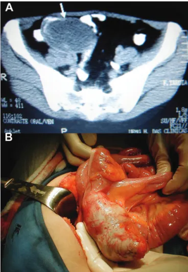
A 40-year-old female patient presented with abdominal pain lasting 3 months. The patient underwent an abdominal CT scan that showed a 12-cm well-encapsulated cystic mass in the right lower quadrant (Figure 1A). However, there was no mucinous ascites, and MA of malignant origin was suspected.
A median laparotomy, rather than a laparoscopic approach, was indicated to avoid rupturing the tumor. A right hemicolectomy (RHC) was performed due to tumor size . The surgical field was protected with pads to prevent the contamination of tumor cells into the peritoneal cavity. The specimen was removed intact (Figure 1B). The pathologic exam showed mucinous cystadenocarcinoma of
the appendix without rupture. No metastases were found in 31 tested lymph nodes.
One year later, the patient was found to have an elevated serum carcinoembryonic antigen. On the pelvic CT, a tumor mass involving the right ovary and salpinx was discovered. A cytoreductive surgery was indicated. During laparotomy, four-quadrant peritoneal dissemination of the tumor was observed along with a large tumor involving the right ovary. An extensive 10-hour cytoreductive surgery, as
*This work was presented at 6thInternational Workshop on Peritoneal Surface
Malignancy, Lyon, France, 2008.
*This work was supported by FAPEMIG (Fundac¸a˜o de Amparo a` Pesquisa do
Estado de Minas Gerais).
Copyrightß2010CLINICS– This is an Open Access article distributed under the terms of the Creative Commons Attribution Non-Commercial License (http:// creativecommons.org/licenses/by-nc/3.0/) which permits unrestricted non-commercial use, distribution, and reproduction in any medium, provided the original work is properly cited.
Figure 1- A) CT scan showing a well-encapsulated cystic mass in the right lower quadrant (arrow); B) Mucocele of the appendix.
CLINICS 2010;65(8):817-818 DOI:10.1590/S1807-59322010000800015
recommended by Sugarbaker,4 was performed; addition-ally, the patient received 90 minutes of intraperitoneal chemotherapy with mitomycin C. During the postoperative period, the patient developed pneumonia in the ICU and died 30 days after the procedure.
DISCUSSION
During procedures involving mucoceles caused by mucinous cystadenomas and cystadenocarcinomas, the most important concern for the surgeon is that a possible rupture of the mucocele, either spontaneous or accidental, may result in PP.5
Considering these concerns, an oncological RHC was performed to prevent both tumor cell spillage into the peritoneal cavity and rupture of the mucocele. Unfortunately, this patient presented with metastasis of malignant cells throughout the peritoneal cavity in the form of multiple mucinous deposits at 12 months after the initial surgery.
MA of malignant origin may result in the PP syndrome even after an intact tumor resection has been performed. We believe that intraperitoneal chemotherapy should be used in addition to the primary surgical treatment of MA even for intact tumor resections.
REFERENCES
1. Pitiakoudis M, Tsaroucha AK, Mimidis K, Polychronidis A, Minopoulos G, Simopoulos C. Mucocele of the appendix: a report of five cases. Tech Coloproctol. 2004;8:109–12, doi: 10.1007/s10151-004-0067-3.
2. Andersson A, Bergdahl L, Boquist L. Primary carcinoma of the appendix. Ann Surg. 1976;183:53–7, doi: 10.1097/00000658-197601000-00011. 3. Gonzalez Moreno S, Shmookler BM, Sugarbaker PH. Appendiceal
mucocele. Contraindication to laparoscopic appendectomy. Surg Endosc 1998;12:1177–9, doi: 10.1007/s004649900811.
4. Sugarbaker PH. Peritonectomy procedures. Ann Surg. 1995; 221:29–42, doi: 10.1097/00000658-199501000-00004.
5. Landen S, Bertrand C, Maddern GJ, Herman D, Pourbaix A, de Neve A, et al. Appendiceal mucoceles and pseudomyxoma peritonei. Surg Gynecol Obstet. 1992;175:401–4.
Pseudomyxoma peritonei syndrome
da Fonseca LM et al. CLINICS 2010;65(8):817-818