Key words:
MicroRNAs; MIRN145 microRNA, human [Supple-mentary Concept]; Urinary Bladder; Diagnostic Techniques, Urological; Biological Markers
Int Braz J Urol. 2013; 39: 95-102
__________________
Submitted for publication: January 01, 2011
__________________
Accepted after revision: November 09, 2011 Purpose: Bladder cancer (BC) is the second most common malignancy of the urinary
tract, with high mortality. The knowledge of the molecular pathways associated with BC carcinogenesis is crucial to identify new diagnostic and prognostic biomarkers. MicroRNAs (miRNAs) are short non-coding RNA molecules that play important roles in the regulation of gene expression by acting directly on mRNAs. miR-145 has been considered as a tumor suppressor, which targets the c-MYC, MUC-1 and FSCN1 genes. Our aim was to evaluate the expression profile of miR-145 in low-grade non-invasive and high-grade invasive bladder urothelial carcinomas.
Materials and Methods: We studied 30 specimens of low-grade, non-invasive pTa and 30 of pT2/pT3 high-grade invasive UC obtained by transurethral resection or radical cys-tectomy, followed over a mean time of 16.1 months. Normal controls were represented by five samples of normal bladder biopsy from patients who underwent retropubic prostatec-tomy to treat BPH. miRNA extraction and cDNA generation were performed using com-mercial kits. Analysis was performed by qRT-PCR, and miR-145 expression was calculated using the 2-ΔΔct method; we used RNU-43 and RNU-48 as endogenous controls.
Results: miR-145 was under-expressed in 73.3% and 86.7% of pTa and pT2/pT3, res-pectively, with expression means of 1.61 for the former and 0.66 for the last. There were no significant differences in miR-145 expression and histological grade, tumor stage, angiolymphatic neoplastic invasion and tumor recurrence.
Conclusion: miR-145 is under-expressed in low-grade, non-invasive and high-grade invasive urothelial bladder carcinoma and may play an important role in the carcino-genesis pathway, being an interesting candidate diagnostic marker.
INTRODUCTION
Bladder cancer (BC) is the second most common malignancy of the urinary tract, and ap-proximately 383,300 new cases are estimated to be diagnosed in 2011 (1). Ninety percent of BC are urothelial carcinomas (UC), previously known as transitional cell carcinomas, and the majority are papillary low-grade, non-muscle invasive cancers
that recur in up to 80% of cases but rarely progress to muscle invasion (2,3). In contrast, 10 to 20% of tumors are muscle invasive at diagnosis, and 50% of patients die from metastatic disease (4).
The molecular pathways underlying the two main distinct types of UC, low-grade non--muscle invasive UC and high-grade muscle in-vasive UC (2,5) have been investigated to identify new potential markers for diagnosis, disease
mo-Expression profile of microrna-145 in urothelial bladder
cancer
_______________________________________________
Nelson Dip, Sabrina T. Reis, Miguel Srougi, Marcos F. Dall´Oglio, Katia R. M. Leite
Laboratory of Medical Investigation, Urology Department – LIM55, University of Sao Paulo Medical School (ND, STR, MS, KRML) and Uro-Oncology Group, Urology Department, Sao Paulo Cancer Institute (ICESP) (MFD), Sao Paulo, Brazil
ABSTRACT
ARTICLE
INFO
nitoring, prognosis and the development of new targeted therapies (2,6).The most common genetic alteration of BC associated with low-grade and low-stage is an activating mutation of the fibro-blast growth factor receptor 3 (FGFR3) gene (6,7), whereas mutations in p53, retinoblastoma (RB1) and PTEN have been identified as being charac-teristic of the carcinogenesis pathway for high--grade invasive BC (7-9).
The FGFR3 gene belongs to the growth factor receptor family related to the tyrosine ki-nase signaling pathway, which plays an important role in embryogenesis, development, angiogenesis, wound healing, tissue homeostasis and tumorige-nesis, by regulating the processes of cellular pro-liferation, migration and apoptosis (7). Mutations are the primary phenomenon related to FGFR3 dysfunction by allowing ligand-independent ope-ration (7,10).
P53, RB1 and PTEN are traditional tumor suppressor genes that work together and trigger BC carcinogenic pathways through several me-chanisms, ranging from missense mutation and subsequently loss-of-function (9). Moreover, PTEN gene is another tumor suppressor and play roles in cell proliferation, migrations and invasion trou-gh PI3K/AKT/mTOR carcinogenic pathway (3,9). Despite PTEN is related to development of non--invasive tumors, it is much more associated with promoters pathways and progression of invasive neoplasias. Other events are also related to alte-rations in protein expression, including DNA me-thylation, histone acetylation and abnormalities in the expression of microRNAs that contribute to the development and progression of BC.
MicroRNAs (miRNAs) are members of small single-stranded regulatory RNAs (21-25 nucleotides) that can suppress translation or pro-mote degradation of mRNAs, thereby regulating the expression of target genes, including trans-cription factors, oncogenes and tumor suppres-sor genes. MicroRNAs have been reported to be differentially expressed in several types of can-cers. Currently, there are more than 1400 miRNAs identified in humans, and up to 30% of genes are thought to be regulated by miRNAs (11). MicroR-NAs are involved in cell development, differen-tiation, apoptosis, tissue homeostasis and several
metabolic pathways (12-15) and have been related to carcinogenesis by acting as negative regulators of genes related to cancer, as exemplified by the effects of miR-15a and miR-16-1 on BCL2 mRNA, miR-143 and miR-let7c on RAS mRNA as well as miR-21 on p53 mRNA (16-19).
miR-145 is located in chromosome 5q32 and is widely established as a tumor suppressor. Several studies have validated miR-145 as an inhibitor of cellular proliferation, apoptosis, in-vasiveness and metastasis, as it is down-regula-ted in an array of cancers including those of the colorectum, lung, breast and prostate (20-22). In colorectal cancer, it was firstly demonstrated that down-regulation of miR-145 is involved with malignancies where miR-145 may play a role in tumor initiation (23,24). In BC, a Japanese group previously showed under-expression of miR-145 that normally targets the oncogenes KRT-7 and FSCN1 (25,26). However, they have not related miR-145 expression with important prognostic factors or performed any follow-up. Our aim is to study the expression profile of miR-145 in low--grade, non-invasive and high-grade invasive BC, speculating on its potential role as a prognostic or diagnostic marker.
MATERIALS AND METHODS
Patients
Specimens of 30 low-grade, non-invasive, staged pTa and 30 high-grade invasive, staged pT2/pT3 urothelial carcinomas obtained from pa-tients who underwent transurethral resection or radical cystectomy were the subject of the study. Seventy-three percent of patients were male, and the mean age was 66.5 years, ranging from 41 to 86 years. As a control, we used normal bladder tis-sue from five patients who underwent retropubic prostatectomy to treat benign prostatic hyperpla-sia. All patients provided informed consent, and the study design was approved by the Institutional Board of Ethics, Protocol #0176/10.
A 1 cm2 fragment of tumor tissue was
Only low-grade, non-invasive pTa and high-grade invasive pT2/pT3 tumors were included in the stu-dy. For tumor grading, we used the WHO/ISUP 2004 and for staging AJCC/TNM 2010 classifica-tions. The mean follow-up time was 16.1 months.
miRNA extraction and amplification
MiR-145 was isolated using a mirVana Kit® (Applied Biosystems, CA, USA) according to the manufacturer’s instructions, and the concentra-tion was determined by 260/280 nm absorbance using a Nanodrop® ND-1000 spectrophotometer (Thermo Scientific). miRNA cDNA was generated using a Taqman MicroRNA Reverse Transcription Kit® (Applied Biosystems, CA, USA). miRNA reac-tions were incubated at 16°C for 30 min, 42°C for 30 min and 85°C for 5 min. The cDNA was stored at -20°C until further use.
For miRNA amplification, a Taqman Rea-gent Kit® (Applied Biosystems, CA, USA) and the 7500 Fast Real-Time PCR System® (Applied Bio-systems, CA, USA) were used.
Expression profiles of miR-145 were obtai-ned by relative quantification determiobtai-ned by the 2-ΔΔct method. The formula includes the following
mathematic sentences: ΔΔCT = dCT1 - dCT2, where dCT1 = CT of miRNA-target, (tumor sample) - CT of mean of endogenous control (tumor sample), and dCT2 = CT of mean of normal controls (nor-mal bladder samples) - CT of mean of endogenous control (normal bladder samples). The final result is obtained by application of 2-ΔΔct method and fin-dings greater and smaller than 1 are considerate over and under-expressed, respectively.
The reactions were conducted in duplica-te, and RNU-43 and RNU-48 were used as endo-genous controls. Endoendo-genous RNUs are miRNAs produced by cell machinery and they do not play roles over cellular functions and show a stable behavior. Thus, they were used to stardardize the values in mathematic formula.
Statistical analysis
Mann-Whitney U tests, ANOVA and T tests were used to compare miR-145 expression levels with tumor grade, stage and angiolymphatic
in-vasion, respectively. The distribution of the ex-pression levels of miR-145 was skewed; therefore, the data were log-transformed for analyses. The results are presented as the geometric means at a 95% confidence interval (95% CI). The number 1 in logarithmic graphics represent normal bladder samples and the expression levels are represented at times, for overexpression (greater than 1) or un-derexpression (smaller than 1).
RESULTS
miR-145 was under-expressed in 73.3% (22/30) and 86.7% (26/30) of pTa and pT2/pT3, respectively, as presented in Figure-1. Although both groups showed miR-145 under-expressed, we can observe that levels of under-expression were more evident in pT2/pT3 group. Further-more, when we consider samples over-expressed (8/30 for pTa and 4/30 for pT2/pT3), the levels of over-expression were greater in low-grade, non--invasive pTa group, when compared with high--grade invasive pT2/pT3 tumors.
Expression values of miR-145 in pTa, pT2/ pT3 and normal bladder controls are represented in Table-1. The mean and median expression of
Figure 1 - Expression levels (fold-change) of miR-145 in pTa and pT2/pT3 tumors.
pTa
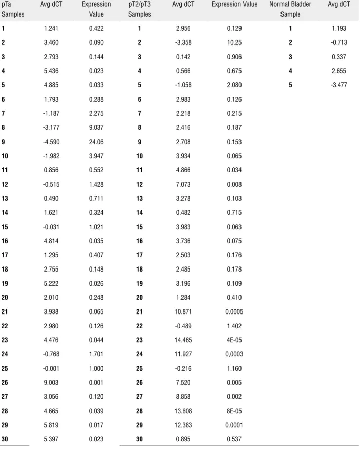
Table 1 - pTa, pT2/pT3 and controls dCTs and expression values for both tumor groups. dCT = miR-145 amplification value - mean of RNU-43 and RNU-48 amplification value. Final expression values were obtained by 2-ΔΔct method .
pTa Samples
Avg dCT Expression Value
pT2/pT3 Samples
Avg dCT Expression Value Normal Bladder Sample
Avg dCT
1 1.241 0.422 1 2.956 0.129 1 1.193
2 3.460 0.090 2 -3.358 10.25 2 -0.713
3 2.793 0.144 3 0.142 0.906 3 0.337
4 5.436 0.023 4 0.566 0.675 4 2.655
5 4.885 0.033 5 -1.058 2.080 5 -3.477
6 1.793 0.288 6 2.983 0.126
7 -1.187 2.275 7 2.218 0.215
8 -3.177 9.037 8 2.416 0.187
9 -4.590 24.06 9 2.708 0.153
10 -1.982 3.947 10 3.934 0.065
11 0.856 0.552 11 4.866 0.034
12 -0.515 1.428 12 7.073 0.008
13 0.490 0.711 13 3.278 0.103
14 1.621 0.324 14 0.482 0.715
15 -0.031 1.021 15 3.983 0.063
16 4.814 0.035 16 3.736 0.075
17 1.295 0.407 17 2.503 0.176
18 2.755 0.148 18 2.485 0.178
19 5.222 0.026 19 3.196 0.109
20 2.010 0.248 20 1.284 0.410
21 3.938 0.065 21 10.871 0.0005
22 2.980 0.126 22 -0.489 1.402
23 4.476 0.044 23 14.465 4E-05
24 -0.768 1.701 24 11.927 0,0003
25 -0.001 1.000 25 -0.216 1.160
26 9.003 0.001 26 7.520 0.005
27 3.056 0.120 27 8.858 0.002
28 4.665 0.039 28 13.608 8E-05
29 5.819 0.017 29 12.383 0.0001
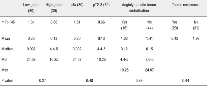
miR-145 was 1.61 and 0.2 (0.002 - 24.07) in pTa and 0.66 and 0.13 (4.4E-05 - 10.25) in pT2/pT3, respectively. We observed no significant differen-ces when we compared miR-145 expression in pTa and pT2/pT3 when considering grade (p = 0.27), stage (p = 0.48) and angiolymphatic invasion (p = 0.89). Additionally, there was no difference regar-ding tumor recurrence (Table-2).
DISCUSSION
Although we did not detect any differences in miR-145 expression between low- and high--grade tumors, our results show that this miRNA is under-expressed in both urothelial carcinomas; the lowest miRNA levels were detected in the se-cond group (86.7%). We did not include pT1 and Cis tumors because they have genetic molecular profiles that harbor both the carcinogenic pa-thway of pTa, as of the pT2/pT3.
In BC, miR-145 can silence a large num-ber of genes and has been involved in bladder carcinogenesis. Ichimi et al. (27) have previously shown miR-145 under-expression in BC by micro-array and qRT-PCR, but they did not characterize the relationship between miR-145 expression with histological grading, staging or tumor behavior.
MiR-145 is located on chromosome 5q32 and is considered a tumor suppressor miRNA
be-Table 2 - Comparison of miR-145 by grade, stage, angiolymphatic invasion and tumor recurrence.
Low grade (30)
High grade (30)
pTa (30) pT2-3 (30) Angiolymphatic tumor embolization
Tumor recurrence
miR-145 1.61 0.66 1.61 0.66 Yes (16)
No (44)
Yes (29)
No (31)
Mean 0.20 0.13 0.20 0.13 1.03 1.41 0.43 1.93
Median 0.002 4.4-5 0.002 4.4-5 0.12 0.15
Min 24.07 10.25 24.07 10.25 4.4-5 8.0-5
Max 10.25 24.07
P value 0.27 0.48 0.89 0.44
cause its negative regulation of target mRNAs in-volved in control of cell cycle, apoptosis, inva-siveness and metastasis, exerting a fundamental role in cellular and tissue homeostasis (22-24,28).
The first report of miR-145 under-expres-sion was performed by Michael et al. (23) in a stu-dy suggesting that these alterations could be in-volved in the initiation of colorectal cancer. These
findings were confirmed by Shi et al. (24) who showed that miR-145 under-expression was asso-ciated with malignant tumors.
directly related to the high-grade invasive BC car-cinogenesis pathway. In 2009, Sachdeva et al. (22) observed that, under physiological conditions, hi-gher levels of p53 due to cellular stress leads to increased levels of miR-145 through p53 response element (p53RE). P53 can also trigger the enzyma-tic machinery of microRNAs, mainly the RNase III Drosha, promoting higher expression levels of se-veral microRNAs including miR-145. Loss of p53 function through mutations could generate a lack of production and consequent under-expression of miR-145 (28).
Spizzo et al. (29) demonstrated a tumor suppressor role of miR-145, preventing cell gro-wth and inducing apoptosis through stimulation of p53, by transfecting miR-145 into breast cancer cell lines. This experiment could suggest a thera-peutic use of miR-145 in tumors.
The FSCN1 gene is another target of miR-145. The protein product of this gene is required to form protrusions of the cellular membrane and cytoplasmic movements related to migration. In malignant neoplasms, FSCN1 activity has been correlated to high-grade disease, extensive inva-sion, metastasis and poor prognosis (30). Chiyo-maru et al. (26) concluded that there is an asso-ciation between FSCN1 oncogene over-expression due to miR-145 under-expression in BC, which leads to a more aggressive phenotype. The authors found a positive correlation between high tumor stage and low levels of miR-145. However, we did not find a relationship between miR-145 expres-sion and tumor grade, stage or angiolymphatic tu-mor embolization.
We also investigated the levels of miR-145 with tumor recurrence and found no association between both groups. Here, we can make the case that miR-145 is important to tumor carcinogenesis and triggers low-grade, non-invasive pTa tumori-genesis through the lack of control of AKT. We also speculate that miR-145 is involved in our cases of high-grade invasive pT2/pT3 carcinomas through MUC-1, c-MYC, p53 and FSCN1 deregulation.
The casuistic was limited, and groups gre-ater than 30 samples are wished. Maybe this fact could really be related to absence of statistical di-fferences. Another limitation was a short follow-up.
CONCLUSIONS
MiR-145 is a well-characterized tumor su-ppressor miRNA. We hypothesize that lack of pro-tector role promoted by this miRNA over probable target genes PI3K/AKT, FSCN1, MDM2, c-Myc and MUC-1 could be involved in carcinogenic process of low-grade, non-invasive and high-grade inva-sive urothelial carcinomas, triggering their carci-nogenesis. Since we found miR-145 widely under--expressed in both tumor groups, we speculate its use as a possible diagnostic marker. These findings should be tested in experimental models.
ABREVIATIONS
BC: Bladder Cancer
miR and miRNA: micro RNA
qRT-PCR: quantitative transcriptase reverse poly-merase chain reaction
cDNA: complementar DNA
UC(s): urothelial carcinoma(s)
mRNA: messenger RNA
FGFR3: fibroblast growth factor receptor 3
ISUP: International Society of Urological Pathology
RB1: retinoblastoma gene
PTEN: phosphatase and tensin homolog gene
WHO: World Health Organization
ACKNOWLEDGEMENTS
Sponsor by: FAPESP 10/50824-1
CONFLICT OF INTEREST
None declared.
REFERENCES
1. Jemal A, Bray F, Center MM, Ferlay J, Ward E, Forman D: Global cancer statistics. CA Cancer J Clin. 2011; 61: 69-90. Epub 2011 Feb 4. Erratum in: CA Cancer J Clin. 2011; 61: 134. 2. Jebar AH, Hurst CD, Tomlinson DC, Johnston C, Taylor CF,
Knowles MA: FGFR3 and Ras gene mutations are mutually exclusive genetic events in urothelial cell carcinoma. Onco-gene. 2005; 24: 5218-25.
S, et al.: Molecular genetics of bladder cancer: Emerging mechanisms of tumor initiation and progression. Urol On-col. 2010; 28: 429-40.
4. Borden LS Jr, Clark PE, Hall MC: Bladder cancer. Curr Opin Oncol. 2005; 17: 275-80.
5. van Rhijn BW, van der Kwast TH, Vis AN, Kirkels WJ, Boevé ER, Jöbsis AC, et al.: FGFR3 and P53 characterize alterna-tive genetic pathways in the pathogenesis of urothelial cell carcinoma. Cancer Res. 2004 15; 64: 1911-4.
6. Castillo-Martin M, Domingo-Domenech J, Karni-Schmidt O, Matos T, Cordon-Cardo C: Molecular pathways of uro-thelial development and bladder tumorigenesis. Urol Oncol. 2010; 28: 401-8.
7. Pandith AA, Shah ZA, Siddiqi MA: Oncogenic role of fibro-blast growth factor receptor 3 in tumorigenesis of urinary bladder cancer. Urol Oncol. 2010; 3. [Epub ahead of print] 8. Bakkar AA, Wallerand H, Radvanyi F, Lahaye JB, Pissard S,
Lecerf L, et al.: FGFR3 and TP53 gene mutations define two distinct pathways in urothelial cell carcinoma of the blad-der. Cancer Res. 2003; 63: 8108-12.
9. Wu XR: Biology of urothelial tumorigenesis: insights from genetically engineered mice. Cancer Metastasis Rev. 2009; 28: 281-90.
10. Ornitz DM, Xu J, Colvin JS, McEwen DG, MacArthur CA, Coulier F, et al.: J Biol Chem. 1996 21; 271: 15292-7. 11. Lewis BP, Burge CB, Bartel DP: Conserved seed pairing,
often flanked by adenosines, indicates that thousands of human genes are microRNA targets. Cell. 2005 14; 120: 15-20.
12. Ambros V: MicroRNA pathways in flies and worms: growth, death, fat, stress, and timing. Cell. 2003 13; 113: 673-6. Review. Erratum in: Cell. 2003; 114: 269.
13. Bartel DP: MicroRNAs: genomics, biogenesis, mechanism, and function. Cell. 2004; 116: 281-97.
14. Croce CM, Calin GA: miRNAs, cancer, and stem cell divi-sion. Cell. 2005; 122: 6-7.
15. Blenkiron C, Miska EA: miRNAs in cancer: approaches, ae-tiology, diagnostics and therapy. Hum Mol Genet. 2007; 16 Spec No 1: R106-13.
16. Cimmino A, Calin GA, Fabbri M, Iorio MV, Ferracin M, Shi-mizu M, et al.: miR-15 and miR-16 induce apoptosis by tar-geting BCL2. Proc Natl Acad Sci U S A. 2005; 102: 13944-9. Erratum in: Proc Natl Acad Sci U S A. 2006; 103: 2464. 17. Lin T, Dong W, Huang J, Pan Q, Fan X, Zhang C, et al.:
MicroRNA-143 as a tumor suppressor for bladder cancer. J Urol. 2009; 181: 1372-80.
18. Johnson SM, Grosshans H, Shingara J, Byrom M, Jarvis R, Cheng A, et al.: RAS is regulated by the let-7 microRNA family. Cell. 2005; 120: 635-47.
19. Catto JW, Miah S, Owen HC, Bryant H, Myers K, Dudziec E,
et al.: Distinct microRNA alterations characterize high- and low-grade bladder cancer. Cancer Res. 2009; 69: 8472-81. 20. Ozen M, Creighton CJ, Ozdemir M, Ittmann M: Widespread
deregulation of microRNA expression in human prostate cancer. Oncogene. 2008; 27: 1788-93.
21. Izzotti A, Calin GA, Arrigo P, Steele VE, Croce CM, De Flora S: Downregulation of microRNA expression in the lungs of rats exposed to cigarette smoke. FASEB J. 2009; 23: 806-12.
22. Sachdeva M, Zhu S, Wu F, Wu H, Walia V, Kumar S, et al.: p53 represses c-Myc through induction of the tumor sup-pressor miR-145. Proc Natl Acad Sci U S A. 2009; 106: 3207-12.
23. Michael MZ, O’ Connor SM, van Holst Pellekaan NG, Young GP, James RJ: Reduced accumulation of specific microRNAs in colorectal neoplasia. Mol Cancer Res. 2003; 1: 882-91.
24. Shi B, Sepp-Lorenzino L, Prisco M, Linsley P, deAngelis T, Baserga R: Micro RNA 145 targets the insulin receptor substrate-1 and inhibits the growth of colon cancer cells. J Biol Chem. 2007 9; 282: 32582-90.
25. Ichimi T, Enokida H, Okuno Y, Kunimoto R, Chiyomaru T, Kawamoto K, et al.: Identification of novel microRNA tar-gets based on microRNA signatures in bladder cancer. Int J Cancer. 2009; 125: 345-52.
26. Chiyomaru T, Enokida H, Tatarano S, Kawahara K, Uchida Y, Nishiyama K, et al.: miR-145 and miR-133a function as tu-mour suppressors and directly regulate FSCN1 expression in bladder cancer. Br J Cancer. 2010; 102: 883-91.
27. Sachdeva M, Mo YY: miR-145-mediated suppression of cell growth, invasion and metastasis. Am J Transl Res. 2010; 2: 170-80.
28. Suzuki HI, Yamagata K, Sugimoto K, Iwamoto T, Kato S, Miyazono K: Modulation of microRNA processing by p53. Nature. 2009; 460: 529-33.
29. Spizzo R, Nicoloso MS, Lupini L, Lu Y, Fogarty J, Rossi S, et al.: miR-145 participates with TP53 in a death-pro-moting regulatory loop and targets estrogen receptor-alpha in human breast cancer cells. Cell Death Differ. 2010; 17: 246-54.
30. Vignjevic D, Kojima S, Aratyn Y, Danciu O, Svitkina T, Bori-sy GG: Role of fascin in filopodial protrusion. J Cell Biol. 2006; 174: 863-75.
_____________________
Correspondence address:
EDITORIAL COMMENT
Recently discovered, the micro RNAs have been extensively studied in recent years in several malignancies. This paper brings relevant new infor-mation about the role of a micro RNA in two ex-tremes of urothelial BC: the low grade pTa and the high grade invasive ones (pT2/3). The result shows a reduced action of this miR as in low risk pTa, as in invasive BC. Theorically, target therapies that im-prove or regenerate the actions of miR145 could be planned in the future if these findings are validated.
The authors did not investigate pT1 and in situ (CIS) BC, justifying that these tumors have di-fferent carcinogenic pathways.
Meanwhile, I think it would be interesting to test miR145 in pT1 and in CIS, to be sure that this micro RNA is really not involved in the car-cinogenic process of these high risk non-muscle invasive bladder tumors. Some of pT1 tumors e.g., presents as recurrence after the treatment of pTa lesions and CIS can co exist side by side of some papillary lesions. Are theses lesions independent?
In the future, other interesting issue con-cerning recurrent non-muscle invasive BCs might to investigate if intravesical BCG instillations could influence (restore?) the miR145 expression.