Evaluation of estrogen receptor expression in
low-grade and high-low-grade astrocytomas
Cléciton Braga Tavares1,2 Francisca das Chagas Sheyla Gomes-Braga1 Emerson Brandão Sousa2 Umbelina Soares Borges1 Carla Solange Escórcio-Dourado3 João Paulo da Silva-Sampaio1 Benedito Borges da Silva1,3
1. Federal University of Piauí, Teresina, PI, Brasil
2. Department of Oncology, Sao Marcos Hospital, Teresina, PI, Brasil
3. Northeast Network of Biotechnology (RENORBIO), Teresina, PI, Brasil
http://dx.doi.org/10.1590/1806-9282.64.12.1129
SUMMARY
OBJECTIVE: This study aims to compare estrogen receptor expression between low and high-grade astrocytomas.
METHOD: A study using paraffin blocks of glial tumors from the Anatomy Pathology archives of São Marcos Hospital was carried out and began after approval by the Review Board of the Federal University of Piaui. Specimens were histochemically marked with an an-ti-ER alpha antibody. Brown-stained nuclei were considered positive, regardless of reaction intensity. Data were statistically analyzed using the Mann-Whitney test and Spearman’s correlation. Statistical significance was established at p<0.05.
RESULTS: The mean percentage of nuclei stained with anti-ER alpha in low- and high-grade astrocytomas was 0.04 and zero,
respec-tively, while Spearman’s correlation showed a strong negative association between low and high-grade tumors (p<0.001) and (r= -0.67), respectively.
CONCLUSION: In the current study, estrogen receptor expression was positive only in low-grade astrocytomas and nil in high-grade astrocytomas, showing that ER expression declines with the grade of tumor malignancy.
KEYWORDS: Astrocytoma. Glioma. Glioblastoma. Estrogen receptor beta. Estrogen receptor alpha.
DATE OF SUBMISSION: 04-Mar-2018
DATE OF ACCEPTANCE: 03-Apr-2018
CORRESPONDING AUTHOR: Cléciton Braga Tavares Rua Acre, 251, Teresina, PI, Brasil – CEP 64001-625 Phone: +55 86 99925-9772
E-mail: [email protected]
INTRODUCTION
Gliomas are the most common primary tumors of the central nervous system. These tumors have four histologic subtypes, and astrocytoma is the most
prevalent type 1. According to the World Health
Or-ganization (WHO), astrocytomas may be classified as grades 1 and 2 (low-grade or benign) and grades 3 and 4 (high-grade or malignant). High- grade astrocyto-mas are highly aggressive tumors. Despite adequate
surgical resection, chemotherapy, and radiotherapy,
high-grade tumors have a poor prognosis 1-4.
Estrogen exerts essential effects on the re-productive and gastrointestinal tracts, mammary glands, skeleton, immune system and even on the central nervous system. The majority of its effects are mainly mediated by its interaction with estro-gen receptors alpha and beta (ERα and ERβ) 8,9.
However, although the primary mechanism of ac-tion of estrogen occurs through the interacac-tion with the estrogen receptor, evidence from an in vitro study showed that substances with anti-estrogenic activity, such as tamoxifen, reduced cell
prolifera-tion through protein kinase C (PKC) 6,8-10.
The presence of estrogen receptors alpha and beta in gliomas is related to tumor aggressiveness,
according to some authors 8,9. These receptors are
present in healthy brain tissue, and its expression decreases significantly with increasing histolog-ic malignancy 11. In contrast, a study showed an
inverse result regarding ER beta expression. The study described that ER beta expression was higher in high-grade malignant neoplasms than in normal
tissues and benign gliomas 12. Therefore, the
exist-ing controversy and paucity of studies comparexist-ing estrogen receptor expression among benign and malignant astrocytomas, motivated the current study design.
METHODS Study Design
This study used paraffin blocks of gliomas ob-tained from Pathology archives of São Marcos Hos-pital, Teresina, Brazil and began after approval by the Review Board of the Federal University of Piaui. Only astrocytomas were selected that were not sub-mitted to any treatment before the primary surgery and stored for a maximum of five years (histopatho-logical exams were collected between June 2012 and June 2017).
Forty cases were histologically divided into two groups (low-grade and high-grade astrocytomas). Each group had 20 cases and were chosen in a simple random manner among tumors that met the inclu-sion criteria.
Immunohistochemical Method
Samples of tumor tissue were fixed in buffered formalin for a period of 12-24 h and cut into 3-µm-thick sections. Tissue sections were then processed and stained with hematoxylin and eosin. Slides
were deparaffinized with xylene for 15 minutes at a temperature of 60ºC, dehydrated with graded eth-anol concentration 100, 95, 80 and 70% for 30 sec-onds each and rinsed with distilled water. For the performance of antigen retrieval, the slides were immersed in citrate buffer solution and heated in a microwave for 15 minutes at maximum power. Then, the slides were treated with 3% hydrogen peroxide in buffer solution for 10 minutes in each immersion. Slides were washed with distilled wa-ter and phosphate buffer saline solution. The slides were then placed in a BenchMark Ultra staining in-strument (Ventana Medical Systems®), which used NCL-ER-6F11 monoclonal antibody (Novocastra Laboratories Ltd.) as immunohistochemical mark-ers for estrogen receptors. Cells that displayed brown-stained nuclei, whether intensely brown or not, were considered positive.
Quantitative Method
A microscope (Nikon Eclipse E-400, optical mi-croscope, Tokyo, Japan) attached to a color video camera (Samsung digital camera CHC-370N, Seul, Korea) was used to capture an image and transmit it to a computer equipped with the Imagelab software, version 2.3, developed by Softium Informática Ltda. (São Paulo, Brazil) for image analysis.
For estrogen receptor expression, 600 stained or non-stained cells were counted, using a magnifica-tion of 400x, starting with areas that had a higher expression of marked cells. In each slide, the per-centage of cells was obtained from the ratio between the number of cells with stained nuclei and the total number of cells, multiplied by 100.
Statistical Analysis
RESULTS
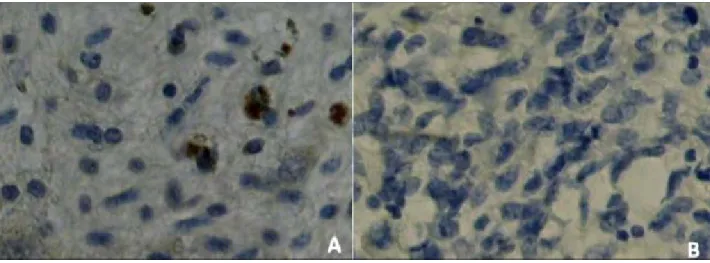
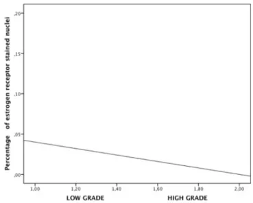
Light microscopy showed a greater concentration of stained nuclei for estrogen receptors in the low-grade astrocytomas group compared to the group of high-grade astrocytomas (Fig. 1). The mean percent-age of nuclei stained for estrogen receptors was 0.04 and zero in low-grade and high-grade astrocytomas, respectively (Table 1 and Fig. 2), while there was a strong negative correlation between high-grade tu-mors and estrogen nuclear receptor expression (r=-0.67) that was statistically significant (p<0.01) (Fig. 3).
DISCUSSION
Estrogen is a steroid hormone that exerts essen-tial effects on various organs and tissues, including the central nervous system. It acts mainly by inter-action with estrogen receptors. Estrogen may even influence the development and control of the growth
of brain tumors, such as astrocytomas 4,8,13.
Estrogen receptors are intracellular proteins with two different subtypes (alpha and beta). Despite be-ing produced by separate genes, these highly homol-ogous receptors are located in chromosome 6q25.1 and 14q22-24, respectively. Although these two re-ceptors share 97% homology in their DNA binding
domains, they exhibit contradictory biological
func-tions. ERα gene is generally believed to be an
onco-gene and promotes cell proliferation, whereas ERβ
gene is anti-proliferative and acts as a putative tumor suppressor9,14. It is very well-known that ER exists
and has a function in various tissues and neoplasms. However, the pathophysiology of ER is not fully un-derstood, since few studies have shown its expres-sion in breast, ovarian, prostate, colon cancers and astrocytic tumors 1,4.
The current study showed estrogen receptor al-pha expression only in low-grade astrocytomas. In contrast, the majority of studies demonstrate the presence of estrogen receptor expression in low-grade and high-low-grade astrocytomas, although there is a lower proportion in high-grade astrocytoma
8,9,11,15. Although nuclear ER expression was
evaluat-ed, the result of this study was similar to findings by
Fujimoto et al.13 which showed the presence of
cyto-solic estrogen receptor only in benign astrocytomas. Despite few studies in the literature concerning ER expression in brain tissue, it is known that ER is present in hippocampal neurons, pituitary tumors, glial cells, and astrocytomas. ER expression declines with increasing malignancy, as observed in the find-ings of the current study. However, its specific
func-FIGURE 1: PHOTOMICROGRAPH OF THE HISTOLOGIC SECTION OF GLIOMAS, SHOWING SOME NUCLEI STAINED BROWN FOR ESTROGEN RECEPTORS IN LOW-GRADE ASTROCYTOMAS (A) AND ABSENCE OF NUCLEI STAINED IN HIGH-GRADE ASTROCYTOMAS (B).
TABLE 1: MEAN PERCENTAGE OF STAINED NUCLEI OF ESTROGEN RECEPTOR PER GROUP.
Groups N Mean SE Mean Minimum Maximum Median
High Grade 20 0 0 0 0 0
Low Grade 20 0.04* 0.0536 0 0.1894 0.0164
FIGURE 2: MEAN PERCENTAGE OF NUCLEI STAINED WITH ESTROGEN RECEPTORS IN HIGH-GRADE AND LOW-GRADE GLIOMAS.
FIGURE 3: DISPERSION GRAPH BETWEEN THE EXPRESSION OF ESTROGEN RECEPTORS AND GRADE OF TUMOR MALIGNANCY IN ASTROCYTOMAS.
tion in the pathogenesis, progression, and prognosis
of these neoplasms remains unknown 8,9,16.
A negative correlation was shown between ER expression and grade of astrocytoma malignan-cy, which is in agreement with the literature. ER is mainly expressed in normal astrocytic cells and low-grade gliomas, promoting neuroprotective role
8,9,11,16-18.
Estrogen receptors may also be used as prognos-tic biomarkers since a positive correlation between
ER α and survival time of glioma patients has been
shown 8. Nevertheless, regression models, using
the Kaplan-Meier curve have demonstrated a better prognosis and longer survival in patients with ERβ
positive tumors 12,17.
Therefore, findings in the current study showed that estrogen receptor expression was positive only in low-grade and zero astrocytomas in high-grade
as-trocytomas, consistent with the benign glial tumor marker. However, due to the limitations of our work such as the number of paraffin blocks used and the non-assessment of estrogen receptor subtypes, fur-ther research involving a larger sample size and with
immunohistochemical markers for ERα and ERβ are
required.
CONCLUSIONS
In the current study, estrogen receptor expression was positive only in low-grade astrocytomas and nil in high-grade astrocytomas, showing that expression declines with increasing grade of tumor malignancy.
Conflict of interest statement
There is no conflict of interest of any of the au-thors with this work.
RESUMO
OBJETIVO: O objetivo deste estudo é comparar a expressão do receptor de estrogênio entre astrocitomas de baixo e alto grau.
MÉTODO: Foi realizado um estudo usando blocos de parafina de tumores gliais dos arquivos de Anatomia Patológica do Hospital São
Marcos e iniciado após aprovação pelo Comitê de Ética da Universidade Federal do Piauí. Os espécimes foram marcados histoquimi-camente com anticorpo anti-ER alpha. Os núcleos corados em marrom foram considerados positivos, independentemente da inten-sidade da reação. Os dados foram analisados estatisticamente utilizando o teste de Mann-Whitney e a correlação de Spearman. A significância estatística foi estabelecida em p<0,05.
RESULTADOS: A porcentagem média de núcleos corados com anti-ER alfa em astrocitomas de baixo e alto grau foi de 0,04 e zero, re-spectivamente, enquanto a correlação de Spearman mostrou uma forte correlação negativa entre tumores de baixa e alta qualidade (p<0,001) e (r=-0,67), respectivamente.
CONCLUSÕES: No presente estudo, a expressão do receptor de estrogênio foi positiva apenas em astrocitomas de baixo grau e nula em astrocitomas de alto grau, mostrando que a expressão de ER diminui com o grau de malignidade tumoral.
REFERENCES
1. Pollack IF, Randall MS, Kristofik MP, Kelly RH, Selker RG, Vertosick FT Jr. Effect of tamoxifen on DNA synthesis and proliferation of human malig-nant glioma lines in vitro. Cancer Res. 1990;50(22):7134-8.
2. Uematsu M, Ohsawa I, Aokage T, Nishimaki K, Matsumoto K, Takahashi H, et al. Prognostic significance of the immunohistochemical index of sur-vivin in glioma: a comparative study with the MIB-1 index. J Neurooncol. 2005;72(3):231-8.
3. Tang P, Roldan G, Brasher PM, Fulton D, Roa W, Murtha A, et al. A phase II study of carboplatin and chronic high-dose tamoxifen in patients with recurrent malignant glioma. J Neurooncol. 2006;78(3):311-6.
4. Patel S, DiBiase S, Meisenberg B, Flannery T, Patel A, Dhople A. Phase I clinical trial assessing temozolomide and tamoxifen with concomitant radiotherapy for treatment of high-grade glioma. Int J Radiat Oncol Biol Phys. 2012;82(2):739-42.
5. Gerdes J, Schwab U, Lemke H, Stein H. Production of a mouse monoclonal antibody reactive with a human nuclear antigen associated with cell prolif-eration. Int J Cancer. 1983;31(1):13-20.
6. Hsu DW, Louis DN, Efird JT, Hedley-White ET. Use of MIB-1 (Ki-67) Im-munoreactivity in differentiating grade II and grade III gliomas. J Neuro-pathol Exp Neurol. 1994;56(8):857-65.
7. Johannessen AL, Torp SH. The clinical value of Ki-67/MIB-1 labeling index in human astrocytomas. Pathol Oncol Res. 2006;12(3):143-7.
8. Batistatou A, Stefanou D, Goussia A, Arkoumani E, Papavassiliou AG, Ag-nantis NJ. Estrogen receptor beta (ERbeta) is expressed in brain astrocytic tumors and declines with dedifferentiation of the neoplasm. J Cancer Res Clin Oncol. 2004;130(7):405-10.
9. Batistatou A, Kyzas PA, Goussia A, Arkoumani E, Voulgaris S, Polyzoidis K, et al. Estrogen receptor beta (ERβ) protein expression correlates with BAG-1 and prognosis in brain glial tumours. J Neuro-Oncol. 2006;77(1):17-23.
10. Robins HI, Won M, Seiferheld WF, Schultz CJ, Choucair AK, Brachman DG, et al. Phase 2 trial of radiation plus high-dose tamoxifen for glioblas-toma multiforme: RTOG protocol BR-0021. Neuro Oncol. 2006;8(1):47-52.
11. Liu C, Zhang Y, Zhang K, Bian C, Zhao Y, Zhang J. Expression of estrogen receptors, androgen receptor and steroid receptor coactivator-3, is nega-tively correlated to the differentiation of astrocytic tumors. Cancer Epide-miol. 2014;38(3):291-7.
12. Li W, Winters A, Poteet E, Ryou MG, Lin S, Hao S, et al. Involvement of estrogen receptor b5 in the progression of glioma. Brain Research. 2013;1503:97-107.
13. Fujimoto M, Yoshino E, Hirakawa K, Fujimoto J, Tamaya T. Estrogen recep-tors in brain tumors. Clin Neuropharmacol. 1984;7(4):357-62.
14. Cao L, Qu D, Wang H, Zhang S, Jia C, Shi Z, et al. Toosendanin exerts an anti-cancer effect in glioblastoma by inducing estrogen receptor β and p53-mediated apoptosis. Int J Mol Sci. 2016;17(11). Pii: E1928.
15. Lan YL, Zou S, Wang X, Lou JC, Xing JS, Yu M, et al. Update on the thera-peutic significance of estrogen receptor beta in malignant gliomas. Onco-target. 2017;8(46):81686-96.
16. Sareddy GR, Nair BC, Gonugunta VK, Zhang QG, Brenner A, Brann DW, et al. Therapeutic significance of estrogen receptor β agonists in gliomas. Mol Cancer Ther. 2012;11(5):1174-82.
17. Dueñas Jiménez JM, Candanedo Arellano A, Santerre A, Orozco Suárez S, Sandoval Sánchez H, Feria Romero I, et al. Aromatase and estrogen re-ceptor alpha mRNA expression as prognostic biomarkers in patients with astrocytomas. J Neurooncol. 2014;119(2):275-84.