Urethral pressure variation: a neglected contributing factor
in patients with overactive bladder syndrome?
_______________________________________________
Ruth Kirschner-Hermanns
1, Ralf Anding
1, Nariman Gadzhiev
2, Ing Goping
3, Adele Campbell
3,
Nadine Huppertz
11 Departament of Neuro-urology, University Hospital Friederich Wilhelms, University Bonn, Germany;
² Department of Urology, The Federal State Institute of Public Health, The Nikiforov Russian Center of
Emergency and Radiation Medicine, Saint-Petersburg, Russian Federation; ³ Laborie, Mississauga, ON, Canada
ABSTRACT
ARTICLE
INFO
______________________________________________________________ ______________________
Objective: To study urethral pressure variations during the whole filling phase among different groups of patients.
Material and Methods: We investigated 79 consecutive patients from January 2011 to June 2012. All patients were recruited within our routine practice in our continence clinic and were evaluated with urodynamic exam according to the standards of the International Continence Society (ICS) with an additional continuous measurement of the urethral pressure profile (cUPP) that was done in a supine position. Patients with genital prolapse >grade I, as well as patients with impaired cognitive function or neu-rogenic disorders were excluded. Bacteriuria at the time of investigation was excluded by urine analysis. Urethral pressure changes higher than 15cmH
2O were considered as
‘urethral instability’.
Results: From 79 investigated patients, 29 were clinically diagnosed with OAB syn-drome, 19 with stress urinary incontinence (SUI) and 31 with mixed (OAB and SUI) in-continence. The prevalence of ‘urethral instability’ as defined in this study was 54.4% (43/79). The mean Δp in patients with OAB (36.5cmH2O) was significantly higher (p<0.05) than in groups with pure stress (14.9cmH2O) and mixed urinary incontinence (19.3cmH2O).
Conclusions: Etiology of ‘urethral instability’ is unknown, but high prevalence among patients with overactive bladder syndrome, especially concomitant with detrusor activ-ity can raise a fair question and direct further diagnostic as well as treatment efforts.
Keywords:
Urinary Bladder, Overactive; Urodynamics; Urethra
Int Braz J Urol. 2017; 43: 272-9
_____________________ Submitted for publication: May 30, 2016
_____________________ Accepted after revision: August 03, 2016
_____________________ Published as Ahead of Print: November 03, 2016
INTRODUCTION
Overactive bladder (OAB) syndrome has a great impact on health-related quality of life with a high prevalence of up to 13%, in the female po-pulation over 18 years (1). According to the In-ternational Urogynecological Association (IUGA) and the International Continence Society (ICS), the term OAB describes the combination of
pres-sure profile meapres-surement catheter withdrawal along the urethra gives only a little information concer-ning external urethral sphincter behavior during the whole filling phase, which is according to “guarding reflex” theory by Park et al. can have a bladder--modulating role (4), as well as low efficacy of the existing drugs (anti-muscarinic, beta3-mimetics) targeting at the detrusor wall (5) – all that encoura-ged us to look closer if there is any consistent pat-tern of expat-ternal sphincter functioning in different group of patients. In this regard, we investigated urethral sphincter pressure variations and in OAB, SUI and MUI patient’s continuously measuring ure-thral pressure profile during the whole filling phase.
MATERIALS AND METHODS
Study Design
Ethical approval (EK 085/11-Universityclinic Aachen) and patient consent was obtained and stu-dies were done in accordance to the declaration of Helsinki. The study evaluated 79 consecutive female patients with lower urinary tract symptoms (LUTS), including signs of overactive bladder syndrome (OAB), stress (SUI) and mixed urinary incontinence (MUI). Patients were referred to the Continence cli-nic of the University Hospital (RWTH) Aachen, an interdisciplinary unit for incontinence diagnostics. Period of enrollment was from January 2012 throu-gh June 2012.
Patients with genital prolapse >grade I, as well as patients with impaired cognitive function or neurogenic disorders were excluded. Bacteriuria at the time of investigation was excluded by uri-ne analysis. All patients underwent pressure/flow studies followed by a conventional urethral pres-sure profile meapres-surement with a triple lumen 9Fr catheter and a continuous urethral pressure profile (cUPP) registration for 60s during a second comple-te filling phase with the cathecomple-ter positioned at the site of the maximum urethral closure pressure. The bladder was filled with medium filling speed of 15 to 30mL/min. The cUPP was done in a supine position to reduce movement artifacts and the pa-tient was asked to lie relaxed without movement if possible. Throughout the investigation, pelvic floor electromyography (EMG) was registered by surface electrodes.
Evaluation of cUPP
The difference between the highest and lo-west urethral pressure during cUPP was calcula-ted. A urethral pressure drop with urgency to void before micturition was neglected, because focus of analysis was on the cUPP during filling phase. Urethral pressure variations exceeding 15cmH2O were defined as ‘urethral instability’ (6-12). Ure-thral pressure variation (cmH2O), maximum ure-thral closing pressure (cmH2O), minimum urethral closing pressure (cmH20) and functional urethral length were determined for each patient group (OAB, MUI, SUI).
Area Under the Curve (AUC)
The 60s of raw data was obtained through the ASCII-exportation of Laborie UDS data into a standard spreadsheet program and included time and corresponding urethral pressure with a sample rate of 10Hz. Area under the curve (AUC) was cal-culated with the trapezoidal rule and correspon-ding minimum pressure values. Data were pooled for every patient group (OAB, MUI, SUI).
Fast Fourier Transform (FFT)
Raw data were used for the application of Fast Fourier Transform (FFT) and graphic functions. Basic control data (eg: known sinusoidal data) was also used to validate the FFT process to ensure that the frequency domain transformation was correct. Applying a Fast Fourier Transform (FFT) converted the temporal information of Pura into the frequency domain and thereby allowed the reviewer to visua-lize, from the spectral density perspective, the fre-quency content of the signal under review. While no specific frequency component was expected to be identified during a ‘urethral instability’, we were looking for large clusters of frequency content to be uniquely differentiated from normal Pura mea-surements. The FFT samples were calculated and plotted for a visual review.
Statistics
Shapiro-Wilk normality test and non-parametric Mann-Whitney u-test. Relationship between ‘ure-thral instability’ and functional ure‘ure-thral length was quantified by Spearman-rank test and Pear-son r-squared correlation. A p-value of <0.05 was considered statistically significant. All statistical tests reported in this study were two tailed.
RESULTS
Of the 79 female patients, 29 presented with OAB syndrome, 19 with SUI and 31 with mixed (OAB and SUI) symptoms. Patient’s charac-teristics are shown in Table-1.
The prevalence of ‘urethral instability’ (pressure variance over 15cmH2O) in this cohort was 54.4% (43/79). For those with OAB syndrome, ‘urethral instability’ occurred in 23 of 29 (79.3%) patients, in 9 of 19 (47.4%) of those with stress incontinence and in 11 of 31 (36.6%) with mixed incontinence. Typical urodynamic traces of a pa-tient with OAB and DO and a papa-tient with SUI without DO are shown in Figure-1.
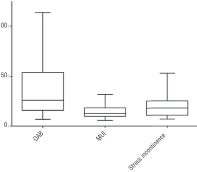
The mean Δp (delta pressure) in patients with a sensory component related to OAB syn-drome (36.5cmH2O) was significantly higher (p<0.05) than in groups with pure stress (14.9cm-H2O) and mixed urinary incontinence (19.30cm-H2O) (Figure-2). Patients of the OAB group were divided into sub-groups ‘with DO’ and ‘without DO’. Mean urethral pressure of OAB patients with DO (85.5±37.0cmH2O (mean±SD) was
signifi-cantly higher than in OAB patients without DO (45.2±30.65cmH2O) (p<0.05) (Figure-3).
The analysis of the area under the curve showed that urethral pressure curves of OAB pa-tients are in sum either longer or higher than those of mixed or stress urinary incontinence (1195.7±985.6 (OAB) vs. 429.3±214.5 (MUI) vs. SUI 549.4±273.0 (SUI) (Figure-4).
Statistical analysis revealed significant dif-ferences with p<0.005 (OAB vs. MUI) and p<0.01 (OAB vs. SUI). Fast Fourier Transformation gener-ated FFT-Plots for every patient and visual review revealed higher magnitudes and more peaks in the range of 0-0.5Hz within the group of patients with OAB (Figure-5).
DISCUSSION
Our study revealed that patients with OAB have statistically significant higher range of ure-thral pressure variation than in SUI and MUI pa-tient groups, moreover we found that in papa-tients with OAB and concomitant DO urethral pressure variation range was even higher than without DO (p<0.05) (Figure-3).
During urodynamic investigations (UDS), the focus regularly lies on the bladder pressure changes neglecting the fact that in the process of urine storage, the external urethral sphincter may play an important role. In 1959, the “guarding reflex” by the external urethral sphincter was brought into discussion by Garry et al. and
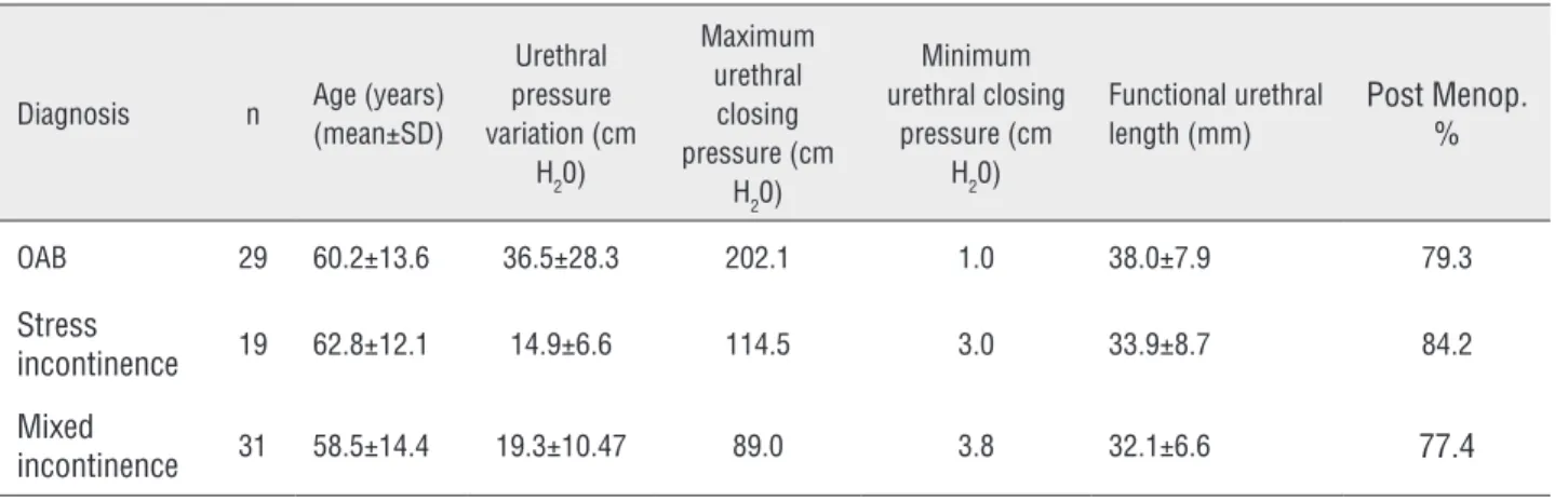
Table 1 - Main patient’s characteristics.
Diagnosis n Age (years)
(mean±SD)
Urethral pressure variation (cm
H20)
Maximum urethral
closing pressure (cm
H20)
Minimum urethral closing
pressure (cm H20)
Functional urethral length (mm)
Post Menop. %
OAB 29 60.2±13.6 36.5±28.3 202.1 1.0 38.0±7.9 79.3
Stress
incontinence 19 62.8±12.1 14.9±6.6 114.5 3.0 33.9±8.7 84.2
Mixed
Figure 1 - Urodynamic evaluation in a patient with DO (left) and SUI (right).
Figure 2 - Diagram of variance of delta pressure between the groups.
100
50
0
Figure 3 - Urethral Pressure in OAB patients with (left) or without (right) DO, single asterisk displays signifi cance difference between the two groups determined with two-tailed t-test.
Figure 4 - Area under the curve of cUPP in patients with OAB, MUI and SUI, applied trapezoidal rule calculated from min. pressure.
4000
3000
2000
1000
0
250
200
150
100
50
0
-50
*P=0.0149
OAB with DO OAB w/o DO
Pclo
Pressure [cm H
2
0]
OAB
AUC
was attributed a main role in prohibiting stress incontinence (13). Referring to the “guarding refl ex”, Park et al. later stipulated a theory that the external sphincter has a bladder-modulating role (4). McGuire and Sørensen in the 70s and 80s concluded that urethral pressure variations seem to play an important role in normal urethral physiology, possibly contributing to continence and prevention of urinary tract infection (14, 15). In contrast, Vereecken et al. failed to demonstrate a difference between patients with urge and stress incontinence with regard to ‘urethral instability’, except urethral pressure variations of more than
35cmH2O, which provoked urge (9). No difference in terms of prevalence or severity of urgency/ frequency, nocturia or urge incontinence was reported between patients with or without ‘urethral instability’, and the symptom of stress incontinence was more common in women with ‘urethral instability’ (9, 16). Nevertheless, Matthiason et al. also came to the conclusion that women with stress, urge, and mixed urinary incontinence seem to have a primary neuromuscular disorder in the urethra. They described urethral pressure variation as an overactive opening mechanism with a fall in urethral pressure instead of a pressure increase
Figure 5 - Representative FFT-Transformation of urethral pressure measures in a patient with OAB (above) and MUI (bottom).
Frequency (Hz)
MUI OAB
Frequency (Hz)
Ma
gnitude
10
8
6
4
2
0
0 0,5 1 1,5 2 2,5 3 3,5 4 4,5 5
Ma
gnitude
10
8
6
4
2
0
on provocation during the filling phase of the bladder, and during bladder emptying a more efficient opening of the bladder outlet than in normal women. They suggested that one and the same pathophysiological mechanism participates in female stress, urge, and mixed incontinence (17). Moreover in 2009, Groenendijk et al. stated that it makes sense to measure and register detrusor and urethral function during filling and voiding (18). Despite the fact that most authors consider urethral pressure variations, exceeding an amplitude of 15cmH2O, as an abnormal finding (6-12) the significance of these fluctuations is still unclear, and the terms ‘urethral instability’ or ‘unstable urethra’ lack clarity and are not accurately defined by the ICS today.
In accordance with our own data, Sørensen et al. demonstrated that mean maximum urethral pressure and the mean maximum urethral closure pressure are significantly reduced in women with stress incontinence compared to women with de-trusor overactivity (19).
Our study is not without limitations. First limitation is in the study design-small sample size, absence of assessors blinding, absence of a healthy patients group. Second is that the neurophysiolo-gic explanation of urethral pressure variation is unclear. Urethral pressure variation may be caused by diminished sympathetic influence or by increa-sed parasympathetic activity. It has been reported that neuronal nitric oxide (NO) synthase, known for its significant role in nociceptive pathways in the bladder has also been found in human fema-le striated urethra sphincter (20). It may be that contraction of the detrusor is caused by a fall in urethral pressure (18). However, looking at the traces, it is very difficult to determine (Figure-1) which came first: detrusor overactivity, then ure-thral pressure drop or vice versa, or both appeared at the same time. Whatever concomitant finding of detrusor overactivity and urethral pressure va-riations may suggest a combined pathophysiology being a cofactor in some OAB patients. Conside-ring that some DO might not have been detected, correlation should be even higher.
Further investigation of urethral pressure variation with high-speed urethral pressure urody-namics will let to sample at up to 1000Hz and not
10 to 50Hz used today in conventional equipment and possibly shed some more light on that issue.
CONCLUSIONS
Given the growing relevance of OAB syn-drome and widely expanding armamentarium of treatment modalities and drugs as well the outco-mes are still quite disappointing (21) and it see-ms that we are missing something important in patients with OAB. Possibly we should stress our attention on both: detrusor overactivity and ure-thral instability.
ACKNOWLEDGMENTS
We would like to thank Ellen Dicks from our research group in Bonn and Veronica Ciolfi from Laborie Medical Technologies for editing the manuscript.
CONFLICT OF INTEREST
None declared.
REFERENCES
1. Irwin DE, Milsom I, Hunskaar S, Reilly K, Kopp Z, Herschorn S, et al. Population-based survey of urinary incontinence, overactive bladder, and other lower urinary tract symptoms in five countries: results of the EPIC study. Eur Urol. 2006;50:1306-14
2. Abrams P, Cardozo L, Fall M, Griffiths D, Rosier P, Ulmsten U, et al. The standardisation of terminology of lower urinary tract function: report from the Standardisation Sub-committee of the International Continence Society. Neurourol Urodyn. 2002;21:167-78.
3. Al-Ghazo MA, Ghalayini IF, Al-Azab R, Hani OB, Matani YS, Haddad Y. Urodynamic detrusor overactivity in patients with overactive bladder symptoms. Int Neurourol J. 2011;15:48-54.
4. Park JM, Bloom DA, McGuire EJ. The guarding reflex revisited. Br J Urol. 1997;80:940-5.
5. Schwantes U, Grosse J, Wiedemann A. Refractory overactive bladder: a common problem? Int Urogynecol J. 2015;26:1407-14.
7. Penders L, de Leval J, Petit R. Enuresis and urethral instability. Eur Urol. 1984;10:317-22.
8. Venema PL, Kramer AE. The clinical implication of continuous urethral pressure measurement in women with incontinence. Acta Urol Belg. 1984;52:207-15.
9. Vereecken RL, Das J. Urethral instability: related to stress and/or urge incontinence? J Urol. 1985;134:698-701. 10. Weil A, Miège B, Rottenberg R, Krauer F. Clinical significance
of urethral instability. Obstet Gynecol. 1986;68:106-10. 11. Klein LA. Urge incontinence can be a disease of bladder
sensors. J Urol. 1988;139:1010-4.
12. Clarke B. Urethral instability. Aust N Z J Obstet Gynaecol. 1992;32:270-5.
13. Garry RC, Roberts TD, Todd JK. Reflexes involving the external urethral sphincter in the cat. J Physiol. 1959;149:653-65. 14. McGuire EJ. Reflex urethral instability. Br J Urol.
1978;50:200-4.
15. Søresen S, Kirkeby HJ, Stødkilde-Jørgensen H, Djurhuus JC. Continuous recording of urethral activity in healthy female volunteers. Neurourol Urodyn. 1986;5:5–16.
16. Wise BG, Cardozo LD, Cutner A, Benness CJ, Burton G. Prevalence and significance of urethral instability in women with detrusor instability. Br J Urol. 1993;72:26-9.
17. Mattiasson A, Teleman P. Abnormal urethral motor function is common in female stress, mixed, and urge incontinence. Neurourol Urodyn. 2006;25:703-8.
18. Groenendijk PM, Heesakkers JP, Ouwerkerk TJ, Lycklama A Nijeholt AA. Urethral instability: current pathophysiological concept. Urol Int. 2009;83:125-33.
19. Sørensen S. Urethral pressure and pressure variations in stress incontinent women and women with unstable detrusor. Neurourol Urodyn. 1991;10:483–92.
20. Ho KM, Borja MC, Persson K, Brading AF, Andersson KE. Expression of nitric oxide synthase immunoreactivity in the human female intramural striated urethral sphincter. J Urol. 2003;169:2407-11.
21. Flynn MK, Amundsen CL, Perevich M, Liu F, Webster GD. Outcome of a randomized, double-blind, placebo controlled trial of botulinum A toxin for refractory overactive bladder. J Urol. 2009;181:2608-15.
_______________________ Correspondence address:
Nariman Gadzhiev, MD Department of Urology, Saint-Petersburg, 194044 St.Petersburg, Acad.Lebedev St., 4/2