Obesity may influence the relationship between sex
hormones and lower urinary tract symptoms
_______________________________________________
Alberto A. Antunes, Luiz Henrique Araújo, Elcio Nakano, Eduardo Muracca, Miguel Srougi
Division of Urology, University of Sao Paulo Medical School, SP, Brazil
ABSTRACT
ARTICLE
INFO
______________________________________________________________ ______________________
Purpose: The effects of serum testosterone in the lower urinary tract symptoms (LUTS) in patients with benign prostatic hyperplasia (BPH) are not well established. The objec-tive of the study is to evaluate the association of sex hormones with LUTS and control the results by patient weight.
Materials and Methods: The study comprised a cross-sectional analysis of 725 men included in a prostate cancer screening program at University of Sao Paulo Medical School. The serum concentrations of total testosterone (TT), free testosterone (FT) and sex hormone binding globulin (SHBG) were measured. Variables analyzed were age, American Urological Association (AUA) symptom score, storage symptoms, voiding symptoms, quality of life score, prostate specific antigen levels and prostate volume. Obesity was measured through the calculation of body mass index (BMI). A regression analysis model was performed.
Results: Median patient age was 65 years (48 to 94). A higher TT level was significantly associated with a severe AUA symptom score only among patients with a BMI ≥ 25. Median TT was 371, 370 and 427ng/dL (p = 0.017) in patients with mild, moderate and severe LUTS respectively. The multivariate regression analysis in patients with BMI ≥ 25 showed that only age, TT and sex score were related to LUTS.
Conclusions: A higher TT is associated with a severe AUA score symptom index only in obese patients. Further analysis are necessary to evaluate the mechanisms through which testosterone may influence LUTS in these patients.
Key words:
Prostatic Hyperplasia; Lower Urinary Tract Symptoms; Testosterone; Obesity
Int Braz J Urol. 2014; 40: 240-6
_____________________
Submitted for publication: July 16, 2013
_____________________
Accepted after revision: February 29, 2014
INTRODUCTION
Benign prostatic hyperplasia (BPH) is the main cause of lower urinary tract symptoms (LUTS) in the aging man. About 90% of men in their 70s have some LUTS related to BPH and the main consequence is impairment in their health-related quality of life (1). The lifetime probability among men in their fifties to receive treatment for LUTS secondary to BPH is estimated to reach 35% of cases (2). Less commonly, LUTS related to BPH may also progress to acute urinary
reten-tion, need for surgery, urinary incontinence or recurrent urinary tract infection (3).
androgen deprivation therapy (8). However, it is unclear whether altered serum concentrations of androgens are associated with increased risk of LUTS or clinical BPH.
Thus far, published studies regarding the association between androgens and LUTS or BPH have reported controversial results (9-12). Addi-tionally, studies that have found positive asso-ciations have analyzed different hormones. These discrepancies may be attributed to small sample sizes, important selection bias, inadequate assess-ment of BPH, and failure to control by other po-tentially confounding factors that may influence serum testosterone levels, such as obesity (13,14).
As it’s well known, obesity in men is com-monly accompanied by a decline of serum testos-terone (TT) levels through different mechanisms (15,16). The objective of the study was to evaluate the association of sex hormones with LUTS and control the results by the body mass index (BMI).
MATERIALS AND METHODS
The study comprised a cross-sectional analysis of 725 men who participated in a prostate cancer screening program. Patients who were us-ing 5-alpha reductase inhibitors, alpha adrenergic blockers, had history of prostatic surgery or endo-crine diseases, were not considered for analysis. Patients with abnormal digital rectal examination (DRE) findings or an elevated serum PSA level (i.e. > 4.0ng/mL) were referred for prostate biopsy to exclude the possibility of prostate cancer.
Variables analyzed were age, American Urological Association (AUA) symptom score, AUA storage symptoms, AUA voiding symptoms, nocturia, AUA quality of life score (17), inter-national index of erectile function (IIEF) score, prostate specific antigen levels and prostate vol-ume. We measured the serum concentrations of total testosterone (TT), free testosterone (FT) and sex hormone binding globulin (SHBG) in all men. These hormones were measured through the fluo-rimetric, fluoroimmunoassay, and immunofluori-metric methods respectively (Wallac Perkin Elmer, Finland). We considered the following normal ranges: 271 to 965ng/dL for TT, 131 to 640pmol/L for FT and 12 to 75nmol/L for SHBG. Patients
were submitted to DRE, which was performed only by urologists with experience in prostate ex-amination. An enlarged prostate was defined as prostate volume > 30 grams. Obesity was defined through calculation of the BMI, which was calcu-lated as weight in kilograms divided by the square of height in meters (Kg/m2) and categorized as
underweight (< 18.5Kg/m2), normal weight (18.5
to 24.9Kg/m2), overweight (25 to 29.9Kg/m2) and
obese (≥ 30Kg/m2). Waist circumference (WC) and
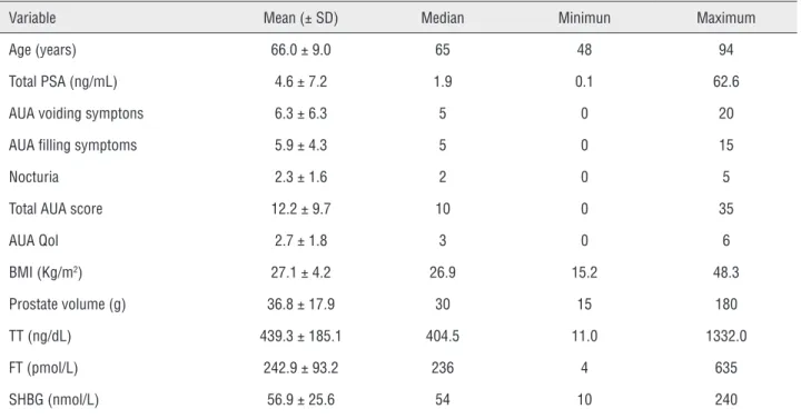
hip circumference (HC) were also measured. De-mographic characteristics of the men are described in Table-1. Median patient age was 65 years (48 to 94). Median serum PSA level was 1.9ng/mL (0.1 to 62.6) and median AUA symptom score was 10 (0 to 35). Median total testosterone, free testoster-one and SHBG levels were 404.5, 236 and 54ng/ dL respectively. Median BMI was 27 (15 to 48). TT, FT and SHBG were correlated with all clinical parameters. For analysis of obesity, patients were divided in two groups according to the BMI ≥ 25 or < 25. Sex hormones were correlated with all urinary symptoms among both groups.
For statistical analysis we used the Chi-square, ANOVA and Kruskal Wallis tests to corre-late the sex hormones with clinical parameters. A multivariate regression analysis model was used. Statistical analysis was performed using the SPSS 12.0 for Windows software and significance was set as a p ≤ 0.05.
RESULTS
Table-2 shows correlation of patient char-acteristics with urinary symptoms. Patients with older age, higher PSA levels and lower IIEF pre-sented worse LUTS with statistically significant results. Patients with a higher TT level and great-er prostate weight also presented worse LUTS, but with marginally significant results. Mean TT among patients with mild, moderate and severe symptoms was 430.2, 430.3 and 468.0ng/dL re-spectively. When we analyzed filling symptoms, voiding symptoms and nocturia separately, no statistically significant associations were found.
lower levels of TT (p < 0.001). Median FT levels were statistically lower among patients with 65 years or older (211 vs 254ng/dL) (p < 0.001), and with a higher BMI (p = 0.006). SHBG levels were statistically higher among patients with 65 years or older (p < 0.001) and statistically lower among
patients with a higher BMI. These two hormones failed to show any significant correlation with urinary symptoms or other clinical parameters (data not shown).
Due to associations between serum hor-mone levels and surrogate measures of obesity, Table 1 - Demographic characteristics of the study population.
Variable Mean (± SD) Median Minimun Maximum
Age (years) 66.0 ± 9.0 65 48 94
Total PSA (ng/mL) 4.6 ± 7.2 1.9 0.1 62.6
AUA voiding symptons 6.3 ± 6.3 5 0 20
AUA filling symptoms 5.9 ± 4.3 5 0 15
Nocturia 2.3±1.6 2 0 5
Total AUA score 12.2±9.7 10 0 35
AUA Qol 2.7±1.8 3 0 6
BMI (Kg/m2) 27.1 ±4.2 26.9 15.2 48.3
Prostate volume (g) 36.8± 17.9 30 15 180
TT (ng/dL) 439.3 ± 185.1 404.5 11.0 1332.0
FT (pmol/L) 242.9 ± 93.2 236 4 635
SHBG (nmol/L) 56.9 ±25.6 54 10 240
Table 2 - Correlation of AUA symptom score categories with clinical variables.
Variable (± SD) Mild Moderate Severe p
Age (years) 64.5 (± 8.7) 66.4 (± 9.2) 68.3 (± 8.6) 0.00
Total PSA (ng/mL) 4.7 (± 9.1) 4.4 (± 6.7) 7.4 (± 27.2) 0.01
TT (ng/dL) 430.2 (± 179.5) 430.3 (± 177.1) 468.0 (± 202.9) 0.06
FT (pmol/L) 239.8 (± 97.2) 241.6 (± 88.7) 250.2 (± 92.7) 0.48
SHBG (nmol/L) 56.8 (± 24.4) 55.7 (± 27.4) 58.9 (± 25.0) 0.45
IIEF 16.4 (± 7.5) 14.1 (± 7.6) 11.7 (± 7.6) 0.00
BMI (kg/m2) 27.1 (± 4.0) 27.0 (± 4.3) 27.3 (± 4.5) 0.72
WC (cm) 95.0 (± 11.6) 96.3 (± 13.1) 95.8 (± 13.0) 0.35
HC (cm) 99.2 (± 9.3) 98.1 (± 14.7) 98.7 (± 10.2) 0.59
we analyzed the associations between hormone levels and urinary symptoms among patients with BMI < 25 (underweight or normal weight) and ≥ 25 (overweight or obese) separately. The results showed that TT levels were significantly associat-ed with AUA symptom score among patients with a BMI ≥ 25. Median TT was 371, 370 and 427ng/dL (p = 0.017) in patients with mild, moderate and se-vere LUTS respectively. FT and SHBG were not as-sociated with urinary symptoms in this BMI group (Table-3). Analysis of voiding symptoms, filling symptoms and nocturia separately failed to show any association with hormone levels according to this BMI category.
No associations were found among pa-tients with a BMI < 25. Median TT was 453, 450 and 467ng/dL (p = 0.952) among patients with mild, moderate and severe LUTS respectively. FT and SHBG were not associated with urinary toms in this BMI group. Analysis of voiding symp-toms, filling symptoms and nocturia separately also failed to show any association.
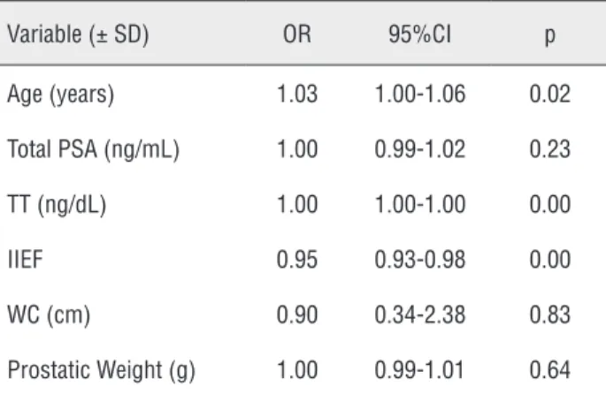
Finally, we performed a multivariate re-gression analysis for the occurrence of severe LUTS among patients with a BMI ≥ 25 and < 25. An in-creased age, a higher TT level and a worse IIEF were independently associated with the presence of se-vere LUTS among patients from the former group. Only a worse IIEF was independently associated with LUTS among patients with a BMI < 25.
Table 3 - Logistic regression analysis for the occurrence of severe LUTS among patients with BMI ≥ 25.
Variable (± SD) OR 95%CI p
Age (years) 1.03 1.00-1.06 0.02
Total PSA (ng/mL) 1.00 0.99-1.02 0.23
TT (ng/dL) 1.00 1.00-1.00 0.00
IIEF 0.95 0.93-0.98 0.00
WC (cm) 0.90 0.34-2.38 0.83
Prostatic Weight (g) 1.00 0.99-1.01 0.64
DISCUSSION
In the present study, we found that serum TT and FT levels were significantly lower, and SHBG levels were significantly higher in obese patients when compared to patients with lower weights. When the entire cohort was analyzed, TT levels were higher in patients with severe LUTS, however, these results reached only marginal sta-tistical significance. When patients with a BMI
≥ 25 were analyzed, TT levels were significantly higher in patients with severe LUTS when com-pared to patients with moderate or mild LUTS. These findings suggest that sexual hormones may play a role in the pathogenesis of LUTS related to BPH. Multivariate analysis confirmed TT as an independent variable associated to a severe LUTS. No correlations were observed among patients with a BMI < 25.
The reasons why sex hormones may influ-ence LUTS related to BPH are not well described. Despite the lack of knowledge regarding several aspects of BPH pathogenesis, its well recognized that the development of the histological features of the disease is dependent on the bioavailability of TT and its metabolite, dihydrotestosterone (18). A congenital lack of 5α-reductase results in a ves-tigial prostate gland (19) and castration in a man leads to glandular atrophy and regression of LUTS (20). It’s also reasonable to think that systemic ef-fects of TT in bone and muscle mass, fat mass, body energy, and physical, sexual, and cognitive function may also be reflected on LUTS. Finally, there may be some direct effects of such hormones on bladder muscle function.
Some limitations were the small sample size, the homogenous character of the community cohort (mostly white and upper middle class) limiting the external validity, and the 20-year gap between hormone measurement and LUTS assessment that precluded analysis of changes in serum sex ste-roid levels during this period. In another study, the authors performed a nested case-control analysis using the placebo arm of the Prostate Cancer Pre-vention Trial. The authors reported a decreased risk of incident BPH with higher baseline total testosterone and total testosterone : 17β -diol-glucuronide (21). Litman et al. (9), analyzing data from the Boston Area Community Health (BACH) Survey, reported an inverse correlation of AUA-SI with both TT and bioavailable testosterone in non-age adjusted models and bioavailable testos-terone in age-adjusted models. No significant as-sociation between LUTS and FT were found in the present analysis. Interestingly, some authors have also reported inverse relationships between LUTS and FT levels (22,23).
Conversely, other studies have reported positive relationships. Favilla et at. (2010) investi-gated a possible association between the severity of LUTS and the serum levels of sex hormones in 122 men with symptomatic BPH. In accordance to our results, on statistical analysis they found that the total IPSS was significantly associated with age and TT but not with free testosterone or the serum levels of the other sex hormones. While men with a IPSS > 19 presented a median testos-terone of 425.6ng/mL, median testostestos-terone level among men with a IPSS < 19 was 346.8ng/mL). However, the limitations of the study were again the small sample size and the fact that they only included men with severe LUTS who were candi-dates to surgery.
Reasons that may help to explain the lack of associations in some studies may be attributed to determination of sexual hormones levels at dif-ferent points in the natural history of BPH. Ad-ditionally, it’s possible that plasma levels of these hormones are not representative of intra-prostatic levels. Regarding this fact, the dosage of sexual hormones metabolites may be more representa-tive of the intra-prostatic environment. Platz et al. (12), demonstrated the association between
an SHBG metabolite, androstanediol glucuronide (AAG), severe LUTS and the risk of BPH surgery. Patients with higher serum levels of AAG were at increased risk of having either BPH surgery or se-vere LUTS.
The unique strength of the present analy-sis is that we demonstrated a clear influence of BMI on the relationship between testosterone and LUTS, a variable that has been neglected by most studies. According to some reports, obesity may be associated with BPH. Giovannucci et al. (24), followed men enrolled in the Health Professionals Follow-up Study for the incidence of prostatecto-my due to BPH and for the frequency and severity of symptoms of urinary obstruction. After adjust-ment for age, smoking, and BMI, only abdomi-nal obesity showed a relationship with the rate of prostatectomy. Similar results were observed regarding frequency of urinary symptoms among those without prostatectomy (OR 2.0). However, these results should be analyzed carefully, since only a minority of patients with BPH undergoes surgical treatment for relieving LUTS.
Conversely, associations between obesity and BPH were not reproduced by other authors. Meigs et al. (25), defined risk factors for a clini-cal diagnosis of BPH among subjects of the pop-ulation-based Massachusetts Male Aging Study. They found that BMI did not individually predict clinical BPH. More recently, in the study of 1,206 participants in the comparison arm of the Air Force Health Study with a median follow-up of 15.6 years, no relation was seen between weight or BMI and BPH (26). In another recent study of a cohort of healthy caucasian men aged 40-79 years randomly selected from the Olmsted County, Min-nesota, few significant associations of anthropo-metric measures with the presence or progression of components of BPH were found (27).
as-CONFLICT OF INTEREST
None declared.
REFERENCES
1. Walsh PC: Treatment of benign prostatic hyperplasia. N Engl J Med. 1996; 335: 586-7.
2. Oesterling JE: Benign prostatic hyperplasia: a review of its histogenesis and natural history. Prostate Suppl. 1996; 6: 67-73.
3. Cockett AT, Barry MJ, Holtgrewe HL, Sihelnick S, Williams R, McConnell J: Indications for treatment of benign prostatic hyperplasia. The American Urological Association Study. Cancer. 1992; 70: 280-3.
4. Guess HA: Benign prostatic hyperplasia: antecedents and natural history. Epidemiol Rev. 1992; 14: 131-53.
5. Steiner MS: Review of peptide growth factors in benign prostatic hyperplasia and urological malignancy. J Urol. 1995; 153: 1085-96.
6. Soulitzis N, Karyotis I, Delakas D, Spandidos DA: Expression analysis of peptide growth factors VEGF, FGF2, TGFB1, EGF and IGF1 in prostate cancer andbenign prostatic hyperplasia. Int J Oncol. 2006; 29: 305-14.
7. Smith P, Rhodes NP, Ke Y, Foster CS: Upregulation of estrogen and androgen receptors modulate expression of FGF-2 and FGF-7 in human,cultured, prostatic stromal cells exposed to high concentrations of estradiol. Prostate Cancer Prostatic Dis. 2002; 5: 105-10.
8. Peters CA, Walsh PC: The effect of nafarelin acetate, a luteinizing-hormone-releasing hormone agonist, on benign prostatic hyperplasia. N Engl J Med. 1987; 317: 599-604. Erratum in: N Engl J Med. 1988; 318: 580.
9. Litman HJ, Bhasin S, O’Leary MP, Link CL, McKinlay JB, BACH Survey Investigators: An investigation of the relationship between sex-steroid levels and urological symptoms: results from theBoston Area Community Health survey. BJU Int. 2007; 100: 321-6.
10. Trifiro MD, Parsons JK, Palazzi-Churas K, Bergstrom J, Lakin C, Barrett-Connor E: Serum sex hormones and the 20-year risk of lower urinary tract symptoms in community-dwelling oldermen.BJU Int. 2010; 105: 1554-9.
11. Platz EA, Kawachi I, Rimm EB, Longcope C, Stampfer MJ, Willett WC, et al.: Plasma steroid hormones, surgery for benign prostatic hyperplasia, and severe lower urinary tractsymptoms. Prostate Cancer Prostatic Dis. 1999; 2: 285-289.
12. Favilla V, Cimino S, Castelli T, Madonia M, Barbagallo I, Morgia G: Relationship between lower urinary tract symptoms and serum levels of sex hormones in men withsymptomatic benign prostatic hyperplasia. BJU Int. 2010; 106: 1700-3.
sess BPH, including clinical (LUTS), physiologic (urinary flow rates), anatomic (prostatic volume) and biochemical (PSA levels) measures (28). For this reason, in the present study we investigated specifically the associations with LUTS.
Some advantages of the present study over the others should be mentioned. We analyzed a rel-ative large series of patients who were not candi-dates for surgery, thus all LUTS categories could be analyzed. Additionally, patients were not using any placebo, which could have influenced LUTS. Limi-tations of the present study should also be noted. We didn’t analyze other important sex hormones such as dihydrotestosterone and oestradiol. Other clinical conditions that could influence sex hor-mone levels such as metabolic syndrome, diabetes or tobacco use were not included in the analysis.
Finally, if the results of the present series could be confirmed by further clinical analyses, there will be a role for the clinical use of serum testosterone, at least in obese patients with BPH. Basic research studies addressing the mecha-nisms of action of testosterone over the bladder and prostate tissues on a molecular basis are also necessary for the understanding of LUTS physio-pathology and for the development of new thera-peutic targets.
CONCLUSIONS
Serum TT and FT levels are significantly lower, and SHBG levels is significantly higher in obese patients when compared to patients with lower weights. A higher TT is independently as-sociated with a worse AUA symptom index only in obese patients.
ABBREVIATIONS
LUTS = Lower Urinary Tract Symptoms BPH = Benign Prostatic Hyperplasia TT = Total Testosterone
FT = Free Testosterone
AUA = American Urological Association BMI = Body Mass Index
GFs = Growth Factors
13. Selvin E, Feinleib M, Zhang L, Rohrmann S, Rifai N, Nelson WG, et al.: Androgens and diabetes in men: results from the Third National Health and Nutrition Examination Survey(NHANES III). Diabetes Care. 2007; 30: 234-8. Erratum in: Diabetes Care. 2007; 30: 1683.
14. Li C, Ford ES, Li B, Giles WH, Liu S: Association of testosterone and sex hormone-binding globulin with metabolic syndrome and insulin resistance in men. Diabetes Care. 2010; 33: 1618-24. 15. Couillard C, Gagnon J, Bergeron J, Leon AS, Rao DC, Skinner
JS, et al.: Contribution of body fatness and adipose tissue distribution to the age variation in plasma steroidhormone concentrations in men: the HERITAGE Family Study. J Clin Endocrinol Metab. 2000; 85: 1026-31.
16. Gautier A, Bonnet F, Dubois S, Massart C, Grosheny C, Bachelot A, et al.: Associations between visceral adipose tissue, inflammation and sex steroid concentrations in men. Clin Endocrinol (Oxf). 2013; 78: 373-8.
17. Barry MJ, Fowler FJ Jr, O’Leary MP, Bruskewitz RC, Holtgrewe HL, et al.: The American Urological Association symptom index for benign prostatic hyperplasia. The MeasurementCommittee of the American Urological Association. J Urol. 1992; 148: 1549-57; discussion 1564.
18. Bartsch G, Rittmaster RS, Klocker H: Dihydrotestosterone and the concept of 5alpha-reductase inhibition in human benign prostatic hyperplasia. Eur Urol. 2000; 37: 367-80.
19. Imperato-McGinley J, Guerrero L, Gautier T, Peterson RE: Steroid 5alpha-reductase deficiency in man: an inherited form of male pseudohermaphroditism. Science. 1974; 186: 1213-5. 20. Huggins C, Clark PJ: Quantitative studies of prostatic secretion:
II. The effect of castration and of estrogeninjection on the normal and on the hyperplastic prostate glands of dogs. J Exp Med. 1940; 72: 747-62.
21. Kristal AR, Schenk JM, Song Y, Arnold KB, Neuhouser ML, Goodman PJ, et al.: Serum steroid and sex hormone-binding globulin concentrations and the risk of incident benign prostatichyperplasia: results from the prostate cancer prevention trial. Am J Epidemiol. 2008; 168: 1416-24.
22. Miwa Y, Kaneda T, Yokoyama O: Association between lower urinary tract symptoms and serum levels of sex hormones in men. Urology. 2008; 72: 552-5.
23. Tan MO, Karabiyik I, Uygur MC, Diker Y, Erol D: Serum concentrations of sex hormones in men with severe lower urinary tract symptoms and benign prostatic hyperplasia. Int Urol Nephrol. 2003; 35: 357-63.
24. Giovannucci E, Rimm EB, Chute CG, Kawachi I, Colditz GA, Stampfer MJ, et al.: Obesity and benign prostatic hyperplasia. Am J Epidemiol. 1994; 140: 989-1002.
25. Meigs JB, Mohr B, Barry MJ, Collins MM, McKinlay JB: Risk factors for clinical benign prostatic hyperplasia in a community-based population of healthy agingmen. J Clin Epidemiol. 2001; 54: 935-44.
26. Gupta A, Gupta S, Pavuk M, Roehrborn CG: Anthropometric and metabolic factors and risk of benign prostatic hyperplasia: a prospective cohort studyof Air Force veterans. Urology. 2006; 68: 1198-205.
27. Burke JP, Rhodes T, Jacobson DJ, McGree ME, Roberts RO, Girman CJ, et al.: Association of anthropometric measures with the presence and progression of benign prostatichyperplasia. Am J Epidemiol. 2006; 164: 41-6. 28. Jacobsen SJ, Girman CJ, Lieber MM: Natural history of
benign prostatic hyperplasia. Urology. 2001; 58: 5-16; discussion 16.
_______________________ Correspondence address: Alberto A. Antunes, MD Rua Frei Caneca, 558 - 2501/02