Complete supine PCNL: ultrasound vs. fluoroscopic guided:
a randomized clinical trial
_______________________________________________
Siavash Falahatkar
1, Aliakbar Allahkhah
1, Majid Kazemzadeh
1, Ahmad Enshaei
1, Maryam Shakiba
1,
Fahimeh Moghaddas
11 Urology Research Center, Guilan University of Medical Sciences, Guilan, Iran
ABSTRACT
ARTICLE
INFO
______________________________________________________________ ______________________
Introduction and Hypothesis: To compare complications and outcomes of complete supine percutaneous nephrolithotomy (csPCNL) with ultrasound guided and fluorosco-pically guided procedure.
Materials and Methods: In this randomized clinical trial study from January 2009 to September 2010, 26 of 51 patients with renal stones underwent csPCNL with ultraso-nographic guidance in all steps of the procedure (group A), and the other 25 patients underwent standard fluoroscopically guided csPCNL (group B). All of the patients un-derwent PCNL in the complete supine position. Statistical analysis was performed with SPSS16 software.
Results: Mean BMI was 28.14 in group A and 26.31 in group B (p=0.30). The mean stone burden was 26.48 and 30.44 in groups A and B, respectively (p=0.20). The stone free rate was 88.5% in group A and 75.5% in group B, that was no significant (p=0.16).Overall 2 patients (7.7%) in group A and 6 patients (24%) in group B had complications (p=0.11). Mean operative time in group A was 88.46 minutes, and in group B it was 79.58 minutes (p=0.39). Mean hospital stay was 69.70 and 61.79 hours in group A and B, respectively (p=0.22). There was no visceral injury in groups.
Conclusions: This randomized study showed that totally ultrasonic had the same outcomes of fluoroscopically csPCNL. Ultrasonography can be an alternative rather than fluorosco-py in PCNL. We believe that more randomized studies are needed to allow endourologists to use sonography rather than fluoroscopy in order to avoid exposition to radiation.
Keywords:
Nephrostomy, Percutaneous; Ultrasonography; Fluoroscopy; Supine Position
Int Braz J Urol. 2016; 42: 710-6
_____________________
Submitted for publication: June 15, 2014
_____________________
Accepted after revision: October 12, 2015
INTRODUCTION
Percutaneous nephrolithotomy (PCNL) is a common method for treatment of kidney stones (1, 2). All of the steps in PCNL should be perfor-med with proper image guidance. The imageless PCNL should never be applied because it is dange-rous to vital structures (3).
The popular imaging of PCNL is fluorosco-py, so the patient and surgical team are exposed
to some level of radiation by fluoroscopy during PCNL. The side effects of extensive radiation are well known. Thus, the ultrasound-guided PCNL can be an alternative method to decrease the ra-diation exposure hazard to the surgeon (4-6).
ultrasound-guided csPCNL procedure with the urological community.
MATERIAL AND METHODS
In this randomized clinical trial stu-dy from January 2009 to September 2010, 51 patients with renal stones were selected for csPCNL. All participants were informed about the surgical method and consent. We used to-tally ultrasonographic guidance in all steps of the procedure during csPCNL in 26 of our pa-tients (group A), whereas the other 25 papa-tients underwent standard fluoroscopically guided csPCNL (group B). All patients in both groups performed PCNL in the complete supine position without any towel under the patient’s flank and with no change in leg position. For all patients, routine blood and urine tests, coagulation pro-file and imaging series, including intravenous urogram and ultrasonography, were carried out and medical conditions were studied.
Inclusion criteria were patients with sin-gle large pelvic stone, lower caliceal stone, sto-nes in the pelvis and lower calyx, middle cali-ceal stones, and non-opaque stones (staghorn stones) with hydronephrosis.
Exclusion criteria’s in this study were multiple stones in multiple calyxes, staghorn stones (except non-opaque stones), urinary tract anomalies, single kidney and morbid obesity and non-opaque stones (staghorn stones) wi-thout hydronephrosis.
All of the patients underwent general anesthesia, and a 5F ureteral catheter was pla-ced transurethrally for injection of saline or contrast media. Injection of saline obtained mild dilatation of collecting system and this was use-ful especially for the totally ultrasound-guided PCNL group.
In group A, ultrasonography was used to observe the location of the kidney, needle entrance point, urinary tract dilatation and to check for residual stone at the end of csPCNL. Because the Rouch guidewire is more rigid, and in order to not miss the access, we used this type of guidewire, although the guidewire was clearly visible but the Amplatz dilatators and
the Amplatz sheath were not exactly visible by ultrasonography.
In group B, we performed all the above steps of csPCNL with the guidance of fluorosco-py. Our technique was a one-shot dilatation in both groups.
In this study, the items including side of renal unit, stone burden, stone-free rate, compli-cations (extravasation, colon injury, fever, etc.), and the history of previous open renal surgery or previous ESWL, mean hospital stay, mean operati-ve time, body mass index (BMI), serum creatinine before the operation, and hemoglobin before and after the csPCNL were studied.
In group A, after removal of the stone(s), ultrasonography was used to detect any residual stones, hematoma, or extravasation of urine out-side of the kidney.
In the fluoroscopic group, residual stones and extravasation were checked by fluoroscopy. We performed tubeless PCNL except in patients with severe extravasation, ureteral obstruction, severe hemorrhage, or large residual stone.
Statistical analysis was performed with SPSS16 software. A P value of less than 0.05 was considered statistically significant.
This study was approved by ethical review committee of Guilan University of Medical Scien-ce and the trial registered at http://www.irct.ir (IRCT138805251853N3).
RESULTS
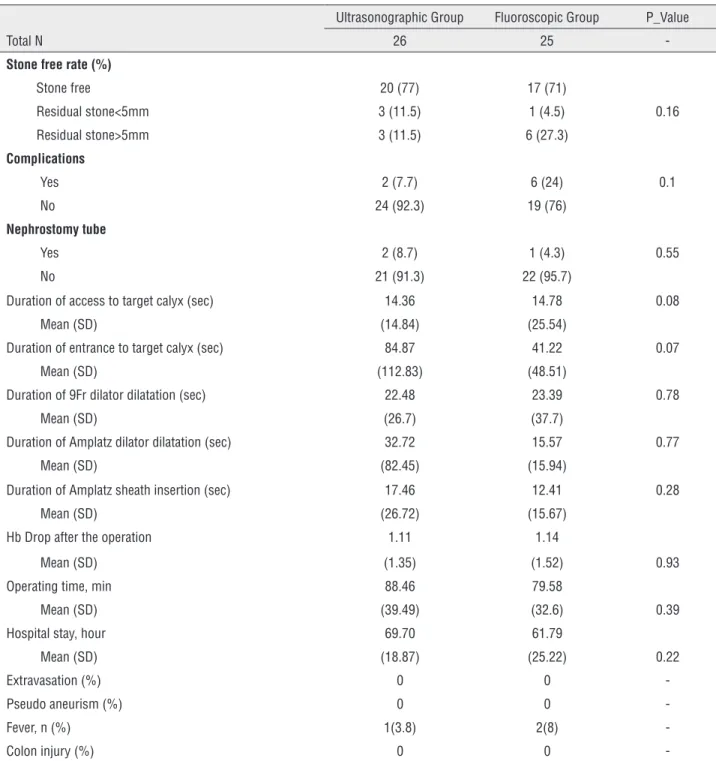
the patients underwent general anesthesia and the access was sub costal in all patients. Intra and postoperative parameters of the two groups are shown in Table-2. The stone free rate was 88.5% in group A and 75.5% in group B, that was no significant (p=0.16). Overall, 2 patients (7.7%) in group A and 6 patients (24%) in group B had com-plications (p=0.11). In group A, 1 patient (3.8%) had fever, and in group B, 4 patients (16%) needed
transfusion and 2 patients (8%) had fever (Grade I and II of the Clavien Classification of Surgical Complications). Mean operation time in group A was 88.46 minutes, and in group B, it was 79.58 minutes (p=0.39). Mean hospital stay was 69.70 and 61.79 hours in groups A and B, respectively (p=0.22). There was no complications compatible with Grade III to V of the Clavien Classification of Surgical Complications in both groups.
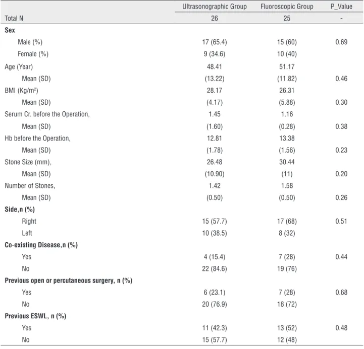
Table 1 - This table showed the demographic data of two groups according to method of study.
Ultrasonographic Group Fluoroscopic Group P_Value
Total N 26 25
-Sex
Male (%) 17 (65.4) 15 (60) 0.69
Female (%) 9 (34.6) 10 (40)
Age (Year) 48.41 51.17
Mean (SD) (13.22) (11.82) 0.46
BMI (Kg/m2) 28.17 26.31
Mean (SD) (4.17) (5.88) 0.30
Serum Cr. before the Operation, 1.45 1.16
Mean (SD) (1.60) (0.28) 0.38
Hb before the Operation, 12.81 13.38
Mean (SD) (1.78) (1.56) 0.23
Stone Size (mm), 26.48 30.44
Mean (SD) (10.90) (11) 0.20
Number of Stones, 1.42 1.58
Mean (SD) (0.50) (0.50) 0.26
Side,n (%)
Right 15 (57.7) 17 (68) 0.51
Left 10 (38.5) 8 (32)
Co-existing Disease,n (%)
Yes 4 (15.4) 7 (28) 0.44
No 22 (84.6) 19 (76)
Previous open or percutaneous surgery, n (%)
Yes 6 (23.1) 7 (28) 0.68
No 20 (76.9) 18 (72)
Previous ESWL, n (%)
Yes 11 (42.3) 13 (52) 0.48
DISCUSSION
The scope of endourology despite its short age has been widened. The first step in percutaneous procedures is to access to the col-lecting system, usually performed by
fluorosco-py, ultrasonography, or computed tomography (CT) guidance (7-9).
To reduce the risk of radiation exposure,
using ultrasonography for PCNL can be an
al-ternative imaging method to fluoroscopy as the
first and standard imaging technique (10, 11).
Table 2 - This table showed the comparison of results after the procedure between two groups.
Ultrasonographic Group Fluoroscopic Group P_Value
Total N 26 25
-Stone free rate (%)
Stone free 20 (77) 17 (71)
Residual stone<5mm 3 (11.5) 1 (4.5) 0.16
Residual stone>5mm 3 (11.5) 6 (27.3)
Complications
Yes 2 (7.7) 6 (24) 0.1
No 24 (92.3) 19 (76)
Nephrostomy tube
Yes 2 (8.7) 1 (4.3) 0.55
No 21 (91.3) 22 (95.7)
Duration of access to target calyx (sec) 14.36 14.78 0.08
Mean (SD) (14.84) (25.54)
Duration of entrance to target calyx (sec) 84.87 41.22 0.07
Mean (SD) (112.83) (48.51)
Duration of 9Fr dilator dilatation (sec) 22.48 23.39 0.78
Mean (SD) (26.7) (37.7)
Duration of Amplatz dilator dilatation (sec) 32.72 15.57 0.77
Mean (SD) (82.45) (15.94)
Duration of Amplatz sheath insertion (sec) 17.46 12.41 0.28
Mean (SD) (26.72) (15.67)
Hb Drop after the operation 1.11 1.14
Mean (SD) (1.35) (1.52) 0.93
Operating time, min 88.46 79.58
Mean (SD) (39.49) (32.6) 0.39
Hospital stay, hour 69.70 61.79
Mean (SD) (18.87) (25.22) 0.22
Extravasation (%) 0 0
-Pseudo aneurism (%) 0 0
-Fever, n (%) 1(3.8) 2(8)
-Some studies
reported that PCNL under ultrasonography guidance in the flank or prone position has high success rates and limited com-plications andcan be a safe and effective
al-ternative to fluoroscopy in experienced hands
(
11-18
).Ultrasound-guided PCNL without fluoros-copy has some advantages and disadvantages. Advantages: Avoidance of X-ray exposure, no necessity of lead shield, all organs on the way of access are visible, search for residual stones at the end of the procedure especially for non-opaque stones. Disadvantages: Endourologists are unfa-miliar with ultrasonography, and poor echogeni-city of the Amplatz dilatator and Amplatz sheath (11, 12, 19, 20).
Nowadays, PCNL is considered a generally safe management option with a low incidence of complications and is the method of choice for tre-atment of renal stones (11, 21, 22).
PCNL is done in the prone, flank, semi-su-pine, and csPCNL positions. We performed csPCNL in our patients due to better control of the airway, better tolerance for patients especially with car-diopulmonary disease, easier to perform ureteros-copy or TUL, better drainage and evacuation of stones by the Amplatz sheath, possibility to chan-ge regional anesthesia to chan-general anesthesia, and probably less risk of colon injury. These are some advantages of csPCNL (13).
Because of levitation of the colon in the abdominal cavity in the supine position it is less affected to injury when puncture site is in the pos-terior axillary line (23).
The access to the kidney is important in PCNL and usually is performed by fluoroscopy, ultrasonography, or computed tomography gui-dance with rates of success of 86.7-100% (9, 11, 20, 24-26). The success rate in achieving access in our study was 100% in both groups. The gaining access to the collecting system under ultrasono-graphic guidance was similar to the fluoroscopic--guided access.
Some studies showed that the stone-free rate in percutaneous nephrolithotomy with ultra-sonography guidance varied from 66.6 to 94.7% (5, 7, 12, 18, 20, 27). Other studies showed that primary stone-free rate and total stone-free rate
with ultrasound-guided percutaneous nephro-lithotomy were 45.7 - 69.6% and 82.6 - 96.5%, respectively (21, 26). In our study, similar to the others, the stone-free rate was 88.46% and 72%, without any significant statistical difference in groups A and B, respectively (p=0.16).
The mean operative time was 120±68 mi-nutes (range 45-350) in one study. The real-time ultrasound can be used to guide the percutaneous puncture (26). In another study mean (range) of operative time was 111 (70-180) minutes. They emphasized that ultrasonographic - guided PCNL is feasible but fluoroscopy must be present in the operating room (27). Mean operative time was re-ported as 88.92 and 79.28 minutes in sonographic and fluoroscopic groups, respectively (20). In the current study mean operative time was similar to other studies without any significant statistical di-fference (p-value=0.39).
Hospital stay was 3.6 days (range 2-8 days) in one study and other studies reported 2.7 to 4.1 days (5, 12, 20 24, 27). In our study, hospital stay was similar to other studies without any signifi-cant difference (p=0.22).
Although we had seen more complications in fluoroscopic group, they were not significant.
In this study we found no extravasation in both groups. This result was similar to others (20, 21). Some studies reported 4-9% with pos-toperative fever (12, 18). Other studies reported 26.3-27.6% postoperative fever and the patients responded to antibiotics (21, 27). In this study fe-ver had no effect on the results of our study. All of the patients with fever were cured with appropria-te antipyretics and antibiotics. Septic shock was not a major complication in our patients.
In other studies, like ours, no severe com-plications such as colon damage, pneumothorax or hydrothorax or any adjacent injuries occurred (24, 26). We had no patient with injury to adjacent organs during csPCNL till now.
Totally ultrasound-guided PCNL is feasible and safe in patients with a history of renal surgery (28).
study. We achieved access in all patients and we believe that ultrasound-guided csPCNL in obese patients is more difficult but it is safe and feasible. Sometimes it was imperative to draw up the fatty abdomen with a strip band for preventing any en-cumbrance during the procedure.
PCNL is feasible and safe in the supine po-sition (13, 29-32).
CONCLUSIONS
This randomized study showed that totally ultrasonic csPCNL had the same outcomes of fluo-roscopically-guided csPCNL. We believe that more randomized studies are needed to allow endouro-logists to use sonography rather than fluoroscopy to avoid exposing the radiation.
CONFLICT OF INTEREST
None declared.
REFERENCES
1. Fernström I, Johansson B. Percutaneous pyelolithotomy. A new extraction technique. Scand J Urol Nephrol. 1976;10:257-9.
2. Steele D, Marshall V. Percutaneous nephrolithotomy in the supine position: a neglected approach? J Endourol. 2007;21:1433-7.
3. Kalogeropoulou C, Kallidonis P, Liatsikos EN. Imaging in percutaneous nephrolithotomy. J Endourol. 2009;23:1571-7.
4. Rao PN, Faulkner K, Sweeney JK, Asbury DL, Sambrook P, Blacklock NJ. Radiation dose to patient and staff during percutaneous nephrostolithotomy. Br J Urol. 1987;59:508-12.
5. Karami H, Arbab AH, Rezaei A, Mohammadhoseini M, Rezaei I. Percutaneous nephrolithotomy with ultrasonography-guided renal access in the lateral decubitus flank position. J Endourol. 2009;23:33-5.
6. Majidpour HS. Risk of radiation exposure during PCNL. Urol J. 2010;7:87-9.
7. Etemadian M, Amjadi M, Simforoosh N. Transcutaneous ultrasound guided nephrolithotomy: the first report from Iran. Urol J. 2004;1:82-4.
8. Inglis JA, Tolley DA, Law J. Radiation safety during percutaneous nephrolithotomy. Br J Urol. 1989;63:591-3.
9. Lee WJ. Advances in percutaneous nephrostomy. Yonsei Med J. 1990;31:285-300.
10. Grasso M: Techniques for percutaneous renal access. Textbook of Endourology. Philadelphia, WB Saunders. 1997; pp.101-2.
11. Basiri A, Ziaee AM, Kianian HR, Mehrabi S, Karami H, Moghaddam SM. Ultrasonographic versus fluoroscopic access for percutaneous nephrolithotomy: a randomized clinical trial. J Endourol. 2008;22:281-4.
12. Hosseini MM, Hassanpour A, Farzan R, Yousefi A, Afrasiabi MA. Ultrasonography-guided percutaneous nephrolithotomy. J Endourol. 2009;23:603-7.
13. Falahatkar S, Moghaddam AA, Salehi M, Nikpour S, Esmaili F, Khaki N. Complete supine percutaneous nephrolithotripsy comparison with the prone standard technique. J Endourol. 2008;22:2513-7.
14. Fu YM, Chen QY, Zhao ZS, Ren MH, Ma L, Duan YS, Ultrasound-guided minimally invasive percutaneous nephrolithotomy in flank position for management of complex renal calculi. Urology. 2011;77:40-4.
15. Alan C, Koçoğlu H, Ateş F, Ersay AR. Ultrasound-guided X-ray free percutaneous nephrolithotomy for treatment of simple stones in the flank position. Urol Res. 2011;39:205-12. 16. Agarwal M, Agrawal MS, Jaiswal A, Kumar D, Yadav H,
Lavania P. Safety and efficacy of ultrasonography as an adjunct to fluoroscopy for renal access in percutaneous nephrolithotomy (PCNL). BJU Int. 2011;108:1346-9. 17. Yan S, Xiang F, Yongsheng S. Percutaneous nephrolithotomy
guided solely by ultrasonography: a 5-year study of >700 cases. BJU Int. 2013;112:965-71.
18. Basiri A, Ziaee SA, Nasseh H, Kamranmanesh M, Masoudy P, Heidary F, et al. Totally ultrasonography-guided percutaneous nephrolithotomy in the flank position. J Endourol. 2008;22:1453-7.
19. Falahatkar S, Allahkhah A: Recent developments in percutaneous nephrolithotomy: benefits of the complete supine position. Urotoday Int J. 2010;3(2).
20. Osman M, Wendt-Nordahl G, Heger K, Michel MS, Alken P, Knoll T. Percutaneous nephrolithotomy with ultrasonography-guided renal access: experience from over 300 cases. BJU Int. 2005;96:875-8.
21. Saxby MF, Sorahan T, Slaney P, Coppinger SW. A case-control study of percutaneous nephrolithotomy versus extracorporeal shock wave lithotripsy. Br J Urol. 1997;79:317-23.
22. Steele D, Marshall V. Percutaneous nephrolithotomy in the supine position: a neglected approach? J Endourol. 2007;21:1433-7.
24. Karami H, Rezaei A, Mohammadhosseini M, Javanmard B, Mazloomfard M, Lotfi B. Ultrasonography-guided percutaneous nephrolithotomy in the flank position versus fluoroscopy-guided percutaneous nephrolithotomy in the prone position: a comparative study. J Endourol. 2010;24:1357-61.
25. Montanari E, Serrago M, Esposito N, Rocco B, Kartalas-Goumas I, Del Nero A, et al. Ultrasound-fluoroscopy guided access to the intrarenal excretory system. Ann Urol (Paris). 1999;33:168-81. 26. Zhou X, Gao X, Wen J, Xiao C. Clinical value of minimally
invasive percutaneous nephrolithotomy in the supine position under the guidance of real-time ultrasound: report of 92 cases. Urol Res. 2008;36:111-4.
27. Basiri A, Mohammadi Sichani M, Hosseini SR, Moradi Vadjargah A, Shakhssalim N, Kashi AH, et al. X-ray-free percutaneous nephrolithotomy in supine position with ultrasound guidance. World J Urol. 2010;28:239-44.
28. Falahatkar S, Panahandeh Z, Ashoori E, Akbarpour M, Khaki N. What is the difference between percutaneous nephrolithotomy in patients with and without previous open renal surgery? J Endourol. 2009;23:1107-10.
29. Shoma AM, Eraky I, El-Kenawy MR, El-Kappany HA. Percutaneous nephrolithotomy in the supine position: technical aspects and functional outcome compared with the prone technique. Urology. 2002;60:388-92.
30. Rana AM, Bhojwani JP, Junejo NN, Das Bhagia S. Tubeless PCNL with patient in supine position: procedure for all seasons?--with comprehensive technique. Urology. 2008;71:581-5.
31. Valdivia Uría JG, Valle Gerhold J, López López JA, Villarroya Rodriguez S, Ambroj Navarro C, Ramirez Fabián M, et al. Technique and complications of percutaneous nephroscopy: experience with 557 patients in the supine position. J Urol. 1998;160:1975-8.
32. Ng MT, Sun WH, Cheng CW, Chan ES. Supine position is safe and effective for percutaneous nephrolithotomy. J Endourol. 2004;18:469-74.