Minimally invasive percutaneous nephrolithotomy guided
by ultrasonography to treat upper urinary tract calculi
complicated with severe spinal deformity
_______________________________________________
Zhaohui He
1, Caixia Zhang
2, Guohua Zeng
11 Department of Urology, Minimally Invasive Surgery Center, the first affiliated Hospital of Guangzhou
Medical University. Guangdong Key laboratory of Urology Guangzhou, China; 2 Department of Urology, Sun Yat-sen Memorial Hospital, Sun Yat-sen University, China
ABSTRACT
ARTICLE
INFO
______________________________________________________________ ______________________
Objective: To report our experience of minimally invasive percutaneous nephrolithotomy(MPCNL) in managing upper urinary tract calculi complicated with severe spinal deformity.
Materials and Methods: Between August 2001 to December 2012, 16 upper urinary cal-culi in 13 patients with severe spinal deformity were treated by MPCNL. Preoperative investigation of the respiratory function, evaluation of anatomy by intravenous uro-graphy (IVU) and CT scan, and preoperative kidney ultrasonauro-graphy with simulation of the percutaneous puncture were performed in all patients. The percutaneous puncture was guided by ultrasonography.
Results: A total of 19 MPCNL procedures were performed in 16 kidneys, with an ave-rage 1.2 procedures in each kidney. Three kidneys needed two sessions of MPCNL, and 2 kidneys needed combined treatment with retrograde flexible ureterscopic lithotripsy. All procedures were successfully completed with no major complications during or after surgery. The mean (range) operative duration was 67 (20-150) min and the mean postoperative haemoglobin drop was 1.0 (0.2-3.1) g/dL. Complete stone-free status was achieved in 14 kidneys. At a mean follow-up of 48(3-86) months, recurrence of small lower calyx stone was detected in one patient. Recurrent UTI was documented by urine culture in two patients and managed with sensitive antibiotics.
Conclusion: PCNL for patients with severe spinal deformities is challenging. Ultra-sonography-assisted puncture can allow safe and successfully establishment of PCN tract through a narrow safety margin of puncture and avoid the injury to the adjacent organs. However, the operation should be performed in tertiary centers with significant expertise in managing complex urolithiasis.
Keywords:
Nephrostomy, Percutaneous; Minimally Invasive Surgical Procedures; Ultrasonography; Urinary Tract
Int Braz J Urol. 2016; 42: 960-6
_____________________
Submitted for publication: July 24, 2015
_____________________
Accepted after revision: November 06, 2015
INTRODUCTION
Management of upper urinary tract cal-culus complicated with severe spinal deformity remains a challenge. For anesthesiologists, the skeletal deformity may compromise pulmonary
considered. Moreover, for the urologist, altered urinary anatomy and transposition of visceral or-gans increase the difficulties of access tract crea-tion and endoscopic manipulacrea-tion. Due to the po-tential complications mentioned above and being relatively uncommon in clinics, reports about en-doscopic management of upper urinary stones in such patients are rare in the literature (2, 3). Here, we report our experience of using Chinese MPCNL in treating patients with upper urinary stones complicated with severe spinal deformity.
MATERIALS AND METHODS
From August 2001 to December 2012, 13 patients with upper urinary tract stones with concomitant spinal deformities were treated by MPCNL. 3 patients had bilateral upper urinary calculi, thus the total number of kidneys treated with MPCNL was 16. The therapy modality was approved by the Hospital ethics committee and
written informed consent from patients was ob-tained prior to surgery. The data were collected retrospectively and evaluated (Table-1).
NSR, no stone recurrence; SRF, stable renal function; UTI, urinary tract infection; Hb, haemo-globin; fURS, flexible ureteroscopy; CaOx, calcium oxalate; UA, uric acid; CaPh, calcium phosphate
The study group included 5 males and 8 females. The average patient age was 53 (ran-ge 36-76). The types of spinal deformity inclu-ded 5 lumbar spinal kyphoscoliosis, 2 cases of thoracolumbar spinal kyphoscoliosis, 3 cases of thoracic spinal kyphoscoliosis, 2 cases of pos-terior lumbar spinal kyphoscoliosis, and 1 case of posterior thoracolumbar spinal kyphoscoliosis complicated with chest deformity. Among them, 3 cases presented also severe hip ankylosis. Cobb angle ranged from 95º to 125º. In eight patients, the affected kidney corresponded to the concave side of the spine, where space for percutaneous access was very limited.
Table 1 - The patient’s characteristics and treatment outcomes.
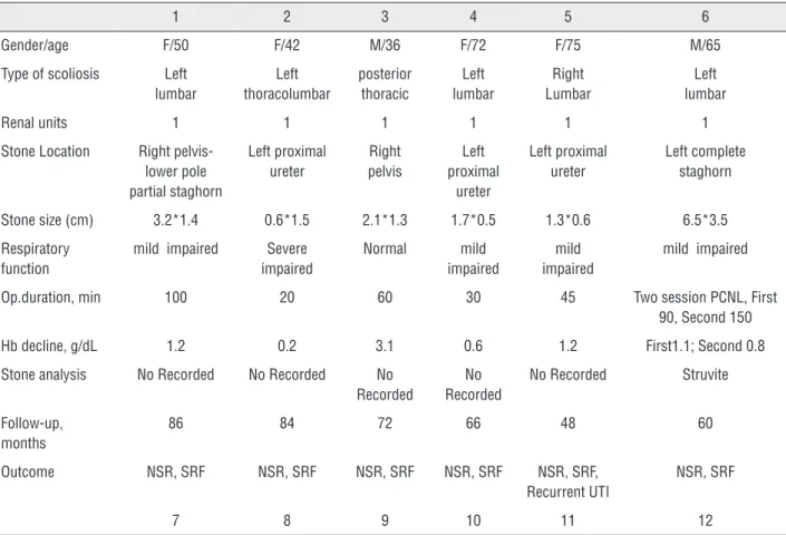
1 2 3 4 5 6
Gender/age F/50 F/42 M/36 F/72 F/75 M/65
Type of scoliosis Left lumbar
Left thoracolumbar
posterior thoracic
Left lumbar
Right Lumbar
Left lumbar
Renal units 1 1 1 1 1 1
Stone Location Right pelvis-lower pole partial staghorn
Left proximal ureter
Right pelvis
Left proximal
ureter
Left proximal ureter
Left complete staghorn
Stone size (cm) 3.2*1.4 0.6*1.5 2.1*1.3 1.7*0.5 1.3*0.6 6.5*3.5
Respiratory function
mild impaired Severe impaired
Normal mild impaired
mild impaired
mild impaired
Op.duration, min 100 20 60 30 45 Two session PCNL, First
90, Second 150
Types of calculi included 4 cases of upper ureteral calculi and 12 renal calculi, among whom 4 had pelvic calculi, 5 had partial staghorn calculi or multiple calculi and 3 complete staghorn calcu-li. The stone size (maximum diameter on the plain film) ranged from 0.7cm×1.1cm to 5.2cm×7.8cm.
All patients were preoperatively submit-ted to complete history, clinical examination, routine laboratory blood investigation, coagu-lation profile, urinalysis and culture, hepato--renal function, and complete evaluation of the respiratory and cardiovascular systems inclu-ding chest radiography or CT scan, pulmonary function tests, arterial blood gas analysis and echocardiography. Preoperative diagnostic eva-luation of the urinary tract consisted of kidneys, ureters, and bladder (KUB) x-ray or intravenous urography (IVU) combined with spiral CT wi-thout enhancement and ultrasonography (US). The renal US was performed with simulation of the percutaneous puncture to confirm the feasi-bility of establishing a percutaneous access.
Respiratory test showed mild restrictive ventilation dysfunction in 8 patients and severe restrictive ventilation dysfunction in 2 patients which were all preoperatively managed with inha-lation of compound ipratropium bromide solution. Urine culture demonstrated the colonization of E.coli in three patients and Pseudomonas aerugi-nosa in one patient and all were managed with sensitive antibiotics.
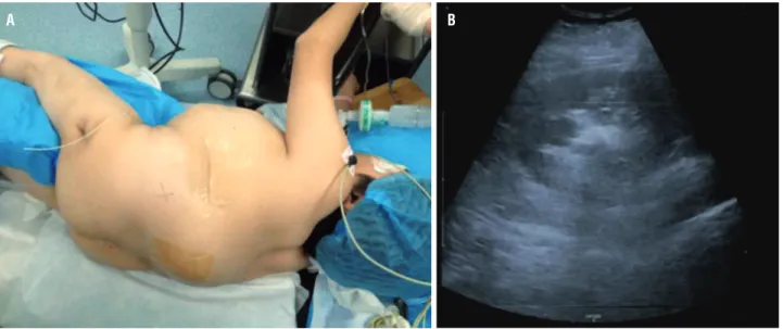
The surgical technique of the Chinese MPCNL has been described in previously publi-shed articles (4-6). Following general anesthesia and intubation, with the patient in a lithotomy position, a 5F open tip ureteral catheter was pla-ced in the ipsilateral ureter under cystoscopy. In 9 cases, the patient was then positioned in prone position with adequate protection to avoid com-pression of any spinal or bony protrusions. Two patient had to be repositioned in the lateral decu-bitus position (Figures 1-3) and two patients were placed in the supine position because decreased blood oxygen saturation in the prone position or
Figures 1a and 1b - A plain abdominal film (KUB) and IVU show a pelvic and lower calycial stone in left kidney with a good function in a posterior thoracolumbar spinal kyphoscoliosis complicated with chest deformity patient.
their spinal deformity prevented them to be placed in the prone position. A US-guided fluoroscopic confirmed puncture was performed to the most ap-propriate calyx based on preoperative imaging and patient positioning. After the puncture was confir-med successful, a 0.035-inch guidewire was inser-ted into the collecting system. The percutaneous tract was then dilated to 18F or 20F with fascial dilators (Cook Urological, Spencer, IN), and a same sized peel-away sheath was placed as the percuta-neous access port. Subsequently, a 8/9.8F semirigid ureteroscope (Richard Wolf, Knittlingen, Germany) or a 8.5/12.5F nephroscope (Lixun Nephroscope, Richard Wolf, Knittlingen, Germany) was used for nephroscopy. The stone was fragmented by pneu-matic lithotripsy or holmium:YAG laser. The larger fragments (0.3cm-0.5cm) were extracted with a 5F forceps (Richard Wolf, Knittlingen, Germany), and the fragments<0.3cm were mainly flushed out with
Figures 2a and 2b - The patient was placed in the lateral decubitus position for the spinal deformity prevented to be placed in the prone position (Figure-2a). A successful US-guided puncture was performed. The dotted line corresponds to the tract of needle (Figure-2b).
Figure 3 - KUB after MPCNL: the stone was removed completely and a ureter stent and a nephrostomy tube were placed in the left kidney.
Finally, a 5F or 6F Double-J stent was in-serted in the ureter, and a same caliber nephros-tomy tube (18F-20F) was left in the collecting sys-tem. If the patient presented with febrile urinary infection or purulent pelvic urine was aspirated during puncture, a nephrostomy tube was placed initially, and the MPCNL was performed after an-tibiotic treatment and drainage for 1 week.
KUB radiography, nephrostography or CT scan were performed 24 to 48 hours after surgery to assess the stone free status. In cases with sig-nificant residual stones, a second-staged MPCNL for stone size >2cm or flexible ureteroscopic li-thotripsy for stone size <2cm was performed 5 to 7 days later. The second stage MPCNL was perfor-med through a new percutaneous access tract if required. If a second-stage surgery was unneces-sary, the nephrostomy tube would be removed on postoperative day 3 to 5 when the drainage was clear, and the Double-J stent would be extracted 2 to 3 weeks postoperatively. The operative time was calculated from puncture to placement of the ne-phrostomy tube. The change in hemoglobin con-centration was estimated by comparing preopera-tive and 48 hours postoperapreopera-tive routine blood test.
RESULTS
A total of 19MPCNLs were performed in 16 kidneys of 13 patients affected by spinal deformi-ty, with an average 1.2 procedures in each kidney. Three kidneys needed two sessions of MPCNL, and 2 kidneys needed combined treatment with retrograde flexible ureterscopic lithotripsy. Multiple tracts were performed in 6 kidneys, with the average 1.38 tracts per kidney. In two patients a nephrostomy tube was placed for 1 week before the MPCNL as they had purulent pelvic urine. The average operative time of each MPCNL procedure was 67 (range 20-150) minutes. The mean hemoglobin drop was 1.0 (0.2-3.1) mg/dL and no blood transfusions were required. Radiography was performed on the second postope-rative day. Complete stone-free status was achieved in 14 kidneys. No complication was noted during or after surgery. Stone analysis was performed in 8 patients and showed calcium oxalate in 3 patients, calcium oxalate with uric acid and calcium phos-phate in two, and struvite in three.
The mean follow-up was 48 (3-86) months. A recurrent stone was detected in lower calyx in one patient but since it was asymptomatic no spe-cific management was done. The serum creatini-ne level was stable at 0.9-1.2mg/dL in all patients (normal 1.3mg/dL at our institutions). Recurrent UTI with the colonization of E. Coli was documen-ted by urine culture in two patients and were ma-naged with sensitive antibiotics.
DISCUSSION
The spinal column has four physiologi-cal curves in the sagittal plane, whereas no cur-ve should be obsercur-ved on the coronal plane. The curve to either side refers to spine scoliosis and the severe scoliosis is generally defined as a Cobb angle above 90º (6). Severe scoliosis may be as-sociated with distortion of the chest cavity, pelvis and compression of peritoneal organs, thus alte-ring the anatomical location of internal organs. In case of severe deformity the altered conforma-tion of the rib cage and restricted lung ventila-tion often cause respiratory dysfuncventila-tion. In this study group, 10 patients (77%) were found to have mild or severe restrictive ventilation dysfunction, so for these patients the preoperative pulmonary function tests and arterial blood gas analysis were performed.
The effect of altered anatomy to the urina-ry system may lie in possibly urinaurina-ry obstruction, further promoting formation of urinary calculi. Theoretically the risk of urinary stone disease is higher in these patients, with reported incidence rates of up to 20%. However, Vetter U et al. repor-ted an incidence of 4.7-6.9% (6/127 and 4/58, res-pectively) in children with osteogenesis imperfec-ta, which did not appear to differ from that seen in the general population (7, 8).
pathway with fluoroscopic guidance alone in such patients is relatively risky or less feasible, requiring the assistance of ultrasound or laparoscopy. The ul-trasound guidance allows the safe establishment of a PCN tract in a narrow safety margin of puncture due to the anatomic alteration or abnormal anato-mic structure and avoidance of injuring the neigh-boring organs. The experience of other authors has supported the superiority of US over fluoroscopy in guiding PCNL in special patients. Desai et al. succes-sfully treated nine patients with ectopic renal calculi by using US assisted PCNL puncture (9).
In the case of less experience in ultraso-nography-assisted percutaneous renal puncture or evident space-occupying organs present around the pre-established PCN pathway, laparoscopy-assisted PCNL can be used. The laparoscopic assistance allo-ws the intentional avoidance or separation of sur-rounding organs, further preventing the injuries of neighboring organs. In 1985, Eshghi et al. first des-cribed the technique of laparoscopy assisted PCNL for ectopic pelvic kidneys (10). Since then, several authors have reported successful experiences with the technique (11, 12). Recently, using the laparosco-pic method, Seref B et al. safely removed a 11.9mm stone from the pelvis of the right kidney in a pa-tient with osteogenesis imperfecta (13). For cases in which there is absence of obvious space-occupying organs around the pre-establishment of PCN access, we prefer ultrasonography-assisted puncture. In our extensive experience with the Chinese MPCNL ho-wever, we prefer US assisted puncture. Not only does this allow visualization of adjacent organs, thus mi-nimizing inadvertent injuries, but it also allows ac-curate puncture through a calyceal fornix, thereby reducing the intraoperative bleeding. Although the laparoscopy-assisted PCNL can be easily performed, it can’t ensure an accurate percutaneous calyceal pathway. In this group all 19 MPCNLs were perfor-med with this procedure and the mean hemoglobin drop for each MPCNL procedure was only 1.6mg/dL
system. In most of the cases the secondary tract was established meanwhile, but through the third puncture we always only put the guidewire in the collecting system without dilating initially. We only dilated the third puncture if the surgery was smoo-th and endoscopic manipulation time was no more than 90 minutes. We preferred that establishment of multiple tracts at the ontset of the surgery because there is no extravasation or bleeding allowing easy and accurate ultrasound-guided puncture. Obviou-sly, the potentially intraoperative leak or hemor-rhage would increase the difficulty in puncturing under the ultrasound and decrease the accuracy in establishing a new percutaneous tract. Furthermore, the simultaneous use of multiple tracts can acce-lerate the removal of stone fragment and shorten the operating time, in addition to reducing the risk of urosepsis by lowering the renal pelvic pressure (4, 14). Finally, it must be admitted that this pa-per is limited to a retrospective study; the collection of data prospectively may evaluate this technique more objectively.
CONCLUSIONS
CONFLICT OF INTEREST
None declared.
REFERENCES
1. Karabiyik L, Parpucu M, Kurtipek O. Total intravenous anaesthesia and the use of an intubating laryngeal mask in a patient with osteogenesis imperfecta. Acta Anaesthesiol Scand. 2002;46:618-9.
2. Goumas-Kartalas I, Montanari E. Percutaneous nephrolithotomy in patients with spinal deformities. J Endourol. 2010;24:1081-9.
3. Argyropoulos AN, Wines M, Tolley D. Case report: endourologic treatment for a ureteral stone in a patient with osteogenesis imperfecta. J Endourol. 2008;22:459-61. 4. Li X, He Z, Wu K, Li SK, Zeng G, Yuan J, et al. Chinese
minimally invasive percutaneous nephrolithotomy: the Guangzhou experience. J Endourol. 2009;23:1693-7. 5. He Z, Li X, Chen L, Zeng G, Yuan J. Minimally invasive
percutaneous nephrolithotomy for upper urinary tract calculi in transplanted kidneys. BJU Int. 2007;99:1467-71.
6. He Z, Zeng G, and Li X. Chinese Minimally Invasive Percutaneous Nephrolithotomy(MPCNL): Overcoming the Difficulties. In Al-Kandari AM, Desai M, Shokeir AA, Shoma AM, Smith AD, eds, Difficult Cases in Endourology.Chapt 10. London: Springer, 2003:97-106.
7. Vetter U, Maierhofer B, Müller M, Lang D, Teller WM, Brenner R, et al. Osteogenesis imperfecta in childhood: cardiac and renal manifestations. Eur J Pediatr. 1989;149:184-7.
8. Vetter U, Pontz B, Zauner E, Brenner RE, Spranger J. Osteogenesis imperfecta: a clinical study of the first ten years of life. Calcif Tissue Int. 1992;50:36-41.
9. Desai MR, Jasani A. Percutaneous nephrolithotripsy in ectopic kidneys. J Endourol. 2000;14:289-92.
10. Eshghi AM, Roth JS, Smith AD. Percutaneous transperitoneal approach to a pelvic kidney for endourological removal of staghorn calculus. J Urol. 1985;134:525-7.
11. Holman E, Tóth C. Laparoscopically assisted percutaneous transperitoneal nephrolithotomy in pelvic dystopic kidneys: experience in 15 successful cases. J Laparoendosc Adv Surg Tech A. 1998;8:431-5.
12. Troxel SA, Low RK, Das S. Extraperitoneal laparoscopy-assisted percutaneous nephrolithotomy in a left pelvic kidney. J Endourol. 2002;16:655-7.
13. Basal S, Ozgok Y, Tahmaz L, Atim A, Zor M, Bilgic S, et al. Extraperitoneal laparoscopy-assisted percutaneous nephrolithotomy in a patient with osteogenesis imperfecta. Urol Res.2011;39:73-6.
14. Guohua Z, Wen Z, Xun L, Wenzhong C, Yongzhong H, Zhaohui H, et al. The influence of minimally invasive percutaneous nephrolithotomy on renal pelvic pressure in vivo. Surg Laparosc Endosc Percutan Tech.2007;17:307-10.
_______________________ Correspondence address:
Zhaohui He, MD Minimally Invasive Surgery Center, The first affiliated hospital of Guangzhou Medical