ABSTRACT
Objective: To determine whether different levels of CPAP improve the lung volumes and capacities of healthy subjects immersed in water. Methods: This was a randomized clinical trial, conducted between April and June of 2016, involving healthy female volunteers who were using oral contraceptives. Three 20-min immersion protocols were applied: control (no CPAP); CPAP5 (CPAP at 5 cmH2O); and CPAP10 (CPAP at 10 cmH2O). We evaluated HR, SpO2, FVC, FEV1, the FEV1/FVC ratio, peak expiratory low rate (PEFR), and FEF25-75%) at three time points: pre-immersion; 10 min after immersion; and 10 min after the end of each protocol. Results: We evaluated 13 healthy volunteers. The CPAP10 protocol reversed the restrictive pattern of lung function induced by immersion in water, maintaining pulmonary volumes and capacities for a longer period than did the CPAP5 protocol. Conclusions: When the hemodynamic change causing a persistent lung disorder, only the application of higher positive pressures is effective in maintaining long-term improvements in the pulmonary proile.
Keywords: Physical therapy modalities; Noninvasive ventilation; Continuous positive airway pressure.
Impact of continuous positive airway
pressure on the pulmonary changes
promoted by immersion in water
Danize Aparecida Rizzetti1, Janayna Rodembuch Borba Quadros1,
Bruna Esmerio Ribeiro1, Letícia Callegaro1, Aline Arebalo Veppo2,
Giulia Alessandra Wiggers1, Franck Maciel Peçanha1
Correspondence to:
Danize Aparecida Rizzetti. BR-472, km 592, Caixa Postal 118, CEP 97500-970, Uruguaiana, RS, Brasil. Tel.: 55 55 3413-4321. E-mail: [email protected]
Financial support: None. INTRODUCTION
Immersion in water results in physiological alterations to various systems, including the musculoskeletal, renal, respiratory, and cardiovascular systems.(1) The hydrostatic
pressure compresses the thoracic cavity, reducing its circumference, which promotes cranial displacement of the diaphragm.(2) In healthy individuals, the cardiovascular
effects, which include increases in venous return, blood volume, and central venous pressure, result in increased cardiac output, greater left atrial diameter, and higher cardiac volume.(3-5)
Cardiorespiratory changes induced by immersion in water reduce the end-expiratory volume and the length-tension ratio of the respiratory muscles, decreasing their capacity to generate and maintain adequate force, thus reducing MIP and MEP.(2) Such changes also increase pulmonary
capillary pressure, which worsens lung function, reducing VC, FEV1, and functional residual capacity, thereby leading to the development of a restrictive pattern of lung function. (2,3,6) In fact, in clinical practice, many
cardiopulmonary diseases induce a restrictive pattern as a consequence of pulmonary congestion, via mechanisms similar to those promoted by immersion in water.
The therapeutic arsenal used by physiotherapists in the treatment of pulmonary congestion and its respiratory complications includes respiratory exercises, incentive spirometry, and noninvasive positive-pressure ventilation
(NPPV).(7,8) In a study involving the induction of restrictive
pulmonary characteristics by immersion in water, Vepo et al.(9) found that neither respiratory exercises nor
incentive spirometry were effective in normalizing lung function during the immersion.
Applying NPPV in the continuous positive airway pressure (CPAP) mode has been shown to improve the hemodynamic status of patients with pulmonary congestion due to heart failure, reducing cardiac preload and afterload, thus increasing lung compliance and lung volumes, as well as reducing the work of breathing.(10) Other studies
have shown that, in patients with heart disease, NPPV reduces venous return, pulmonary capillary pressure, and central venous pressure, consequently normalizing pulmonary function.(11,12)
Some therapeutic approaches do not reverse the restrictive pattern of lung function during the maintenance of hemodynamic changes in this model of pulmonary congestion induced by immersion in water, and there is evidence that NPPV, in CPAP mode, constitutes an important therapeutic tool for the treatment of this condition. It is therefore imperative to investigate the effects that NPPV has on the hemodynamic changes that induce the restrictive pattern characteristic of pulmonary congestion. Therefore, the objective of this study was to determine whether the use of NPPV in CPAP mode at various pressures improves lung volumes and capacities in healthy subjects immersed in water.
1. Curso de Fisioterapia, Universidade Federal do Pampa – UNIPAMPA – Uruguaiana (RS) Brasil.
2. Residência Multiproissional em Saúde, Universidade Federal do Pampa – UNIPAMPA – Uruguaiana (RS) Brasil.
Submitted: 24 March 2017. Accepted: 15 October 2017
METHODS
Study design and subject selection
This was a randomized clinical trial, conducted between April and June of 2016 at a federal university in the Brazilian state of Rio Grande do Sul. All procedures were performed in accordance with Brazilian National Health Council Resolution no. 466/12, and the study was approved by the research ethics committee of the institution (CAEE no. 56861216.7.0000.5323). All participating subjects gave written informed consent.
The sample, determined by sample size calculation for the analysis of quantitative variables of a proportion
of a population of ininite size, considering a 95% conidence level, a 5% maximum error of estimation, and an estimated prevalence of 50%, was composed
of volunteers who met the following criteria: being healthy; being female; being between 18 and 40 years of age; using oral contraceptives during the study period (to ensure homogeneity of the sample, considering that the use of oral contraceptives stabilizes the levels of female sex hormones, thus avoiding possible changes in lung function due to hormonal
luctuations occurring during the various phases of the
menstrual cycle); and having given written informed consent. We excluded subjects who, for any reason, did not participate in all stages of the study; subjects who were unable to perform the maneuvers involved in the assessment and the NPPV; subjects who showed some alteration during spirometry; subjects who presented cardiovascular, respiratory, neurological, or musculoskeletal diseases; subjects who were on medication (except oral contraceptives); and subjects who were smokers or former smokers.
Data collection
The volunteers were submitted to anamnesis (regarding general health status, associated diseases, and medication use) and physical examination (to determine weight, height, and body mass index), performed by a single evaluator according to the standardized criteria established by the International Society for the Advancement of Kinanthropometry. (13)
The subjects also underwent cardiopulmonary evaluation, in which HR, SpO2, systolic blood pressure, and diastolic blood pressure were determined in accordance with the recommendations made in the Fourth Brazilian Guidelines for the Management of Hypertension.(14) We analyzed pulmonary function by
spirometry (Koko Legend®; nSpire Health Inc., Hertford,
UK), in accordance with the criteria established by the American Thoracic Society/European Respiratory Society(15) and the Brazilian Thoracic Association,(16)
using the predicted values for FVC, FEV1, and the FEV1/
FVC ratio, as well as for the peak expiratory low rate
(PEFR) and FEF25-75%, determined by Pereira et al.(17)
We measured slow vital capacity with a respirometer (Wright Mark 8; Ferraris Respiratory, Louisville, CO, USA). We also measured MIP and MEP, using a manometer (MV 120; Comercial Médica, São Paulo,
Brazil), as described by Neder et al.(18) These evaluations
were performed while the subjects were seated, as recommended in the Brazilian Thoracic Association guidelines.(16)
Intervention protocols
Each volunteer was immersed in water up to the level of the xiphoid process, after which NPPV, in CPAP mode (BiPAP®; Respironics, Murrysville, PA, USA), was applied
via face mask. In this immersion model, the reduction in pulmonary function is mainly due to cardiovascular alterations, which, in healthy individuals, simulate a restrictive pattern of lung function, as is typically observed in certain cardiorespiratory complications, such as pulmonary congestion.(9) Vepo et al.(9) employed this
model and reported that, by 10 min after immersion in water, subjects showed reductions in FVC and FEV1 induced by hemodynamic changes, including increased blood volume and higher central venous pressure.
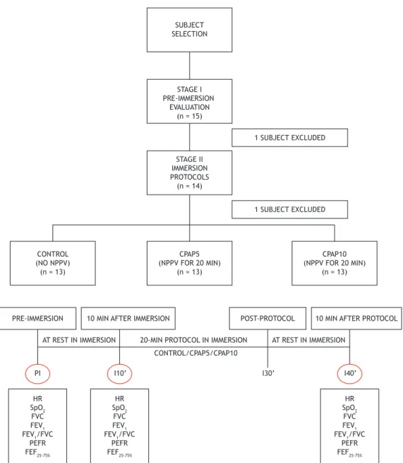
All subjects participated in three NPPV immersion protocols, with an interval of at least 24 h between
each application, in a sequence deined by block
randomization, as shown in Figure 1. The protocols lasted for 20 min each and were given the following designations: control—during spontaneous breathing, at rest and without ventilatory support; CPAP5—CPAP of 5 cmH2O, according to Reis et al.(19); and CPAP10—CPAP
of 10 cmH2O, according to Bellone et al.(20) During the
application of each protocol, the variables FC, SpO2, FVC, FEV1, FEV1/FVC ratio, PEFR, and FEF25-75% were evaluated at the following time points (Figure 1): before immersion in water (pre-immersion); at 10 min after immersion in water; and at 10 min after the end of the protocol (i.e., 40 min after the initiation of the protocol).
Statistical analysis
The results are expressed as means and standard deviations. The Shapiro-Wilk test was used in order to determine the distribution of the data, which was found to be normal for all of the variables analyzed. The data were analyzed by one-way repeated-measures ANOVA and Fisher’s post-hoc test, as appropriate. Values of
p < 0.05 were considered signiicant. The program
GraphPad Prism, version 6.01 (GraphPad Inc., San Diego, CA, USA) was used for the statistical analyses and for the creation of the graphics.
RESULTS
Of the 15 volunteers evaluated, 2 were excluded: for presenting with spirometric changes at the initial evaluation (n = 1); and for not participating in all of
the stages of data collection (n = 1). The inal sample
At 10 min after immersion in water, all of the subjects showed reductions in FVC and FEV1, suggesting a restrictive alteration to the pulmonary pattern, as previously observed by Vepo et al.(9) Those reductions
persisted at 10 min after the end of the control protocol and at 10 min after the end of the CPAP5 protocol. However, the pulmonary changes induced by immersion were normalized after application of the CPAP10 protocol (Figure 2).
The FEV1/FVC ratio also showed a reduction at 10 min after immersion, in all subjects and in all three protocols. In accordance with the reductions in FVC and FEV1, the reduction in the FEV1/FVC ratio persisted at 10 min after the application of the control and CPAP5 protocols, as well as being reversed after the application of the CPAP10 protocol (Figure 3). The
PEFR and FEF25-75% values were also decreased at 10 min after immersion in water. At 10 min after the end of the CPAP5 and CPAP10 protocols, the PEFR values were normalized, although neither protocol was able to normalize the FEF25-75% values (Table 2).
In the cardiorespiratory evaluation, a reduction in HR was observed at 10 min after immersion in water, and that reduction persisted at all of the evaluation time points, for all of the protocols. However, no changes were observed for SpO2, which remained at basal values after immersion and at all other evaluation time points, for all of the protocols (Table 2).
DISCUSSION
In the present study, we demonstrated that the application of CPAP at 10 cmH2O can restore the
SUBJECT SELECTION
STAGE I PRE-IMMERSION
EVALUATION (n = 15)
STAGE II IMMERSION PROTOCOLS (n = 14)
1 SUBJECT EXCLUDED
1 SUBJECT EXCLUDED
CPAP5 (NPPV FOR 20 MIN)
(n = 13)
CPAP10 (NPPV FOR 20 MIN)
(n = 13) CONTROL
(NO NPPV) (n = 13)
10 MIN AFTER IMMERSION
I10’ I30’ I40’
PRE-IMMERSION POST-PROTOCOL 10 MIN AFTER PROTOCOL
PI
HR SpO2 FVC FEV1 FEV1/FVC
PEFR FEF25-75%
HR SpO2
FVC FEV1 FEV1/FVC
PEFR FEF25-75%
HR SpO2
FVC FEV1 FEV1/FVC
PEFR FEF25-75% CONTROL/CPAP5/CPAP10
AT REST IN IMMERSION 20-MIN PROTOCOL IN IMMERSION AT REST IN IMMERSION
Figure 1. Study design and outline of the application of non-invasive positive pressure ventilation protocols (VNIPP).
restrictive pattern of lung function induced by immersion in water in healthy individuals, maintaining pulmonary volumes and capacities at normal levels for a longer period when compared with CPAP at 5 cmH2O. To our
knowledge, this is the irst study suggesting that,
in situations in which a hemodynamic change that causes a restrictive pattern of lung function persists, the application of higher positive pressures is an effective means of maintaining improvements in the
pulmonary proile.
Hemodynamic changes during immersion in water occur in part by hemodilution due to the transfer of
luid—from the interstitial spaces to the intravascular
spaces—within the lower limbs.(5,21) In the respiratory
system, those changes promote reductions in pulmonary volumes and capacities, characteristic of a restrictive pattern.(22)
In clinical practice, pulmonary complications due to cardiovascular changes lead to a restrictive pulmonary pattern, mainly due to the increase in intrathoracic blood volume. In patients with heart disease, pulmonary congestion, which is characterized by elevated pulmonary and systemic venous pressures, is usually related to ventricular dysfunction and a reduction in cardiac output.(23,24) Left untreated, pulmonary
congestion can progress to pulmonary edema, which constitutes a medical emergency.(23-25)
The model of a restrictive pattern of lung function used in the present study, as previously described by
Vepo et al.,(9) uses immersion in water to promote
increased central blood volume in healthy subjects and, consequently, hemodynamic changes similar to those developed in patients with pulmonary congestion. Thus,
this model allows the analysis and clariication of the
effects of CPAP exclusively on changes in lung volumes and capacities promoted by isolated hemodynamic factors, avoiding the risk that associated diseases will
inluence the indings.
After applying CPAP to subjects immersed in water, we found that only the use of a CPAP level of 10 cmH2O provided sustained improvements in lung volumes and capacities. This suggests that when the hemodynamic changes that contribute to congestive pulmonary disease are not controlled, higher positive pressures are required in order to maintain improvements in lung function. Likewise, Vepo et al.(9) observed no
improvements in lung volumes and capacities when respiratory exercises and incentive spirometry were applied in subjects immersed in water, suggesting that these techniques do not provide the expected
beneits when the primary hemodynamic change is
not controlled or reversed.
Although pharmacotherapy, with diuretics and vasodilators, is an essential tool in combating fluid retention and relieving the respiratory symptoms of pulmonary congestion,(24,26,27) the use
of pharmacotherapy in combination with positive pressure has become increasingly more widespread as a means of normalizing the hemodynamic status of patients with pulmonary congestion and of preventing its recurrence. Therefore, CPAP is a mode of ventilation applied in a wide spectrum of clinical situations(28) and
is often indicated for patients in critical condition, such as those with acute cardiogenic pulmonary edema (ACPE). In such cases, the Brazilian Guidelines on Mechanical Ventilation(28) recommend the use of CPAP
at 5-10 cmH2O, which justiies the choice of pressures employed in the present study.
The pulmonary beneits derived from CPAP are
due, in large part, to the elevation of intrathoracic pressure and consequent reduction of left ventricular afterload promoted by the pressurization of the airways. That pressurization consequently increases cardiac output, promoting improvements in lung volumes and capacities, thus reducing respiratory distress.(11,12) The hemodynamic effects of CPAP have
been fully elucidated for the population of patients with pulmonary hypertension due to left ventricular dysfunction. However, our study demonstrated that, when the cause of the hemodynamic imbalance persists, the improvement in pulmonary function is maintained only when higher CPAP is applied to the airways, regardless of the pulmonary improvements observed immediately after application of the technique.
Although numerous studies have involved the application of CPAP for the treatment of cardiogenic pulmonary edema, using the same range of positive pressures employed in the present study, those studies have varied in terms of the effects that such pressures
Table 1. Initial physical, respiratory and cardiovascular evaluation of the subjects evaluated (n = 13).a
Parameter Value
Age, years 23.6 ± 5.5
Height, m 1.60 ± 0.02
Weight, kg 63.7 ± 11.6
BMI, kg/m2 23.6 ± 3.5
HR, bpm 76.2 ± 6.4
SBP, mmHg 120.5 ± 8.6
DBP, mmHg 71.9 ± 7.7
SpO2, % 98.4 ± 0.8
MIP, cmH2O 98.5 ± 26.3
MEP, cmH2O 103.1 ± 26.5
SVC, L 3.3 ± 0.3
SVC, % of predicted 82.2 ± 3.0
FVC, L 3.9 ± 0.6
FVC, % of predicted 98.9 ± 11.4
FEV1, l 3.5 ± 0.5
FEV1,% of predicted 99.9 ± 10.7
FEV1/FVC 0.9 ± 0.1
FEV1/FVC, % 98.0 ± 7.0
PEFR, L/min 8.4 ± 1.7
PEFR, % of predicted 106.2 ± 16.3
FEF25-75%, L/min 4.4 ± 1.1
FEF25-75%, % of predicted 95.2 ± 19.9 BMI: body mass index; SBP: systolic blood pressure; DBP: diastolic blood pressure; SVC: slow vital capacity;
PEFR: peak expiratory low rate. aValues expressed as
were found to have on mortality, ICU admission, and length of hospital stay, as well as in terms of the reported advantages of CPAP over conventional therapy.(20,29,30)
It is known that CPAP increases survival and precludes the need for endotracheal intubation in patients with
ACPE. (29-31) However, in a randomized controlled study
involving patients with ACPE, no differences were found between the patients receiving conventional oxygen therapy and those receiving CPAP in terms of the short- or long-term mortality rates.(32) The
Table 2. Cardiovascular and respiratory parameters evaluated in the immersion protocols.a
Parameter Control CPAP5 CPAP10
PI I10’ I40’ PI I10’ I40’ PI I10’ I40’
HR, bpm 93.5 ±
15.1 78.8 ± 11.2* 79.8 ± 10.5* 101.3 ± 13.9 84.2 ± 10.4* 80.4 ± 13.5* 98.5 ± 14.4 82.2 ± 8.7* 78.4 ± 12.1* SpO2, % 98.8 ± 1.0 98.5 ± 1.1 98.8 ± 0.9 98.3 ± 0.6 98.6 ± 0.9 98.8 ± 0.7 98.5 ± 1.7 98.5 ± 1.2 98.3 ± 1.9 PEFR, L/min 7.06 ± 1.3 7.0 ± 1.3* 6.9 ± 1.2 7.5 ± 1.1 7.2 ± 1.2* 7.3 ± 1.1 7.1 ± 1.0 6.6 ± 1.3* 6.9 ± 1.2 FEF25-75%, L/min 4.1 ± 0.7 3.9 ± 0.8* 3.8 ± 0.7* 4.3 ± 0.7 3.9 ± 0.6* 3.8 ± 0.6* 3.9 ± 0.8 3.9 ± 0.8 3.8 ± 0.7 CPAP5: CPAP of 5 cmH2O; CPAP10: CPAP of 10 cmH2O; PI: pre-immersion; I10’: 10 min after immersion; I40’: 10
min after the end of the (20-min) protocol; and PEFR: peak expiratory low rate. aValues expressed as mean ± SD. *p < 0.05 vs. PI (one-way repeated-measures ANOVA and Fisher’s post-hoc test).
5 4 3 2 1 0
PI I10’ I40’
F V C ( L) F E V1 ( L) F E V1 ( L) F E V1 ( L) A * * 5 4 3 2 1 0
PI I10’ I40’
F V C ( L) B * * 5 4 3 2 1 0
PI I10’ I40’
F V C ( L) C * *
PI I10’ I40’ Control Time point D * * 5 4 3 2 1 0 5 4 3 2 1 0
PI I10’ I40’ CPAP5 Time point E * * 5 4 3 2 1 0
PI I10’ I40’ CPAP10
Control CPAP5 CPAP10
Time point
Time point Time point Time point
F * 1.0 0.8 0.6 0.4 0.2 0.0
PI I10’ I40’
F E V1 / F V C r a ti o Control Time point A
* * 1.0
0.8
0.6
0.4
0.2
0.0
PI I10’ I40’
F E V1 / F V C r a ti o CPAP5 Time point B
* * 1.0
0.8
0.6
0.4
0.2
0.0
PI I10’ I40’
F E V1 / F V C r a ti o CPAP10 Time point C *
Figure 3. FEV1/FVC ratio in the control protocol (in A), the CPAP5 (5 cmH2O) protocol (in B), and the CPAP10 (10 cmH2O) protocol (in C), at the time points evaluated (data expressed as mean ± SD). CPAP: continuous positive airway pressure; PI: pre-immersion; I10’: 10 min after immersion; I40’: 10 min after the end of the (20-min) protocol. *p < 0.05 vs. PI (one-way repeated-measures ANOVA and Fisher’s post-hoc test).
divergences across studies are probably attributable to differences among the patient samples in terms of the associated clinical factors and the pharmacological interventions administered in combination with the CPAP. Both situations prevent the isolated analysis of the effects of CPAP on respiratory changes resulting from hemodynamic disturbances.
The reductions we observed in the other spirometric parameters, such as FEF25-75% and PEFR, after immersion in water, were also reported by Vepo et al.(9) Those
authors reported that FEF25-75% is directly dependent on the FVC values, its reduction therefore being expected during immersion. In addition, this parameter is known to be related to the distal airway permeability,(33)
and it is reduced in proportion with the increase in intrathoracic blood volume and pulmonary capillary pressure observed in this model.
Regarding the cardiorespiratory parameters investigated, we demonstrated a reduction in HR in all
of the protocols applied, which corroborates the indings
of Kaneko et al.,(34) who employed a combination of
CPAP and drug therapy in patients with ACPE. However, in our study, we were unable to associate that change with the application of positive airway pressure, because the same HR behavior was also observed in the control protocol. It has been suggested that the change observed results exclusively from immersion
in water.(35,36) The application of CPAP at 10 cmH 2O
has been shown to be superior to the conventional treatment for improving hypoxemia in patients with ACPE.(37) However, our study did not ind signiicant
differences between the CPAP protocols or the time points evaluated in terms of the SpO2, which could be related to the fact that our subjects were healthy and presented with normal SpO2 values. It should be noted that our subjects reported fatigue after the application of the CPAP10 protocol, which might be attributable to the fact that they had normal pulmonary function and therefore had a lower tolerance to higher positive airway pressures. That might have limited the performance of the study participants during the spirometric evaluation performed immediately after the application of CPAP.
In summary, the indings of the present study suggest
that the use of higher CPAP pressures is effective in maintaining improvements in lung volumes and capacities in situations in which the hemodynamic change causing the restrictive lung pattern persists.
Our indings underscore the importance of adequately
controlling the primary hemodynamic cause of pulmonary congestion, so that the combination of physiotherapeutic techniques and clinical practice will provide the maximum possible improvement of the respiratory changes present in this condition, for a longer period of time.
REFERENCES
1. Sá NC, Banzato TC, Sasseron AB, Ferracini LC, Fregadolli P, Figueiredo LC. Análise comparativa da função respiratória de indivíduos hígidos em solo e na água. Fisioter Pesq. 2010;17(4):337-41. https://doi.org/10.1590/S1809-29502010000400010
2. de Andrade AD, Júnior JC, Lins de Barros Melo TL, Rattes Lima
CS, Brandão DC, de Melo Barcelar J. Inluence of different levels of
immersion in water on the pulmonary function and respiratory muscle pressure in healthy individuals: observational study. Physiother Res Int. 2014;19(3):140-6. https://doi.org/10.1002/pri.1574
3. Yamashina Y, Yokoyama H, Naghavi N, Hirasawa Y, Takeda R, Ota A, et al. Forced respiration during the deeper water immersion causes the greater inspiratory muscle fatigue in healthy young men. J Phys Ther Sci. 2016;28(2):412-8. https://doi.org/10.1589/jpts.28.412
4. Risch WD, Koubenec HJ, Beckmann U, Lange S, Gauer OH. The effect of graded immersion on heart volume, central venous pressure,
pulmonary blood distribution, and heart rate in man. Plügers Arch.
1978;374(2):115-8. https://doi.org/10.1007/BF00581289
5. Johansen LB, Bie P, Warberg J, Christensen NJ, Norsk P. Role of hemodilution on renal responses to water immersion in humans. Am J Physiol. 1995;269(5 Pt 2):R1068-76.
6. Fagundes AA. Efeitos da imersão em água aquecida sobre o sistema respiratório. Fisioter Mov. 2006;19(4):113-8.
7. Lima IN, Fregonezi GA, Rodrigo M, Cabral EE, Aliverti A, Campos TF, et al. Acute effects of volume-oriented incentive spirometry on chest wall volumes in patients after stroke. Respir Care. 2014;59(7):1101-7. https://doi.org/10.4187/respcare.02651
8. Lunardi AC, Porras DC, Barbosa RC, Paisani DM, Marques da Silva
CC, Tanaka C, et al. Effect of volume-oriented versus low-oriented
incentive spirometry on chest wall volumes, inspiratory muscle activity, and thoracoabdominal synchrony in the elderly. Respir Care. 2014;59(3):420-6. https://doi.org/10.4187/respcare.02665
9. Vepo AA, Martinez CS, Wiggers GA, Peçanha FM. Incentive spirometry and breathing exercises were not able to improve restrictive pulmonary characteristics induced by water immersion in healthy subjects. Int J Physiother Res. 2016;4(2):1415-22. https://doi. org/10.16965/ijpr.2016.109
10. Wahab R, Basner RC. Nocturnal non-invasive ventilation for
cardio-respiratory disorders in adults. Expert Rev Respir Med. 2013;7(6):615-29. https://doi.org/10.1586/17476348.2013.839246
11. Naughton MT, Rahman MA, Hara K, Floras JS, Bradley TD. Effect of continuous positive airway pressure on intrathoracic and left ventricular transmural pressures in patients with congestive heart failure. Circulation. 1995;91(6):1725-31. https://doi.org/10.1161/01. CIR.91.6.1725
12. Bento AM, Cardoso LF, Tarasoutchi F, Sampaio RO, Kajita LJ, Lemos Neto PA. Hemodynamic Effects of Noninvasive Ventilation in Patients with Venocapillary Pulmonary Hypertension. Arq Bras Cardiol. 2014;103(5):410-417. https://doi.org/10.5935/abc.20140147
13. International Society for the Advancement of Kinanthropometry (ISAK). International Standards for Anthropometric Assessment. New Zealand: Lower Hutt; 2011.
14. Sociedade Brasileira de Cardiologia; Sociedade Brasileira de Hipertensão; Sociedade Brasileira de Nefrologia. VI Brazilian Guidelines on Hypertension [Article in Portuguese]. Arq Bras Cardiol. 2010;95(1 Suppl):1-51.
15. Miller MR, Hankinson J, Brusasco V, Burgos F, Casaburi R, Coates A, et al. Standardisation of spirometry. Eur Respir J. 2005;26(2):319-38. https://doi.org/10.1183/09031936.05.00034805
16. Pereira CA. Espirometria. J Pneumol. 2002;28(Suppl 3):S1-S82.
17. Pereira CA, Sato T, Rodrigues SC. New reference values for forced spirometry in white adults in Brazil. J Bras Pneumol. 2007;33(4):397-406. https://doi.org/10.1590/S1806-37132007000400008
18. Neder JA, Andreoni S, Lerario MC, Nery LE. Reference values for lung function tests: II. Maximal respiratory pressures and voluntary ventilation. Braz J Med Biol Res. 1999;32(6):719-27. https://doi. org/10.1590/S0100-879X1999000600007
19. Reis HV, Borghi-Silva A, Catai AM, Reis MS. Impact of CPAP on physical exercise tolerance and sympathetic-vagal balance in patients with chronic heart failure. Braz J Phys Ther. 2014;18(3):218-27. https://doi.org/10.1590/bjpt-rbf.2014.0037
21. Larsen AS, Johansen LB, Stadeager CJ, Warberg NJ, Christensen NJ, Norsk P. Volume-homeostatic mechanisms in humans during graded water immersion. J Appl Physiol (1985). 1994;77(6):2832-9.
22. Hsia CC. Cardiopulmonary limitations to exercise in restrictive lung disease. Med Sci Sports Exerc. 1999;31(1 Suppl):S28-32. https://doi. org/10.1097/00005768-199901001-00005
23. Gheorghiade M, Filippatos G, De Luca L, Burnett J. Congestion in acute heart failure syndromes: an essential target of evaluation and treatment. The Am J Med. 2006;119(12 Suppl 1):S3-S10. https://doi. org/10.1016/j.amjmed.2006.09.011
24. Felker GM, Mentz RJ. Diuretics and ultrailtration in acute
decompensated heart failure. J Am Coll Cardiol. 2012;59(24):2145-53. https://doi.org/10.1016/j.jacc.2011.10.910
25. Bitter T, Fox H, Schmalgemeier H, Wellmann B, Zwenke A, Spiesshöfer J, et al. Acute improvement of pulmonary hemodynamics does not alleviate Cheyne-Stokes respiration in chronic heart failure-a randomized, controlled, double-blind, crossover trial. Sleep Breath. 2016; 20(2):795-804. https://doi.org/10.1007/s11325-015-1300-1
26. Vazir A, Cowie MR. Decongestion: Diuretics and other therapies for hospitalized heart failure. Indian Heart J. 2016; 68 Suppl 1:S61-8. https://doi.org/10.1016/j.ihj.2015.10.386
27. Klein L. Treating hemodynamic congestion is the key to prevent heart failure hospitalizations. JACC Heart Fail. 2016;4(5):345-7. https://doi. org/10.1016/j.jchf.2016.03.004
28. Barbas CS, Isola AM, Farias AM, Cavalcanti AB, Gama AM, Duarte AC, et al. Brazilian recommendations of mechanical ventilation 2013. Part I. Rev Bras Ter Intensiva. 2014;26(2):89-121. https://doi. org/10.5935/0103-507X.20140017
29. Masip J, Betbesé AJ, Páez J, Vecilla F, Cañizares R, Padró J, et al. Non-invasive pressure support ventilation versus conventional oxygen therapy in acute cardiogenic pulmonary oedema: a randomised trial. Lancet. 2000;356(9248):2126-32. https://doi. org/10.1016/S0140-6736(00)03492-9
30. Crane SD, Elliott MW, Gilligan P, Richards K, Gray AJ. Randomised controlled comparison of continuous positive airways pressure,
bilevel non-invasive ventilation, and standard treatment in emergency department patients with acute cardiogenic pulmonary oedema. Emerg Med J. 2004;21(2):155-61. https://doi.org/10.1136/ emj.2003.005413
31. Peter JV, Moran JL, Phillips-Hughes J, Graham P, Bersten AD. Effect of non-invasive positive pressure ventilation (NIPPV) on mortality in patients with acute cardiogenic pulmonary oedema: a meta-analysis. Lancet. 2006;367(9517):1155-63. https://doi.org/10.1016/S0140-6736(06)68506-1
32. Liesching T, Nelson DL, Cormier KL, Sucov A, Short K, Warburton R, et al. Randomized trial of bilevel versus continuous positive airway pressure for acute pulmonary edema. J Emerg Med. 2014; 46(1):130-40. https://doi.org/10.1016/j.jemermed.2013.08.015
33. Dompeling E, van Schayck CP, Molema J, Akkermans R, Folgering H, van Grunsven PM, et al. A comparison of six different ways of expressing the bronchodilating response in asthma and COPD; reproducibility and dependence of prebronchodilator FEV1. Eur Respir J. 1992;5(8):975-81.
34. Kaneko Y, Floras JS, Usui K, Plante J, Tkacova R, Kubo T, et al. Cardiovascular effects of continuous positive airway pressure in patients with heart failure and obstructive sleep apnea. N Engl J Med. 2003; 348(13):1233-41. https://doi.org/10.1056/NEJMoa022479
35. Srámek P, Simecková M, Janský L, Savlíková J, Vybíral S. Human physiological responses to immersion into water of different temperatures. Eur J Appl Physiol. 2000;81(5):436-42. https://doi. org/10.1007/s004210050065
36. de Oliveira Ottone V, de Castro Magalhães F, de Paula F, Avelar NC, Aguiar PF, da Matta Sampaio PF, et al. The effect of different water immersion temperatures on post-exercise parasympathetic reactivation. PLoS One. 2014;9(12):e113730. https://doi.org/10.1371/ journal.pone.0113730