Effect of Ventilatory Support on Functional Capacity in Patients with
Heart Failure: a Pilot Study
Eugênia da Silva lima, Cristiano Gonçalves Cruz, Fabiane Costa Santos, Mansueto Gomes-Neto, Hugo Souza
Bittencourt, Francisco José Farias Borges dos Reis, Roque Aras, Armênio Costa Guimarães, Erenaldo de Souza
Rodrigues-Junior
Faculdade Social da Bahia, Hospital Ana Nery, Salvador, BA - Brazil
Mailing address: Eugenia da Silva Lima •
Conj Colinas de Periperi Bl 19 Apt 302 - Periperi - 40725-430 - Salvador, BA - Brazil
E-mail: [email protected]
Manuscript received January 26, 2010; revised manuscript received May 21, 2010; accepted July 05/07/10.
Abstract
Background: Heart failure (HF) is an important public health problem, of which main clinical symptoms are dyspnea and fatigue. Noninvasive ventilatory support has been used as adjuvant therapy in cardiac rehabilitation in order to improve the functional capacity of patients.
Objective: To evaluate the functional capacity of patients with HF submitted to ventilatory support.
Methods: We evaluated the sociodemographic information, as well as data on quality of life, blood pressure (BP), peripheral oxygen saturation (SpO2), dyspnea, lactate concentration before and after the 6-minute walk test (6MWT) and the distance walked by patients of both sexes with chronic heart failure (CHF), with left ventricular ejection fraction
(LVEF) ≤ 45.0% , randomized in two groups: control and CPAP (the group used CPAP - 10 cmH2O for 30 minutes).
Results: A total of 12 patients, of which 8 were males, with CHF functional class II and III (NYHA) participated in the study. The patients had mean LVEF of 35.3 ± 8.7 and mean age was 46.3 ± 10.3 years. When comparing the control group with the CPAP group at the end of the 6th minute, there was a significant difference between the groups regarding SpO2 values (Control: 93.6 ± 1.5 % vs CPAP: 96.1±1.8%; p = 0.027), index of dyspnea (Control: 13.1 ± 1.16 vs CPAP: 11 ± 0.8; p = 0.009), lactate concentration (Control: 3.3 ± 0.7 mmol/l vs CPAP: 2.3 ± 0.5 mmol/l; p = 0.025) and distance walked at the 6MWT (Control: 420.6 ± 73.8 m vs CPAP: 534 ± 89.91 m; p = 0.038).
Conclusion: The previous use of the CPAP had beneficial effects on SpO2, index of dyspnea, lactate concentration, double product and the distance walked at the 6MWT in patients with CHF when performing the 6MWT. (Arq Bras Cardiol 2011;96(3):227-232)
Keywords: Heart failure; cardiac rehabilitation; patient care; vital capacity; respiration, artificial.
In the cardiac rehabilitation (CR) scenario, the individual’s physical conditioning is the objective, aiming at improving the functional capacity and promoting the return to the work and social activities, where the physical performance is a limiting factor5,6. One of the main aggravating factors is the
development of acute respiratory failure due to the decrease in lung compliance, which increases the respiratory workload7.
Such decrease is the result of the circulatory failure caused by the left ventricular (LV) dysfunction, which can cause acute and/or chronic damage to the respiratory function8.
In this scenario, the use of noninvasive ventilatory support (NIVS) has arisen as an adjuvant therapy in cardiac rehabilitation in an attempt to improve the functional capacity of patients, as the NIVS decreases the respiratory workload, improves oxygenation, increases lung compliance associated with the improvement in the ejected volume, due to the increase in the intrathoracic pressure9.
The use of NIVS has been an alternative in the attempt to increase arterial oxygenation and provide better tolerance to physical exercise, due to its significant effect on the cardiorespiratory interaction, resulting in a better cardiac and
Introduction
Heart failure (HF) is an important public health problem1,
of which dyspnea and fatigue during physical exercise or activities of daily living are the main clinical symptoms. It is the leading cause of early interruption of physical activity, resulting in restrictions of the daily activities and consequent limitation in functional capacity2,3.
respiratory response during exercise. However, few studies have taken into account the use of NIVS, associated or not to exercise in HF in the context of CR10.
Therefore, the present study aims to evaluating the functional capacity of patients with HF submitted to NIVS.
Methods
We evaluated patients of both sexes, with a diagnosis of CHF (New York Heart Association [NYHA] Functional Class II and III), referred to the Department of Ergospirometry and Cardiovascular Rehabilitation of Hospital Ana Nery - UFBA. The present study included patients with left ventricular ejection fraction (LVEF) ≤ 45.0% (determined by echocardiography), aged 18 years or older, conscious and neurologically capable of communication and locomotion. Patients that presented limitations to physical activity due to factors other than exertional dyspnea and fatigue, such as intermittent claudication, lower-limb arthrosis and arthritis, psychiatric disease, orovalvular disease of rheumatic origin, anemia, chronic obstructive pulmonary disease (COPD), any febrile condition or infectious disease and those that presented intolerance to NIVS, were excluded from the study.
Initially, an interview was carried out for data collection and sample characterization, taking into account the management of HF and among them, gender, body mass index (BMI), blood pressure (BP) and the functional classification according to the NYHA.
Subsequently, the Minnesota Living with Heart Failure Questionnaire was applied to assess quality of life (QoL), which is a disease-specific tool that evaluates the patient’s perception regarding the influence of HF on the physical, socioeconomic and psychological aspects11.
Another important consideration about this tool is its comprehensive format, the fact that it is easy to apply and also the fact that it has been validated for the Brazilian population12.
The participants answered the 21 items using a 6-point answer scale (0-5). The summary of the total score (global score) can range from 0 to 105; a lower score reflects a better QoL. The scores in the three subscales (dimensions) reflect physical (questions number 2, 3, 4, 5, 6, 7, 12 and 13) and emotional difficulties (questions number 17, 18, 19, 20 and 21) and the other items are associated with financial considerations, side effects of medications and lifestyle (general dimensions). Subsequently, the patients were randomly allocated to two groups by drawing lots, by placing blue and red ribbons inside opaque envelopes. Patients that chose an envelope containing a blue ribbon were allocated in the CPAP group, which underwent continuous positive airway pressure (CPAP) at 10 cmH2O for 30 minutes, before the six-minute walk test
(6MWT) and those that chose the envelopes containing the red ribbon were allocated in the Control group, which was not submitted to CPAP. The patients were not aware of which among them had been submitted to CPAP, as the groups were submitted to the procedures on different days.
A blood sample was collected from the tip of the index finger of the right hand of the patients, using sterile and disposable lancets (Accu-Chek™ Softclix™ Pro, Roche,
Germany), after a 10-minute rest in the sitting position before the 6MWT (for the CPAP group, before the CPAP was applied) and at the end of the 6th minute of the 6MWT for lactate
concentration measurement using Accutrend™ Lactimeter (Roche, Germany).
The 6MWT was carried out according to the criteria established by the American Thoracic Society (ATS)13 and the
guidelines for ergometric evaluation established by the Brazilian Society of Cardiology (SBC)14, on a 30-meter long walking
course, without follow-up and with the patient receiving only the standardized instructions at every minute. The patients were monitored regarding heart rate (HR, bpm), blood pressure (BP, mmHg), peripheral oxygen saturation (SpO2, %), subjective sensation of dyspnea (Borg scale)15 and the values
were recorded at rest and at the 2nd, 4th and 6th minutes.
The study was approved by the Committee of Ethics in Research of Hospital Ana Nery - UFBA, protocol # 37/09 and all patients that agreed to participate signed the Free and Informed Consent Form.
Descriptive and frequency statistics were used to analyze the demographic and clinical data; the Shapiro-Wilks test was used for the normality and homogeneity of the variables, using the SPSS (Statistical Package for the Social Sciences) for Windows software package (release 13.0). The Student’s t test was used to analyze parametric data and for variables with non-parametric distribution. Wilcoxon’s test was used to compare the differences of intragroup means. When comparing between the two groups, for parametric data, Student’s t test was used for independent samples and the Mann-Whitney test was used for the nonparametric ones. The level of significance was set at 5.0%.
Results
A total of 12 patients with NYHA FC II and III CHF participated in the present study. The patients had a mean LVEF of 35.3 ± 8.7 and 8 were males. The mean age was 46.3 ± 10.3 years and the mean BMI was 25.1 ± 5.5 kg/ m2. The data of the clinical characterization of the sample is
shown in Table 1.
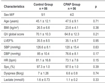
Table 2 shows the comparison of the clinical characteristics of the patients per group (Control group vs. CPAP group) and no significant difference was observed among the variables studied between the groups.
In the control group, when the variables at rest when compared to those at the 6th minute, a significant difference
was observed for the hemodynamic variables (SBP: 126.6 ± 8.1 mmHg vs. 150 ± 8.9 mmHg, with p = 0.003; DBP: 85 ± 10.4 mmHg vs 105 ± 5.4 mmHg, with p = 0.01 and HR: 81.1 ± 166.8 bpm vs 117.8 ± 19.3 bpm, with p = 0.006). The analysis of the respiratory and metabolic variables showed a significant difference regarding SpO2: 97.3 ± 1% vs 93.6 ± 1.5%, p = 0.001; dyspnea 7 ± 1.26 vs 13.1 ± 1.16, with p = 0.000 and the lactate concentration: 1.6 ± 0.73 mmol/l vs 3.3 ± 0.7 mmol/l, p = 0.028 (Table 3).
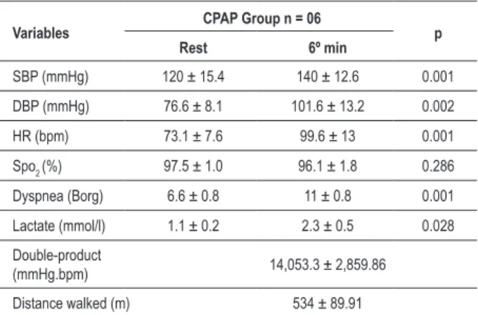
When comparing the variables at rest with those at the 6th minute in the CPAP group, there was also a significant
Table 1 - Clinical characteristics of the study patients
Characteristics n = 12 Mean ± SD
Age (years) 46.3 ± 10.3
BMI (kg/m2) 25.1 ± 5.5
LVEF% 35.3 ± 8.7
SBP (mmHg) 123.3 ± 12.3
DBP (mmHg) 80.8 ± 10
HR (bpm) 77.2 ± 13.2
Spo2 (%) 97.4 ± 1
Dyspnea (Borg) 6.8 ± 1
Lactate (mmol/l) 1.4 ± 0.6
Minnesota Living With Heart Failure Questionnaire (score)
Global score 67.5 ± 11.2
Physical dimension 29.8 ± 6.2
Emotional dimension 13.4 ± 5.4
General dimension 21.2 ± 4.6
N %
Sex
Male 8 66.6
Female 4 33.4
Functional class (NYHA)
II 2 16.6
III 10 83.4
Etiology
Hypertensive 5 41.7
Ischemic 5 41.7
Others* 2 16.6
Medication used
ACE Inhibitor 4 33.4
ARB 6 50
Digitalis 5 41.7
Diuretics 12 100
Betablocker 10 83.4
Vasodilator 2 16.6
Ca2+ channel blocker 1 8.4
Anti-arrhythmic 2 16.6
Anticoagulant agents 0 0
Antiplatelet agents 6 50
n - number of patients; BMI - body mass index; LVEF - left ventricular ejection fraction; SBP - systolic blood pressure; DBP - diastolic blood pressure; HR - heart
rate; SpO2 - peripheral oxygen saturation; NYHA - New York Heart Association;
*others - viral/ peripartum/ Chagas; ACE - angiotensin converting enzyme; ARB -
angiotensin II receptor blockers.
Table 2 - Comparison of the clinical characteristics of patients per group
Characteristics Control Group n = 06
CPAP Group
n = 06 p
Sex M/F 5/1 4/2
Age (years) 45.1 ± 12.1 47.5 ± 9.1 0.71
BMI (kg/m2) 26.5 ± 6.6 23.6 ± 4.1 0.38
QV global score 70.1 ± 10.3 64.8 ± 12.3 0.21
LVEF% 35.5 ± 8.5 35.1 ± 9.7 0.95
SBP (mmHg) 126.6 ± 8.1 120 ± 15.4 0.63
DBP (mmHg) 85 ± 10.4 76.6 ± 8.1 0.17
HR (bpm) 81.1 ± 16.8 73.1 ± 7.6 0.15
Spo2 (%) 97.3 ± 1.0 97.5 ± 1.0 0.39
Dyspnea (Borg) 7 ± 1.26 6.6 ± 0.8 0.74
Lactate (mmol/l) 1.6 ± 0.73 1.1 ± 0.2 0.33
n - number of patients; BMI - body mass index; LVEF - left ventricular ejection fraction; SBP - systolic blood pressure; DBP - diastolic blood pressure; HR -
heart rate; SpO2 - peripheral oxygen saturation.
Table 3 - Comparison of the intragroup variables (control group)
Variables Control Group n = 06 p Rest 6th min
SBP (mmHg) 126.6 ± 8.1 150 ± 8.9 0.003
DBP (mmHg) 85 ± 10.4 105 ± 5.4 0.012
HR (bpm) 81.1 ± 16.8 117.8 ± 19.3 0.006
Spo2 (%) 97.3 ± 1.0 93.6 ± 1.5 0.001
Dyspnea (Borg) 7 ± 1.26 13.1 ± 1.16 0.000
Lactate (mmol/l) 1.6 ± 0.73 3.3 ± 0.7 0.028
Double-product
(mmHg.bpm) 17,758.3 ± 3,623.8
Distance walked (m) 420.6 ± 73.8
n - number of patients; SBP - systolic blood pressure; DBP - diastolic blood
pressure; HR - heart rate; SpO2 - peripheral oxygen saturation.
that there was a significant difference regarding dyspnea 6.6 ± 0.8 vs 11 ± 0.8, with p = 0.001 and lactate concentration: 1.1 ± 0.2 mmol/l vs 2.3 ± 0.5 mmol/l, p = 0.028. However, the analysis of SpO2, did not show a significant difference between the Rest period and the 6th minute (97.5 ± 1.0% vs
96.1 ± 1.8%, p = 0.286), as shown in Table 4.
The comparative analysis between the groups at the end of the 6th minute showed a significant difference in the values
of SpO2% (Control: 93.6 ± 1.5 % vs CPAP: 96.1 ± 1.8%;
p = 0.027), dyspnea (Control: 13.1 ± 1.16 vs CPAP: 11 ± 0.8; p = 0.009), lactate concentration (Control: 3.3 ± 0.7 mmol/l vs CPAP: 2.3 ± 0.5 mmol/l; p = 0.025), double-product (Control: 17,758.3 ± 3,623 mmHg.bpm vs CPAP: 14,035 ± 2,859 mmHg.bpm; p = 0,038) and the distance walked at the 6MWT (Control: 420.6 ± 73.8 m vs CPAP: 534 ± 89.91 m; p = 0.038).
Table 4 - Comparison of intragroup variables (CPAP group)
Variables CPAP Group n = 06 p Rest 6º min
SBP (mmHg) 120 ± 15.4 140 ± 12.6 0.001
DBP (mmHg) 76.6 ± 8.1 101.6 ± 13.2 0.002
HR (bpm) 73.1 ± 7.6 99.6 ± 13 0.001
Spo2 (%) 97.5 ± 1.0 96.1 ± 1.8 0.286
Dyspnea (Borg) 6.6 ± 0.8 11 ± 0.8 0.001
Lactate (mmol/l) 1.1 ± 0.2 2.3 ± 0.5 0.028
Double-product
(mmHg.bpm) 14,053.3 ± 2,859.86
Distance walked (m) 534 ± 89.91
n - number of patients; SBP - systolic blood pressure; DBP - diastolic blood
pressure; HR - heart rate; SpO2 - peripheral oxygen saturation.
Table 5 - Comparison of the variables at the 6th min. between the
Control and CPAP groups
Characteristics Control Group n = 06
CPAP Group
n = 06 p
SBP (mmHg) 150 ± 8.9 140 ± 12.6 0.145
DBP (mmHg) 105 ± 5.4 101.6 ± 13.2 0.583
HR (bpm) 117.8 ± 19.3 99.6 ± 13 0.086
Spo2 (%) 93.6 ± 1.5 96.1 ± 1.8 0.027
Dyspnea (Borg) 13.1 ± 1.16 11 ± 0.8 0.009
Lactate (mmol/L) 3.3 ± 0.7 2.3 ± 0.5 0.025
Double-product
(mmHg.bpm) 17,758.3 ± 3,623.8 14,053.3 ± 2,859.86 0.038
Distance walked
(m) 420.6 ± 73.8 534 ± 89.91 0.038
N - number of patients; SBP - systolic blood pressure; DBP - diastolic blood pressure; HR - heart rate; SpO2 - peripheral oxygen saturation.
Moreover, a difference regarding the hemodynamic data was observed, albeit without statistical significance, as shown in Table 5.
Discussion
The present study shows that the patients submitted to NIVS obtained better results, as demonstrated by the longer distance walked at the 6MWT and consequent better functional capacity16.
HF presents systemic consequences that directly affect patient functional capacity and, consequently, the activities of daily living, as well as the QoL17. This has been corroborated
by the results of the present study.
Cardiac rehabilitation (CR) is considered a set of multidisciplinary therapeutic actions that aim at better clinical evolution, from the physical and psychosocial point of view. During the rehabilitation process, the patients perform several exercises under the supervision of a multiprofessional team. Walking is among the main exercises performed during the treatment of these patients, of which main characteristic is the fact that it is easy to understand and perform on the part of the patients18.
Walking is a physical activity that is limited in patients with HF due to several clinical factors, such as the loss of type I fibers in the skeletal musculature, muscle atrophy, previous hemodynamic condition and metabolic abnormalities19.
Moreover, recent studies have demonstrated the involvement of the respiratory function, with the decrease in the respiratory muscle strength, pulmonary congestion, increase in pulmonary resistance with airflow limitation and ventilation process impairment20.
Schroeder et al21, in 2003, evaluated the level of airflow
limitation in healthy individuals and patients with HF. That study showed an airflow limitation at rest in patients with HF, which was further exacerbated during physical exercise performance. As the NIVS uses positive pressure, it can optimize the cardiac and respiratory function of patients with HF. The positive pressure decreases the large variations in the pleural
pressure and, consequently, reduces the transmural pressure of the LV, improving the contractile performance of the heart22.
The data did not show, regarding the hemodynamic parameters (SBP, DBP and HR), significant effects of the previous performance of the positive pressure, in comparison with the Control group at the end of the 6th minute. That might be due
to the fact that, during the exercise, the patients were no longer using the positive pressure23. However, at the assessment of the
double-product at the end of the exercise, lower values of this physiological parameter were observed in the group of patients submitted to NIVS. That leads us to believe that the NIVS can previously improve the performance of the cardiovascular and respiratory systems, so that during the 6MWT performance, such patients presented a lower cardiac workload22.
In agreement with the findings of the present study, Chermont et al10 studied 12 patients, divided in two groups,
placebo and CPAP for 30 minutes before the 6MWT. That study showed no significant alterations regarding the SBP, DBP and mean arterial pressure (MAP).
Regarding the pulmonary function, as the positive pressure decreases the airway resistance in patients with HF, it increases lung compliance, improves gas exchange, decreases the respiratory workload and reduces airflow limitation24. Similarly,
our results showed that the previous use of CPAP for 30 minutes optimized the patients’ pulmonary function in such a way that they, at the end of the 6th minute of walking, did
not present a significant desaturation.
The comparative analysis of the studied parameters at the end of the 6th minute showed a higher level of SpO
2 and a lower
index of dyspnea in the group previously submitted to CPAP, showing that the beneficial effects of the positive pressure on the pulmonary function of patients with HF persisted until the end of the exercise proposed in this study. Such results demonstrate the capacity of the noninvasive ventilation to improve the pulmonary function parameters and, therefore, allow the more effective performance of the exercise.
Barbas et al25 reported on the beneficial effects of the
cardiopathy, with a decrease in pulmonary congestion and improved gas exchange.
Witmmer et al26 used daily CPAP sessions for 30 minutes
before exercise performance in patients with HF for two weeks and observed an improvement in pulmonary function and the distance walked by the patients that used the CPAP, in agreement with the findings of the present study.
The simplified analysis of the lactate concentrations showed that the patients submitted to NIVS exhibited lower lactate concentrations, suggesting a lower utilization of the anaerobic metabolism. Current scientific evidence demonstrates the association between the anaerobic metabolism and the functional capacity as an important predictor of mortality27,28.
The present study showed some limitations. Initially, it was not possible to define the duration of the effects that were verified, as the administration of the NIVS was interrupted for the performance of the 6MWT. It would be important to determine whether several sessions with NIVS could maintain the effect for a longer period of time and whether these effects are associated with clinical improvement.
Therefore, the continuity of the present study with a larger sample size, as well as other studies on the same subject, is necessary to elucidate the effects of NIVS as
a new alternative in therapeutic support for the CR of patients with HF.
Conclusion
The previous use of CPAP with 10 cmH2O for 30 minutes presented beneficial effects on SpO2, dyspnea index, lactate concentration, double-product and the distance walked by patients with CHF when performing the 6MWT. However, the results of the present study must be taken into account cautiously, as it is a pilot-study carried out with a small sample size.
Potential Conflict of Interest
No potential conflict of interest relevant to this article was reported.
Sources of Funding
There were no external funding sources for this study.
Study Association
This study is not associated with any post-graduation program.
References
1. Barreto ACP, Del Carlo CH, Cardoso JN, Morgado PC, Munhoz RT, Ochiai ME, et al. Re-hospitalizações e morte por insuficiência cardíaca: índices ainda alarmantes. Arq Bras Cardiol. 2008; 91 (5): 335-41.
2. Figueroa MS, Peters JI. Congestive heart failure: diagnosis, pathophysiology, therapy, and implications for respiratory care. Respir Care. 2006; 51 (4): 403-12.
3. Francis GS. Pathophysiology of chronic heart failure. Am J Med. 2001; 110 (Suppl 7A): 37S-46S.
4. Guimarães JI, Mesquita ET, Bocchi EA, Vilas-Boas F, Montera MW, Batlouni M. / Sociedade Brasileira de Cardiologia. Revisão das II Diretrizes para o diagnóstico e tratamento da insuficiência cardíaca. Arq Bras Cardiol. 2002; 79 (4): 1-30.
5. Bocchi EA, Vilas-Boas F, Perrone S, Caamaño AG, Clausell N, Moreira M da C, et al. I Latin American Guidelines for the Assessment and Management of Decompensated Heart Failure. Arq Bras Cardiol. 2005; 85 (Suppl.3): 1-48.
6. Levigner I, Bronks R, Cody DV, Linton I, Davie A. The effect of resistance training on left ventricular function and structure of patients with chronic heart failure. Int J Cardiol. 2005; 105 (2): 159-63.
7. Andrade JP, Montera MW, Almeida DR, Tinoco EM, Rocha RM, Moura LAZ, et al. / Sociedade Brasileira de Cardiologia. II Diretriz brasileira de insuficiência cardíaca aguda. Arq Bras Cardiol. 2009; 93 (3 supl.3): 1-65.
8. Barreto ACP, Ramires JAF. Insuficiência cardíaca. Arq Bras Cardiol. 1998; 71 (4): 635-42.
9. Naughton MT, Liu PP, Bernard DC, Goldstein RS, Bradley TD. Treatment of congestive heart failure and Cheyne-Stokes respiration during sleep by continuous positive airway pressure. Am J Respir Crit Care Med. 1995; 151 (1): 92-7.
10. Chermont S, Quintão MM, Mesquita ET, Rocha NN, Nóbrega AC. Noninvasive ventilation with continuous positive airway pressure acutely improves 6-minute walk distance in chronic heart failure. J Cardiopulm Rehabil Prev. 2009; 29 (1): 44-8.
11. Rector TS, Cohn JN. Assessment of patient outcome with the Minnesota Living With Heart Failure Questionnaire: reliability and validity during a randomized, double-blind, placebo-controlled trial of pimobendan. Am Heart J. 1992; 124 (4): 1017-25.
12. Carvalho VO, Guimarães GV, Carrara D, Bacal F, Bocchi EA. Validação da versão em português do Minnesota Living with Heart Failure Questionnaire. Arq Bras Cardiol. 2009; 93 (1): 39-44.
13. ATS Statement. Guidelines for the six- minute walk test. Am J Respir Crit Care Med. 2002; 166 (1): 111-7.
14. Andrade J, Brito FS, Vilas-Boas F, Castro I, Oliveira JA, Guimarães JI, et al. / Sociedade Brasileira de Cardiologia. II Guidelines on ergometric tests of the Brazilian Society of Cardiology. Arq Bras Cardiol. 2002; 78 (Suppl 2): 1-17.
15. Borg GA. Psychophysical bases of perceived exertion. Med Sci Sports Exerc. 1982; 14 (5): 377-81.
16. Rubim VS, Drumond Neto C, Romeo JL, Monteiro MW. Prognostic value of the six-minute walk test in heart failure. Arq Bras Cardiol. 2006; 86 (2): 120-5.
17. Cipriano GJR, Yuri D, Bernardelli GF, Mair V, Buffolo E, Branco JN. Avaliação da segurança do teste de caminhada dos 6 minutos em pacientes no pré-transplante cardíaco. Arq Bras Cardiol. 2009, 92 (4): 312-9.
18. Andrade JP, Bocchi EA, Braga FGM, Ferreira SMA, Rohde LEP, Oliveira WA, et al. / Sociedade Brasileira de Cardiologia. III Diretriz brasileira de insuficiência cardíaca crônica. Arq Bras Cardiol. 2009; 92 (6 supl.1): 1-71.
19. Hambrecht R, Fiehn E, Yu J, Niebauer J, Weigi C, Hilbrich L, et al. Effects of endurance training on mitochondrial ultrastructure and fiber type destribution in skeletal muscle of patients with stable chronic heart failure. J Am Coll Cardiol. 1997; 29 (5): 1067-3.
20. Gehlbach BK, Geppert E. The pulmonary manifestations of left heart failure. Chest. 2004; 125 (2): 669-82.
22. Meyer EC, Lorenzi Filho G, Schettino GPP, Carvalho RR. Ventilação não-invasiva no cardiopata grave. Rev Soc Cardiol Estado de São Paulo. 1998; 8 (3): 420-7.
23. Furtado EC, Ramos PS, Araújo CGS. Medindo a pressão arterial em exercício aeróbico: subsídios para reabilitação cardíaca. Arq Bras Cardiol. 2009; 93 (1): 45-52.
24. Yan AT, Bradley TD, Liu PP. The role of continuous positive airway pressure in the treatment of congestive heart failure. Chest. 2001; 120 (5): 1675-85.
25. Barbas CSV, Bueno MAS, Amato MBP, Hoelz C, Junior MR. Interação cardiopulmonar durante a ventilação mecânica. Rev Soc Cardiol Estado de São Paulo. 1998; 8 (3): 406-19.
26. Wittmer VL, Simões GM, Sogame LC, Vasquez EC. Effects of continuous positive airway pressure on pulmonary function and exercise tolerance in patients with congestive heart failure. Chest. 2006; 130 (1): 157-63.
27. Arena R, Myers J, Abella J, Peberdy MA, Bensimhon D, Chase P, et al. Development of a ventilatory classification system in patients with heart failure. Circulation. 2007; 115 (18): 2410-7.