Activation of a P2Y
4
-like purinoceptor
triggers an increase in cytosolic [Ca
2+
]
in the red blood cells of the lizard
Ameiva ameiva
(Squamata, Teiidae)
Departamento de Fisiologia, Instituto de Biociências, Universidade de São Paulo, São Paulo, SP, Brasil R. Sartorello and
C.R.S. Garcia
Abstract
An increasing number of pathophysiological roles for purinoceptors are emerging, some of which have therapeutic potential. Erythrocytes are an important source of purines, which can be released under physiological and physiopathological conditions, acting on purinergic receptors associated with the same cell or with neighboring cells. Few studies have been conducted on lizards, and have been limited to ATP agonist itself. We have previously shown that the red blood cells (RBCs) of the lizard Ameiva ameiva store Ca2+ in the endoplasmic
reticulum (ER) and that the purinergic agonist ATP triggers a rapid and transient increase of [Ca2+]
c by mobilization of the cation from
internal stores. We also reported the ability of the second messenger IP3 to discharge the ER calcium pool of the ER. Here we characterize
the purinoceptor present in the cytoplasmic membrane of the RBCs of the lizard Ameiva ameiva by the selective use of ATP analogues and pyrimidine nucleotides. The nucleotides UTP, UDP, GTP, and ATPγS triggered a dose-dependent response, while interestingly 2MeSATP, 2ClATP, α,ß-ATP, and ADP failed to do so in a 1- to 200-µm con-centration. The EC50 obtained for the compounds tested was 41.77 µM
for UTP, 48.11 µM for GTP, 53.11 µM for UDP, and 30.78 µM for ATPγS. The present data indicate that the receptor within the RBCs of
Ameiva ameiva is a P2Y4-like receptor due to its pharmacological
similarity to the mammalian P2Y4 receptor.
Correspondence C.R.S. Garcia
Departamento de Fisiologia Instituto de Biociências, USP Rua do Matão, Travessa 14, 321 05508-900 São Paulo, SP Brasil
Fax: +55-11-3091-7422 E-mail: [email protected]
C.R.S. Garcia was supported by FAPESP.
Received 21 May, 2004 Accepted October 7, 2004
Key words •Red blood cells
•Calcium
•Teiidae
•Ameiva ameiva
•Purinoceptor
•UTP
Introduction
Calcium regulates a myriad of physiologi-cal processes in organisms ranging from pro-tozoa (1-5) to vertebrates (6,7) by an orches-trated cytosolic elevation and/or calcium in-flux (8). Since the early recognition of the potent extracellular actions of ATP and
aden-osine by Drury and Szent-Györgyi, in 1929 (9), considerable knowledge has been accu-mulated about the receptors involved in trans-ducing nucleotide signals (10,11).
receptors on the cell surface, including smooth muscle contraction, neurotransmis-sion, exocrine and endocrine secretion, the immune response, inflammation, plaque ag-gregation, pain, and modulation of cardiac function. These receptors are named puri-nergic receptors (10).
Purinergic receptors are divided into two major families, P1 or adenosine receptors, and P2, which recognize ATP, UTP and UDP. P2 receptors are subdivided into P2Y, the G protein coupled receptors, and P2X, coupled to ionic channels. In mammals, 8 subtypes of P2Y receptors (P2Y1, P2Y2,
P2Y4, P2Y6, P2Y11) (12) and, recently, P2Y12
(13), P2Y13 (14) and P2Y14 (15) have been
identified thus far.
Metabotropic P2Y1-14 receptors are
char-acterized by a subunit topology involving an extracellular N terminus and intracellular C terminus, the latter possessing consensus binding motifs for protein kinases, and seven transmembrane-spanning regions, which help to form the ligand-docking pocket. Each P2Y receptor binds to a single heterotrimeric G protein (typically Gq/11) although P2Y11 can
couple both with Gq/11 and Gs, whereas P2Y12
and P2Y13 couple with Gi and P2Y14 couples
with Gi/0 (16).
The recently cloned P2Y receptors P2Y13
are present mainly in spleen, brain, lymph nodes, and bone marrow, whereas P2Y14
receptors are present in placenta, adipose tissue, stomach, intestine, and discrete brain regions (17).
An increasing number of pathophysiologi-cal roles for purinoceptors are emerging, some of which have therapeutic potential (18-21). Bladder incontinence, contracep-tion and fertility, skin diseases, diabetes, thrombosis, gut motility disorders, cardio-pulmonary diseases, cancer, diseases of the ear, diseases of the eye, behavioral disor-ders, bone disordisor-ders, and pain (19,20) are related in one way or other to responses to purinergic receptors.
Most of the studies concerning the
func-tional distribution of purinoceptors were car-ried out on mammalian preparations even though purinoceptors are widespread throughout the evolutionary scale. Evidence of their presence is found even in protozoa, where ATP was found to have an inhibitory effect on ameboid movement (10).
Few studies have been conducted on liz-ards, and have been limited to ATP agonist itself: an excitatory innervation has been found in the ileum of the lizard Tiliqua rug-osa (11) and in the rectum and portal vein of the lizard Agama agama (22,23). In all cases, the subtype of the receptors involved was unknown.
Erythrocytes are an important source of purines, which can be released in physi-ological and physiopathphysi-ological conditions, acting on purinergic receptors associated with the same cell or with neighboring cells (24). Red blood cells (RBCs) of the lizard Ameiva ameiva loaded with the fluorescent Ca2+ indicator Fluo-3 AM store Ca2+ in the
endoplasmic reticulum and in one or more acidic pools endowed with an H+ pump
sen-sitive to the inhibitors 7-chloro-4-nitrobenz-2-oxa-1,3-diazole and bafilomycin. More-over, the internal Ca2+ pools of the RBCs of
the lizard are sensitive to surface receptor stimulation since the purinergic agonist ATP stimulates the release of Ca2+ in a process
that is hardly affected by the removal of external Ca2+ but is inhibited by suramin and
Material and Methods
Material
2MeSATP, 2-ClATP, ADP, ATP, UDP, UTP, and GTP were purchased from Sigma, St. Louis, MO, USA. Fluo-3 AM was pur-chased from Molecular Probes, Eugene, OR, USA.
Lizards
The lizards Ameiva ameiva were cap-tured with Tomahawk traps in the town of Barretos (20º 33' S, 48º 30' W), State of São Paulo, Brazil, and by hand in the town of Lajeado (10º 43' S, 48º 24' W), State of Tocantins, Brazil. The blood, collected from the lizard’s tail with a syringe, was centri-fuged at 1500 g for 5 min and washed in phosphate-buffered isotonic saline (7.5 mM sodium phosphate and 137 mM NaCl, pH 7.2). Leukocytes were removed from RBCs by blood filtration through a cellulose pow-der column (long fibers; Whatman CF11, Madstone, Kent, UK) according to the method of Homewood and Neame (27).
RBC loading with the calcium indicator Fluo-3 AM
RBCs were washed twice in buffer (116 mM NaCl, 5.4 mM KCl, 0.8 mM MgSO4, 5.5
mM D-glucose, and 50 mM Mops, pH 7.2) and resuspended at 106 cells/ml in the same
buffer containing 1.8 mM probenecid, an in-hibitor of organic anion transport (28), to pre-vent fluorochrome release and sequestration (29). Fluo-3 AM was added at a final concen-tration of 5 µM. The suspension was incu-bated for 1 h at 37ºC followed by three washes with buffer to remove the extracellular dye. In each experiment an aliquot of 100 µl (106 cells) was placed in a thermostated
cu-vette equipped with magnetic stirring. Control experiments with the solvent alone showed no measurable change in fluorescence.
Spectrofluorometric measurements with Fluo-3 AM were performed using a model F-4500 Hitachi spectrofluorometer (Tokyo, Japan) with excitation at 505 nm and emis-sion at 530 nm. The excitation and emisemis-sion slits were 1 mm.
The curves for free Ca2+ concentration
were calculated from fluorescence measured using the Ca2+ software F-4500 Intracellular
Cation Measurement System-Version 1.02 (Copyright© Hitachi, 1994-1995), which
takes into account that [Ca2+] = K
d (F - Fmin/
(Fmax - F), where the Kd utilized for Fluo-3 is
390, F is the fluorescence intensity meas-ured under the conditions of the experiment, Fmax the fluorescence in the presence of
digi-tonin, and Fmin the fluorescence in the
pres-ence of 8 mM EGTA. Unless otherwise speci-fied, all experiments were performed at 37ºC. The results are reported as means ± SEM (N = 3) and data were analyzed statistically by two-way ANOVA.
Results and Discussion
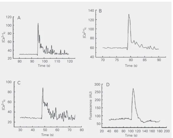
Figure 1 shows that the different concen-trations (100 and 800 µM UTP) were able to trigger the increase of [Ca2+]
c in RBCs of the
lizards Ameiva ameiva loaded with 5 µM Fluo-3 AM.
As shown in Figure 1D, with the aid of confocal microscopy we were able to ob-serve the same effect of calcium mobiliza-tion by a purinergic agonist, in this case, 100 µM UTP. Moreover, the addition of increas-ing concentrations of ATPγS, GTP, UDP, and UTP (from 1 to 800 µM) led to a dose-dependent increase of [Ca2+]
c (data not
shown).
The EC50 was 41.77 µM for UTP (Figure
2A), 48.11 µM for GTP (Figure 2B), 53.11 µM for UDP (Figure 2C), and 30.78 µM for ATPγS (Figure 2D).
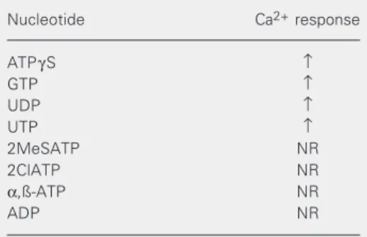
The ATP analogues tested, 2MeSATP,
α,ß-ATP, 2ClATP, as well ADP in a range of 1 to 200 µM failed to promote an increase of [Ca2+]
[Ca
2+
]c
120
100
80
60
40
20
[Ca
2+
]c
140
120
100
80
60
40 80 90 100 110 120
Time (s)
70 75 80 85 90
Time (s)
[Ca
2+
]c
100
80
60
40
20
Fluorescence (AU)
300
250
200
150
100
50
30 40 50 60 70
Time (s)
20 40 60 80 100 Time (s)
80 120 140 160 180 200
A
B
C D
Figure 1. Effect of UTP on lizard red blood cells. Cytosolic cal-cium concentration (A, B, C) was measured fluorometrically with Fluo-3 AM. A, 100 µM UTP; B, 800 µM UTP in nominally cal-cium-free medium; C, 800 µM UTP in 2 mM EGTA medium; D, 100 µM UTP in Fluo-3 AM-loaded lizard red blood cells ob-served by confocal microscopy and reported in arbitrary cence units (AU). The fluores-cence data are qualitative. Note that the ordinate scales of pan-els A, B and C are not equal: B > A > C. Cytosolic calcium con-centration is reported in nM.
[Ca
2+
]c
(nM)
50
40
30
20
10
0
0 1 2 3
Log [UTP] (µM)
A
60
[Ca
2+
]c
(nM)
20
10
0
0 1 2 3
Log [GTP] (µM)
B
30
30
20
10
0
[Ca
2+
]c
(nM)
20
10
0
[Ca
2+
]c
(nM)
0 1 2 3
Log [UDP] (µM)
0 1 2 3
Log [ATPγS] (µM)
C D
Figure 2. Dose-response curves for A, UTP; B, GTP; C, UDP, and
D, ATPγS in Fluo-3 AM-loaded red blood cells of the lizard
Ameiva Ameiva. [Ca2+] c was
measured by loading red blood cells with Fluo-3 AM as de-scribed in Methods. Data are re-ported as means ± SD for three measurements for panels A, B
We have pharmacologically character-ized the P2Y receptor in Ameiva ameiva lizard RBCs. For this purpose, a set of puri-nergic analogues were tested (Table 1). Dose-response curves were obtained for the ago-nists which responded: ATPγS, GTP, UDP, UTP, 2MeSATP, 2ClATP, α,ß-ATP and ADP. The subtype identification has been inferred on the basis of sensitivity to differ-ent types of agonists, the potency relation-ships among them and the ability to inhibit the response. The receptor under study was pharmacologically characterized according to the criteria described below.
A typical trait of P2Y receptors is that they respond differently to natural agonists such as ATP and UDP and their diphosphate and triphosphate analogues (11).
The P2Y1, P2Y11 and P2Y12 receptors are
selective for adenine nucleotides, while the others can be stimulated by uracil nucleo-tides. The P2Y1 and P2Y12 receptors are
equally responsive to 2MeSATP and ATP. The P2Y2 and P2Y4 receptors are equally
responsive to ATP and UTP, and are not responsive to 2MeSATP (21). The P2Y6
re-ceptor is selectively activated by UDP (15). Suramin is a non-selective P2 receptor an-tagonist which competitively antagonizes P2Y1 and P2Y2. The PPADS antagonist
com-petitively antagonizes P2Y1 and P2Y4
recep-tors, but not P2Y2 (30).
The P2Y12 receptor, recently cloned from
human platelets, is stimulated by ADP and ATP and selectively blocked by 2MeSATP (31).
Since addition of the uracil nucleotides UTP and UDP to lizard RBCs elicits a Ca2+
response, the presence of the receptors P2Y1,
P2Y11 and P2Y12 can be eliminated.
Further-more, since similar to adenine nucleotides (ATP), UDP also promotes a Ca2+ response
in these cells, the P2Y6 receptor subtype can
also be ruled out. Since PPADS were able to antagonize the response promoted by ATP addition, we excluded the participation of P2Y2 receptor signaling in these cells.
Fi-nally, P2Y4 seems to be the receptor
in-volved in transducing the signal within the RBCs of Ameiva ameiva lizards.
However, all of these considerations are based on mammalian models, and the de-nomination P2Y4-like receptor seems to be
more appropriate to designate the receptor found on the surface of Ameiva ameiva RBCs until structural information is available.
Acknowledgments
We thank Regina P. Markus, Departa-mento de Fisiologia, USP, São Paulo, SP, Brazil, for helpful criticism, and Miguel T. Rodrigues, Departamento de Zoologia, USP, São Paulo, SP, Brazil, for providing the con-ditions for an expedition to Tocantins to collect lizards.
Table 1. Calcium response of red blood cells of the lizard Ameiva Ameiva to nucleotide agonists.
Nucleotide Ca2+ response
ATPγS ↑
GTP ↑
UDP ↑
UTP ↑
2MeSATP NR
2ClATP NR
α,ß-ATP NR
ADP NR
References
1. Passos AP & Garcia CR (1998). Inositol 1,4,5-trisphosphate induced Ca2+ release from chloroquine-sensitive and insensitive
intracellu-lar stores in the intraerythrocytic stage of the maintracellu-laria parasite P. chabaudi. Biochemical and Biophysical Research Communications, 245: 155-160.
2. Garcia CR, Ann SE, Tavares ES, Dluzewski AR, Mason WT & Paiva FB (1998). Acidic calcium pools in intraerythrocytic malaria para-sites. European Journal of Cell Biology, 76: 133-138.
3. Hotta CT, Gazarini ML, Beraldo FH, Varotti FP, Lopes C, Markus RP, Pozzan T & Garcia CR (2000). Calcium-dependent modulation by melatonin of the circadian rhythm in malarial parasites. Nature Cell Biology, 2: 466-468.
4. Varotti FP, Beraldo FH, Gazarini ML & Garcia CR (2003). Plasmo-dium falciparum malaria parasites display a THG-sensitive Ca2+
pool. Cell Calcium, 33: 137-144.
5. Gazarini ML, Thomas AP, Pozzan T & Garcia CR (2003). Calcium signaling in a low calcium environment: how the intracellular ma-laria parasite solves the problem. Journal of Cell Biology, 161: 103-110.
6. Pozzan T, Rizzuto R, Volpe P & Meldolesi J (1994). Molecular and cellular physiology of intracellular calcium stores. Physiological Re-views, 74: 595-636.
7. Berridge MJ, Bootman MD & Roderick HL (2003). Calcium signal-ling: dynamics, homeostasis and remodeling. Nature Reviews. Mo-lecular Cell Biology, 4: 517-529.
8. Carafoli E (1987). Intracellular calcium homeostasis. Annual Review of Biochemistry, 56: 395-433.
9. Drury AN & Szent-Györgyi A (1929). The physiological activity of adenine compounds with especial reference to their action upon the mammalian heart. Journal of Physiology, 68: 213-237. 10. Ralevic V & Burnstock G (1998). Receptors for purines and
pyrim-idines. Pharmacological Reviews, 50: 413-492.
11. Burnstock G (1996). P2 purinoceptors: historical perspective and classification. Ciba Foundation Symposium 1, 98: 1-28 (Discussion 29-34).
12. Communi D & Boeynaems JM (1997). Receptors responsive to extracellular pyrimidine nucleotides. Trends in Pharmacological Sci-ences, 18: 83-86.
13. Communi D, Motte S, Boeynaems JM & Pirotton S (1996). Pharma-cological characterization of the human P2Y4 receptor. European Journal of Pharmacology, 317: 383-389.
14. Marteau F, Le Poul E, Communi D, Communi D, Labouret C, Pierre Savi, Boeynaems J & Gonzalez NS (2003). Pharmacological charac-terization of the human P2Y13 receptor. Molecular Pharmacology,
64: 104-112.
15. Abbracchio MP, Boeynaems JM, Barnard EA et al. (2003). Charac-terization of the UDP-glucose receptor (re-named here the P2Y14 receptor) adds diversity to the P2Y receptor family. Trends in Phar-macological Sciences, 24: 52-55.
16. Abbracchio MP & Burnstock G (1998). Purinergic signalling: patho-physiological roles. Japanese Journal of Pharmacology, 78: 113-145.
17. Cheung KK, Ryten M & Burnstock G (2003). Abundant and dynamic expression of G protein-coupled P2Y receptors in mammalian de-velopment. Developmental Dynamics, 228: 254-266.
18. Fischer Y, Becker C & Loken C (1999). Purinergic inhibition of glucose transport in cardiomyocytes. Journal of Biological Chemis-try, 274: 755-761.
19. Agteresch HJ, Dagnelie PC, van den Berg WJ & Wilson JH (1999). Adenosine triphosphate: established and potential clinical applica-tions. Drugs, 58: 211-232.
20. Williams M & Jarvis MF (2000). Purinergic and pyrimidinergic recep-tors as potential drug targets. Biochemical Pharmacology, 59: 1173-1185.
21. Communi D, Janssens R, Suarez-Huerta N, Reobaye B & Boey-naems JM (2000). Advances in signalling by extracellular nucleo-tides. The role and transduction mechanisms of P2Y receptors.
Cellular Signalling, 12: 351-360.
22. Ojewole JA (1983). Effects of drugs and electrical stimulation on rainbow lizard (Agama agama Linn.) isolated gastrointestinal tract smooth muscles. Methods and Findings in Experimental and Clini-cal Pharmacology, 5: 299-310.
23. Savage AO & Atanga GK (1985). Effect of adenosine 5’triphosphate (ATP) on the isolated rectum of the rainbow lizard Agama agama.
General Pharmacology, 16: 235-240.
24. Ellsworth ML, Forrester T, Ellis CG & Dietrich HH (1995). The erythrocyte as a regulator of vascular tone. American Journal of Physiology, 269: 2155-2161.
25. Beraldo FH, Sartorello R, Lanari RD & Garcia CR (2001). Signal transduction in red blood cells of the lizards Ameiva ameiva and
Tupinambis merianae (Squamata, Teiidae). Cell Calcium, 29: 439-445.
26. Beraldo FH, Sartorello R, Gazarini ML, Caldeira W & Garcia CR (2002). Red blood cells of the lizards Ameiva ameiva (Squamata, Teiidae) display multiple mechanisms to control cytosolic calcium.
Cell Calcium, 31: 79-87.
27. Homewood CA & Neame KD (1976). A comparison of methods used for the removal of white cells from malaria-infected blood.
Annals of Tropical Medicine and Parasitology, 70: 249-251. 28. Di Virgilio F, Steinberg TH & Silverstein SC (1990). Inhibition of
Fura-2 sequestration and secretion with organic anion transport blockers.
Cell Calcium, 11: 57-62.
29. Di Virgilio F, Steinberg TH & Silverstein SC (1989). Organic-anion transport inhibitors to facilitate measurement of cytosolic free Ca2+
with fura-2. Methods in Cell Biology, 31: 453-462.
30. Charlton SJ, Brown CA, Weisman GA, Turner JT, Erb L & Boarder MR (1996). Cloned and transfected P2Y4 receptors: characteriza-tion of a suramin and PPADS-insensitive response to UTP. British Journal of Pharmacology, 119: 1301-1303.