Relation of cervical length at 22-24
weeks of gestation to demographic
characteristics and obstetric history
1Serviço de Obstetrícia e Ginecologia, Hospital de Clínicas de Porto Alegre,
Universidade Federal do Rio Grande do Sul, Porto Alegre, RS, Brasil
2Clínica de Ultra-som Alpha, Porto Alegre, RS, Brasil
R.S. Palma-Dias1,2,
M.M. Fonseca2,
N.R. Stein1,
A.P. Schmidt1 and
J.A. Magalhães1,2
Abstract
Preterm delivery is the main cause of neonatal death and ultrasono-graphic cervical assessment has been shown to be more accurate than digital examination in recognizing a short cervix. This is a cross-sectional study, involving 1131 women at 22-24 weeks of pregnancy, designed to determine the distribution of cervical length and to examine which variables of demographic characteristics and obstetric history increase the risk of a short cervix (15 mm or less). The distribution of maternal demographic and obstetric history character-istics among patients with cervical length ≤15 mm was analyzed and compared to the findings for the general population. Risk ratios (RR) between subgroups were generated from this comparison. Median cervical length was 37 mmand in 1.5% of cases it was 15 mm or less. The proportion of women with a short cervix (≤15 mm) was signifi-cantly higher among patients with a low body mass index (RR = 3.5) and in those with previous fetal losses between 16-23 weeks (RR = 33.1) or spontaneous preterm deliveries between 24-32 weeks (RR = 14.1). We suggest that transvaginal sonographic measurement of cervical length be performed as part of a routine midtrimester ultra-sound evaluation. There are specific variables of demographic charac-teristics and obstetric history which increase the risk of detecting a short cervix at 22-24 weeks.
Correspondence
R.S. Palma-Dias
Travessa Pedro Redonda 450, Casa 4 Jardim Isabel
91760-630 Porto Alegre, RS Brasil
Fax: +55-51-3316-8148 E-mail: [email protected]
Research supported by the Fetal Medicine Foundation, London (Charity No. 1037116).
Received August 20, 2002 Accepted January 5, 2004
Key words
•Cervical length
•Preterm delivery screening •Transvaginal sonography •Perinatal medicine
Introduction
Preterm delivery is the main cause of neonatal death (1). The risk for this preg-nancy complication varies with maternal char-acteristics, such as ethnic origin, age, body mass index (BMI), cigarette smoking and drug abuse, as well as obstetric history.
Routine assessment of cervical length at 22-24 weeks of gestation provides a
out simultaneously with the fetal anomaly scan at a gestational age just prior to fetal viability. Furthermore, several studies have shown that ultrasonographic cervical assess-ment is more accurate than digital examina-tion for identifying cervical changes (4,5).
Transvaginal cervical length measure-ment has several technical advantages over the transabdominal approach. The main one is that a full bladder, often necessary to visualize the cervix transabdominally, may falsely elongate a short cervix due to the pressure exerted over the lower uterine seg-ment (6).
The aims of the present study were to determine the distribution of cervical lengths in a population of singleton pregnancies at 22-24 weeks of gestation, and to examine the relationship between cervical length and maternal demographic characteristics and obstetric history. Furthermore, this study was planned to identify which variables of these characteristics increase the risk of detecting a cervical length of 15 mm or less, which is associated with a higher risk of spontaneous premature delivery.
Patients and Methods
All women attending the routine prenatal care service at Hospital de Clínicas de Porto Alegre, Porto Alegre, RS, Brazil, are offered the option of having two ultrasound exami-nations: the first at 11-14 weeks of gestation and the second at 22-24 weeks. During a 19-month period (April 1999 to October 2000), women attending the midtrimester scan were offered the option of having transvaginal sonographic assessment of the cervix. Women with in situ cervical cerclage, severe
fetal malformations or polyhydramnios were excluded from the study.
Written informed consent was obtained from women who agreed to participate in the study, which was approved by the hospital Ethics Committee and by the Brazilian Na-tional Ethics Committee. This study was part
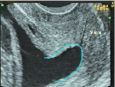
of a multicenter study carried out under the auspices of the Fetal Medicine Foundation, London, in order to find women with a short cervix and to test the effect of different treatment options for these high-risk patients. The women were asked to empty their bladder and were placed in the dorsal litho-tomy position. Transvaginal sonography with a 5-MHz transducer (Aloka1700 II SSD, Aloka Co. Ltd., Tokyo, Japan) was carried out by one of two sonographers who had received the Fetal Medicine Foundation Cer-tificate of competence in cervical assess-ment. The probe was placed in the anterior fornix of the vagina and a sagittal view of the cervix, with the echogenic endocervical mucosa along the length of the canal, was obtained. Care was taken to avoid exerting undue pressure on the cervix. The electronic calipers were used to measure the distance between the triangular area of echodensity at the external os and the V-shaped notch at the internal os (Figure 1) (6). Each examination was performed during a period of about 3 min to observe any cervical changes. Changes that may be due to contractions are observed in less than 1% of patients and, in such cases, the shortest measurement is recorded. The presence or absence of funneling at the inter-nal os was recorded (Figure 2).
Gestational age was determined from the menstrual history and confirmed from the first-trimester scan. When the difference be-tween the two estimates was more than seven days the ultrasound result was used for dat-ing.
The distribution of cervical lengths was tested for normality using the Kolmogorov-Smirnov test. The Tukey-HSD test for mul-tiple range comparisons and the unpaired Student t-test were used to calculate the
sig-nificance of differences in mean cervical length between subgroups. The chi-square test and the Fisher exact test were used to determine the significance of differences between subgroups in cases with cervical length ≤15 mm (Table 4); this cut-off identi-fies a group at higher risk for preterm deliv-ery (3). A 5% alpha error was admitted (P < 0.05).
For analysis of obstetric history, patients were divided into subgroups. Patients with unfavorable obstetric history (miscarriages, spontaneous preterm deliveries) were com-pared to a control group of women with a history of exclusively term deliveries (Table 5). Patients in their first pregnancy were analyzed separately.
Results
During the study period, 1157 women with singleton pregnancies were seen for a scan at 22-24 (median = 23) weeks of gesta-tion and 1131 (97.8%) agreed to participate in the study.
Cervical length was measured success-fully in all cases. Length was approximately distributed normally with some skewness at the lower end (Figure 3). The median (and mean) value was 37 mm, and the 1st and 5th percentiles were 13 and 25 mm, respec-tively. In 11.8, 5.0, 2.4 and 1.5% of cases, the cervical length was ≤30, ≤25, ≤20 and
≤15 mm, respectively (Table 1).
Funneling of the cervical canal at the level of the internal os was observed in 20
Frequency (N)
2 9 16 23 30 37 44 51 58
0 40 80 160
120
Cervical length (mm)
Figure 1. Cervical length-fre-quency distribution. N = 1131.
Figure 2. Normal (36 mm) cervical length measurement.
(1.8%) cases and in 76.5% of the cases with cervical length ≤15 mm.
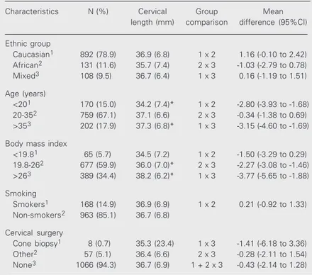
The mean cervical length was shorter in women aged less than 20 years and in those with an unfavorable obstetric history (Tables 2 and 3). In terms of obstetric history, 35% of the patients had no previous pregnancies, 25% had one or more miscarriages and/or termina-tion of pregnancy before 16 weeks of
gesta-tion, 46% had one or more term deliveries, with or without previous fetal losses before 16 weeks, 9% had at least one previous spontane-ous preterm delivery at 33-36 weeks, 4% had at least one previous spontaneous preterm de-livery at 24-32 weeks, 5% had at least one previous miscarriage at 16-23 weeks, and 1% had at least one previous termination at 16-23 weeks. Some patients in the latter four groups may also have had first-trimester losses or term deliveries. The groups are compared for mean cervical length in Table 3.
Women with increased BMI (>26) had increased mean cervical length compared to those with normal or decreased BMI.
In women of African origin the mean cer-vical length tended to be shorter compared to Caucasians, but the difference was not statisti-cally significant. The mean cervical length was not significantly different when women were compared for the use of cigarettes or for past cervical surgery (Table 2).
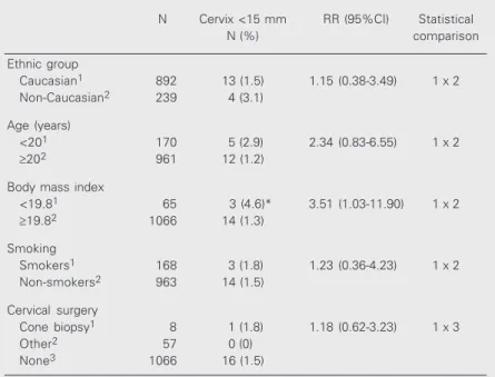
The proportion of women with a very short cervix (≤15 mm) was significantly in-creased in women with a low BMI and in those with previous miscarriages or sponta-neous preterm deliveries (Tables 4 and 5).
Non-Caucasians, patients aged 20 years or less, smokers, those who underwent cer-vical conization and those with first trimes-ter losses were present in a higher than ex-pected proportion in the short cervix (≤15 mm) group. However, the difference be-tween their expected and observed frequency was not statistically significant in these sub-groups (Tables 4 and 5).
Discussion
The present study has demonstrated that the mean cervical length of 37 mm was similar to that of other studies that measured cervical length at 20-24 weeks in low-risk populations in different settings (Table 6) (2,7-19).
The incidence of preterm delivery is higher in all ethnic minorities, particularly in
Table 1. Incidence of short cervixes when differ-ent cervical lengths are used as cut-offs.
Cervical length (mm) Incidence (%)
≤15 mm 1.5
≤20 mm 2.4
≤25 mm 5.0
≤30 mm 11.8
N = 1124. Mean and median cervical length = 37 mm; 1st percentile = 13 mm; 5th percentile = 25 mm.
Table 2. Cervical length at 22-24 weeks of gestation: patient characteristics and comparison of means for each subgroup.
Characteristics N (%) Cervical Group Mean
length (mm) comparison difference (95%CI)
Ethnic group
Caucasian1 892 (78.9) 36.9 (6.8) 1 x 2 1.16 (-0.10 to 2.42)
African2 131 (11.6) 35.7 (7.4) 2 x 3 -1.03 (-2.79 to 0.78)
Mixed3 108 (9.5) 36.7 (6.4) 1 x 3 0.16 (-1.19 to 1.51)
Age (years)
<201 170 (15.0) 34.2 (7.4)* 1 x 2 -2.80 (-3.93 to -1.68)
20-352 759 (67.1) 37.1 (6.6) 2 x 3 -0.34 (-1.38 to 0.69)
>353 202 (17.9) 37.3 (6.8)* 1 x 3 -3.15 (-4.60 to -1.69)
Body mass index
<19.81 65 (5.7) 34.5 (7.2) 1 x 2 -1.50 (-3.29 to 0.29)
19.8-262 677 (59.9) 36.0 (7.0)* 2 x 3 -2.27 (-3.08 to -1.46)
>263 389 (34.4) 38.2 (6.2)* 1 x 3 -3.77 (-5.65 to -1.88)
Smoking
Smokers1 168 (14.9) 36.9 (6.9) 1 x 2 0.21 (-0.92 to 1.33)
Non-smokers2 963 (85.1) 36.7 (6.8)
Cervical surgery
Cone biopsy1 8 (0.7) 35.3 (23.4) 1 x 3 -1.41 (-6.18 to 3.36)
Other2 57 (5.1) 36.4 (6.6) 2 x 3 -0.28 (-2.11 to 1.54)
None3 1066 (94.3) 36.7 (6.9) 1 + 2 x 3 -0.43 (-2.14 to 1.28)
those of African origin, but two large Ameri-can studies have reported that, when social and demographic factors were accounted for, maternal race was not a significant risk factor for preterm delivery (20,21). In our study, women of African origin did not show a significantly shorter cervical length than Caucasian women. This finding, which dis-agrees with other studies with a similar de-sign, may be explained by the high rate of ethnic group mixing found in the Brazilian population, where ethnic group differences may become subtle. Furthermore, all our patients, regardless of ethnic origin, come from lower social classes. This may explain our findings, agreeing with the observation that social and economic status might be more important than ethnic group itself in predicting a poorer pregnancy outcome.
We found that cervical length was shorter in women aged less than 20 years. This is compatible with the well-documented in-creased risk for poorer pregnancy outcome in adolescents. It has been previously sug-gested that this increased risk is probably due to associated social and behavioral fac-tors rather than to intrinsic biological deter-minants of young age (22). This is an impor-tant finding for our population, given the high incidence of adolescents who conceive in Brazil, suggesting that this age group would benefit particularly from cervical assessment as a screening tool for preterm delivery.
The incidence of preterm delivery is higher in women that smoke than in non-smokers (23,24). It has been suggested that smoking may induce labor by increasing the amniotic fluid concentration of the inflam-matory mediator platelet-activating factor since cigarette smoke is a potent inhibitor of the enzyme that degrades platelet-activating factor (25). We found that mean cervical length in cigarette smokers was not signifi-cantly lower than in non-smokers. However, women who smoked were more likely to be part of the very short cervix group (≤15 mm). These findings are in agreement with our
understanding of how preterm labor is trig-gered: both mechanical and biochemical fac-tors might play a role in this process through different mechanisms of action.
The incidence of preterm delivery may
Table 4. Incidence of cervical length of 15 mm or less in each subgroup.
N Cervix <15 mm RR (95%CI) Statistical
N (%) comparison
Ethnic group
Caucasian1 892 13 (1.5) 1.15 (0.38-3.49) 1 x 2
Non-Caucasian2 239 4 (3.1)
Age (years)
<201 170 5 (2.9) 2.34 (0.83-6.55) 1 x 2
≥202 961 12 (1.2)
Body mass index
<19.81 65 3 (4.6)* 3.51 (1.03-11.90) 1 x 2
≥19.82 1066 14 (1.3)
Smoking
Smokers1 168 3 (1.8) 1.23 (0.36-4.23) 1 x 2
Non-smokers2 963 14 (1.5)
Cervical surgery
Cone biopsy1 8 1 (1.8) 1.18 (0.62-3.23) 1 x 3
Other2 57 0 (0)
None3 1066 16 (1.5)
The relative risk was calculated as a fraction of the percentage of women with a cervix <15 mm in the overall population (1.5%). N = 1131. CI = confidence interval; RR = risk ratio.
*P < 0.05 (see statistical comparison; chi-square test/Fisher’s exact test).
Table 3. Cervical length at 22-24 weeks of gestation: obstetric history and comparison of means for each subgroup.
Obstetric history N (%) Cervical Statistical Mean
length comparison difference
(mm) (95%CI)
Primigravidae1 398 (35.2) 35.5 (6.9) 1 x 2 -1.89 (-2.35 to 1.66)
Multigravidae2 733 (64.8) 37.4 (6.7)
Delivery ≥37 weeksA 309 (27.3) 38.7 (6.1)
Delivery 33-36 weeksB 96 (8.5) 36.4 (7.5)* A x B -2.4 (-4.0 to -0.7)
Delivery 24-32 weeksC 44 (3.9) 34.7 (8.9)* A x C -4.1 (-6.9 to -1.3)
Fetal loss 16-23 weeksD 56 (5.0) 34.5 (9.2)* A x D -4.3 (-6.8 to -1.7)
Termination 16-23 weeksE 9 (0.8) 35.5 (6.5) A x E -3.3 (-8.3 to +1.8)
Fetal loss <16 weeksF 239 (21.1) 35.7 (7.1)* A x F -3.1 (-4.2 to -1.9)
Termination <16 weeksG 41 (3.6) 38.5 (7.6) A x G -0.3 (-2.3 to +1.8)
Cervical length is reported as mean and SD in mm for N = 1131. CI = confidence interval.
accordance with most previous studies re-porting on cervical length at midtrimester.
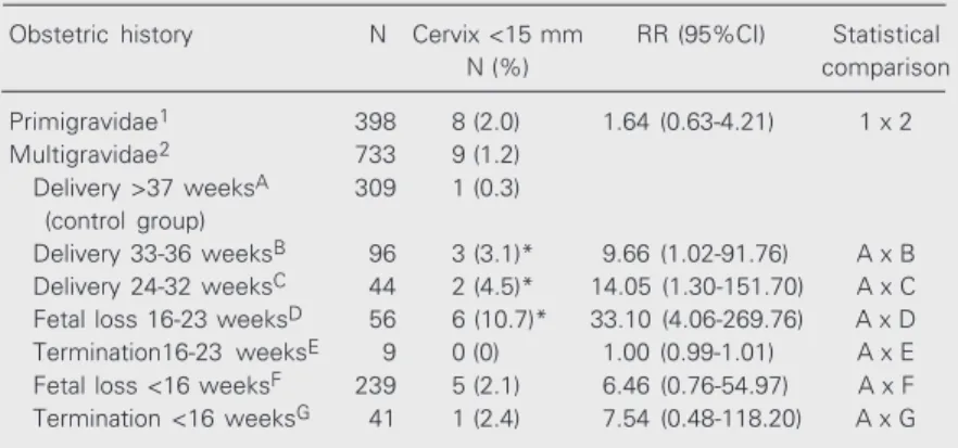
In terms of obstetric history, cervical length was shorter in patients with previous midtrimester losses or previous spontaneous preterm deliveries. Indeed, these findings corroborate classic principles of obstetrics which state that obstetric history is one of the best screening tools for identifying high-risk pregnancies.
However, not all patients with a poor obstetric history will prove to have abnormal cervical findings or a repeated unfavorable pregnancy outcome. Nevertheless, routine cervical assessment at 23 weeks in these patients may be reassuring to patients and obstetricians after a normal result, whereas the confirmation of anatomical changes com-patible with cervical incompetence might justify the early institution of therapeutic strategies based on objective criteria.
Our findings do not corroborate the as-sumption that women with previous cervical surgery, especially cone biopsies, should have a shorter cervix. However, there are several studies showing an increase in preterm de-livery after this type of surgery (26,28,29). Although a significant difference in cervical length has not been found in women who underwent a cone biopsy, it is interesting to notice that the dispersion of this statistic is wide, since a part of these patients has in-deed shown a cervical length more than 2 standard-deviations below the mean.
The possibility that this statistical finding reflects the existence of two different sub-groups in the “cone biopsy” category must be considered. Cone biopsies directed to the ectocervix have a wider diameter and de-creased depth compared to those directed to endocervical lesions, which implicate in deeper resections and therefore are more prone to cause cervical incompetence. The number of patients who underwent cervical assessment after a cone biopsy in our sample was small and, therefore, the above men-tioned potential confounders could not be
Table 5. Incidence of cervical length of 15 mm or less in the obstetric history sub-groups.
Obstetric history N Cervix <15 mm RR (95%CI) Statistical
N (%) comparison
Primigravidae1 398 8 (2.0) 1.64 (0.63-4.21) 1 x 2
Multigravidae2 733 9 (1.2)
Delivery >37 weeksA 309 1 (0.3)
(control group)
Delivery 33-36 weeksB 96 3 (3.1)* 9.66 (1.02-91.76) A x B
Delivery 24-32 weeksC 44 2 (4.5)* 14.05 (1.30-151.70) A x C
Fetal loss 16-23 weeksD 56 6 (10.7)* 33.10 (4.06-269.76) A x D
Termination16-23 weeksE 9 0 (0) 1.00 (0.99-1.01) A x E
Fetal loss <16 weeksF 239 5 (2.1) 6.46 (0.76-54.97) A x F
Termination <16 weeksG 41 1 (2.4) 7.54 (0.48-118.20) A x G
The relative risk was calculated as a fraction of the percentage of women with a cervix <15 mm in the overall population (1.5%). N = 1131. CI = confidence interval; RR = risk ratio.
*P < 0.05 compared to subgroup A (chi-square test/Fisher’s exact test).
Table 6. Studies of low-risk populations providing sufficient data to allow calculation of median or mean cervical length at 20-24 weeks of gestation.
Reference Ultrasonography N Cervical Primigravid
length (mm) vs
multigravida
Ayers et al., 1988 (8) TA 24 52 Similar
Podobnik et al., 1988 (9) TA 80 48
-Andersen et al., 1990 (7) TV 19 40
-TA 21 43
-Kushnir et al., 1990 (10) TV 24 48 Similar
Andersen, 1991 (11) TV 25 41 MG > P
TA 25 42
-Murakawa et al., 1993 (12) TV 44 37 Similar
Zorzoli et al., 1994 (13) TV 121 42 MG > P
Iams et al., 1995 (14) TV 106 37
-Iams et al., 1996 (2) TV 2915 35 MG > P
Cook and Ellwood, 1996 (15) TV 41 42 P > MG
Hasegawa et al., 1996 (16) TV 78 38 Similar
Tongsong et al., 1997 (17) TV 38 42 Similar
Heath et al., 1998 (18) TV 2702 38 Similar
Carvalho et al., 2002 (19) TV 641 39
-Palma Dias et al. (present study) TV 1131 37 Similar
TA = transabdominal; TV = transvaginal. aCervical length of primigravid (P) women
compared to that of multigravid (MG) women.
controlled in our study. Furthermore, most women who undergo a cone biopsy at our hospital are advised to have a first trimester cerclage in future pregnancies, and the pres-ence of a cervical cerclage in situ at 23 weeks
was a reason for exclusion from this study. Low pre-pregnancy maternal weight is associated with an increased risk of preterm delivery, particularly in women who are markedly underweight (less than 80% of recommended weight for height) and have a low weight gain during pregnancy (30,31). In our study, the median cervical length was longer in women with a high BMI and the incidence of a very short cervix (<15 mm) was higher in women with a low BMI (<19.8).
References
1. Guyer B, Martin JA, MacDorman MF, Anderson RN & Strobino DM (1997). Annual summary of vital statistics - 1996. Pediatrics, 100: 905-918.
2. Iams JD, Goldenberg RL, Meis PJ et al. (1996). The length of the cervix and the risk of spontaneous delivery. New England Journal of Medicine, 334: 567-572.
3. Heath VCF, Southall TR, Souka AP, Elisseou A & Nicolaides KH (1998). Cervical length at 23 weeks of gestation: prediction of spontaneous preterm delivery. Ultrasound in Obstetrics and Gyne-cology, 12: 312-317.
4. Lim BH, Mahmood TA, Smith NC & Beat I (1992). A prospective comparative study of transvaginal ultrasonography and digital ex-amination of cervical assessment in the third trimester of preg-nancy. Journal of Clinical Ultrasound, 20: 599-603.
5. Jackson GM, Ludmir J & Bader TJ (1992). The accuracy of digital examination and ultrasound in the evaluation of cervical length.
Obstetrics and Gynecology, 79: 214-218.
6. To MS, Skentou C, Cicero S & Nicolaides KH (2000). Cervical as-sessment at the routine 23-week scan: problems with transabdomi-nal sonography. Ultrasound in Obstetrics and Gynecology, 15: 292-296.
7. Andersen HF, Nugent CE, Wanty SD & Hayashi RH (1990). Predic-tion of risk for preterm delivery by ultrasonographic measurement of cervical length. American Journal of Obstetrics and Gynecology, 163: 859-867.
8. Ayers JW, DeGrood RM, Compton AA, Barclay M & Ansbacher R (1988). Sonographic evaluation of cervical length in pregnancy: diag-nosis and management of preterm cervical effacement in patients at high risk for premature delivery. Obstetrics and Gynecology, 71: 939-944.
9. Podobnik M, Bulie M, Smiljjanie N & Bistricki J (1988). Ultrasonogra-phy in the detection of cervical incompetency. Journal of Clinical Ultrasound, 13: 383-391.
10. Kushnir O, Vigil DA, Izquierdo L, Schiff M & Curet LB (1990). Vaginal ultrasonographic assessment of cervical length changes during
nor-mal pregnancy. American Journal of Obstetrics and Gynecology, 162: 991-993.
11. Andersen HF (1991). Transvaginal and transabdominal ultrasonogra-phy of the uterine cervix during pregnancy. Journal of Clinical Ultra-sound, 19: 77-83.
12. Murakawa H, Utumi T, Hasegawa I, Tanaka K & Fuzimori R (1993). Evaluation of threatened preterm delivery by transvaginal ultrasono-graphic measurement of cervical length. Obstetrics and Gynecol-ogy, 82: 829-832.
13. Zorzoli A, Soliani A, Perra M, Caravelli E, Galimberi A & Nicolini U (1994). Cervical changes throughout pregnancy as assessed by transvaginal sonography. Obstetrics and Gynecology, 84: 960-964. 14. Iams JD, Johnson FF, Sonek J, Sachs L, Gebauer C & Samuels P (1995). Cervical competence as a continuum: a study of ultrasono-graphic cervical length and obstetrical performance. American Jour-nal of Obstetrics and Gynecology, 172: 1097-1106.
15. Cook C-M & Ellwood DA (1996). A longitudinal study of the cervix in pregnancy using transvaginal ultrasound. British Journal of Obstet-rics and Gynecology, 103: 16-18.
16. Hasegawa I, Tanaka K, Takahashi K et al. (1996). Transvaginal ultrasonographic cervical assessment for the prediction of preterm delivery. Journal of Maternal and Fetal Medicine, 5: 305-309. 17. Tongsong T, Kamprapanth P & Pitaksakorn J (1997). Cervical length
in normal pregnancy as measured by transvaginal sonography. In-ternational Journal of Gynecology and Obstetrics, 58: 313-315. 18. Heath VCF, Southall TR, Souka AP, Novakov A & Nicolaides KH
(1998). Cervical length at 23 weeks of gestation: relation to demo-graphic characteristics and previous obstetric hystory. Ultrasound in Obstetrics and Gynecology, 12: 304-311.
19. Carvalho MHB, Bittar RE, Gonzales M, Brizot ML & Zugaib M (2002). Avaliação do risco para parto prematuro espontâneo pelo comprimento do colo uterino no primeiro e segundo trimestre de gravidez. Revista Brasileira de Ginecologia e Obstetrícia, 24: 463-468.
20. Lieberman E, Ryan KJ, Monson RR & Schoenbaym SC (1987). Risk
There is evidence that short cervical length at 22-24 weeks of gestation is associ-ated with increased risk of preterm delivery (2,3). The present study has demonstrated that significant contributions to the explana-tion of the variance in cervical length are provided by ethnic group, BMI, maternal age and obstetric history.
factors accounting for racial differences in the rate of premature birth. New England Journal of Medicine, 317: 743-748.
21. Owen J, Goldenberg RL, Davi RO, Kirk KA & Copper RL (1990). Evaluation of a risk scoring system as a predictor of preterm birth in an indigent population. American Journal of Obstetrics and Gyne-cology, 163: 873-879.
22. Zuckerman BS, Walker DK, Frank DA, Chase C & Hamburg B (1984). Adolescent pregnancy: biobehavioural determinants of outcome.
Journal of Pediatrics, 105: 857-862.
23. Wen SW, Goldenberg RL, Cutter GR, Hoffman HJ & Cliver SP (1990). Intrauterine growth retardation and preterm delivery: prena-tal risk factors in an indigent population. American Journal of Ob-stetrics and Gynecology, 162: 213-218.
24. Wisborg K, Henriksen TB, Hedegaard M & Secher NJ (1996). Smok-ing durSmok-ing pregnancy and preterm birth. British Journal of Obstet-rics and Gynecology, 103: 800-805.
25. Narahara H & Johnston JM (1993). Smoking and preterm labor: effect of cigarette smoke extract on the secretion of platelet acti-vating factor acetylhydrolase by human decidual macrophages.
American Journal of Obstetrics and Gynecology, 169: 1321-1326. 26. Heffner LJ, Sherman CB, Speizer FE & Weiss ST (1993). Clinical and
environmental predictors of preterm labor. Obstetrics and Gynecol-ogy, 81: 750-757.
27. Kaltreider DF & Kohl S (1980). Epidemiology of preterm delivery.
Clinical Obstetrics and Gynecology, 23: 17-32.
28. Kristensen J, Langhoff-Roos J & Kristensen FB (1993). Increased risk of preterm birth in women with cervical conisation. Obstetrics and Gynecology, 81: 1005-1008.
29. Hagen B & Skjeldestad FE (1993). The outcome of pregnancy after CO2 laser conisation of the cervix. British Journal of Obstetrics and Gynecology, 100: 717-720.
30. Mitchell MC & Lerner E (1989). Weight gain and pregnancy out-come in underweight and normal weight women. Journal of the American Dietetic Association, 89: 634-638.