Assessment of Length of Maternal Cervix
between 18 and 24 weeks of Gestation
in a Low-Risk Brazilian Population
Avaliação do comprimento do colo uterino materno
entre 18 e 24 semanas de gestação em uma população
brasileira de baixo risco
Soraya Gomes de Amorim Andrade
1Fernando Moreira de Andrade
1Edward Araujo Júnior
1Cláudio Rodrigues Pires
2Rosiane Mattar
1Antonio Fernandes Moron
11Department of Obstetrics, Escola Paulista de Medicina, Universidade Federal de São Paulo (EPM-UNIFESP), São Paulo, SP, Brazil 2Centro de Referência no Ensino do Diagnóstico por Imagem
(CETRUS), São Paulo, SP, Brazil
Rev Bras Ginecol Obstet 2017;39:647–652.
Address for correspondence Edward Araujo Júnior, PhD, Rua Botucatu, 740, 04023-062, Vila Clementino, São Paulo, SP, Brazil (e-mail: [email protected]).
Keywords
►
gestation
►
uterine cervix
►
biometry
►
morphology
►
transvaginal
ultrasound
Abstract
Purpose
To determine cervical biometry in pregnant women between 18 and
24 weeks of gestation and the ideal mode of measurement of cervical length in cases
of curved and straight cervical morphology.
Methods
The uterine cervices of 752 low-risk pregnant women were assessed using
transvaginal ultrasound in a prospective cross-sectional study. In women with straight
uterine cervices, cervical biometry was performed in a continuous manner. In women
with curved uterine cervices, the biometry was performed using both the continuous
and segmented techniques (in segments joining the cervical os). Polynomial regression
models were created to assess the correlation between the cervical length and
gestational age. The paired Student
t
-test was used to compare measuring techniques.
Results
The cervical biometry results did not vary signi
fi
cantly with the gestational
age and were best represented by linear regression (R
2¼
0.0075 with the continuous
technique, and R
2¼
0.0017 with the segmented technique). Up to the 21
stweek of
gestation, there was a predominance of curved uterine cervix morphology (58.9%),
whereas the straight morphology predominated after this gestational age (54.2%).
There was a signi
fi
cant difference between the continuous and the segmented
measuring methods in all the assessed gestational ages (
p
<
0.001).
Conclusion
Cervical biometry in pregnant women between 18 and 24 weeks was
represented by a linear regression, independently of the measuring mode. The ideal
measuring technique was the transvaginal ultrasound performed at a gestational age
21 weeks.
received April 4, 2017 accepted
September 9, 2017 published online November 27, 2017
DOIhttps://doi.org/ 10.1055/s-0037-1608617. ISSN 0100-7203.
Introduction
Prematurity is a major cause of perinatal morbidity and mortality. A short uterine cervix is a predictor of premature birth; therefore, its morphological and biometric assessment is important.1,2
Cervical assessment is performed by clinical examination and ultrasound.3Bidigital examination requires dilation of the internal cervical os. It is, therefore, a subjective assess-ment of the cervix, and it can underestimate the actual cervical length by up to 12 mm.4The most accurate cervical assessment method is the transvaginal ultrasound, which allows adequate observation of the internal and external cervical os and the endocervical canal, with a biometric precision of100%.5The morphological and biometric study of the uterine cervix should be performed from the 14thweek
of gestation onward because the ultrasonographic differen-tiation between the cervix and the lower uterine segment using ultrasound becomes difficult before this gestational age.6
There is no consensus among authors regarding the value of cervical length that is considered short and the associated risk for premature labor, the reported values varying be-tween 20 and 25 mm.7–9The importance of cervical
biome-try in screening patients for preterm labor has been highlighted in several publications; however, studies that have investigated the techniques of cervical measurement are rare. The evaluation of uterine cervical morphology is
important because it is technically more difficult to measure cervical length in cases of curved uterine cervices, and the best measuring method in these cases is still controversial.10 When the uterine cervix has a straight morphology, its measurement is performed in a continuous manner, that is, along the cervical canal. When the cervix is curved, the biomet-ric measurement may be performed in two ways: (1) in a continuous manner, for example, from the internal os to the external os of the cervix; and (2) in a segmented manner, such as, by dividing the cervix into segments, with the measured cervical length being the sum of these segments.10,11
Routine evaluation of uterine cervical length during the second trimester morphological ultrasound is an impor-tant method to screen for preterm labor because most women do not have risk factors.12,13
The objectives of the present study were to assess the cervical biometry between 18 and 24 weeks of gestation and to analyze the influence of uterine cervical morphology (straight or curved) on the measuring technique for cervical length, for example, segmented or continuous.
Methods
This was a prospective and cross-sectional study of trans-vaginal ultrasound assessment of the uterine cervical biome-try in pregnant women between 18 and 24 weeks of gestation. The patients were recruited at a diagnostic imaging training center and at a fetal medicine outpatient center, both located in Descritores
►
Gestação
►
Colo uterino
►
Biometria
►
Morfologia
►
Ultrassom
transvaginal
Resumo
Objetivo
Determinar a biometria cervical em gestantes entre a 18ª e 24ª semanas,
e ainda a forma ideal de mensuração do comprimento do colo uterino em casos de
morfologias curva e reta.
Métodos
Foram realizadas avaliações ultrassonográ
fi
cas via vaginal dos colos
uterinos de 752 gestantes de baixo risco em um estudo prospectivo transversal.
Nos colos uterinos retos a biometria cervical foi feita de forma contínua, enquanto
nos colos uterinos curvos, a biometria foi realizada de duas formas, contínua e
fracionada (em segmentos unindo os orifícios do colo). Para avaliar a correlação
entre o comprimento do colo uterino e a idade gestacional, foram criados modelos
de regressão polinomial. Para se comparar a técnicas de medida do colo uterino,
utilizou-se o teste t-Student pareado.
Resultados
A biometria do colo uterino não variou de forma signi
fi
cativa com a
idade gestacional, sendo melhor representada por uma regressão linear
(R
2¼
0,0075 na forma contínua, e R
2¼
0,0017 na forma fracionada,
respectiva-mente). Observamos que até a 21ª semana houve predominância de colos curvos
(58,9%), porém após esta idade gestacional a morfologia retilínea predominou
(54,2%). Houve diferença estatisticamente signi
fi
cativa entre a forma de
mensura-ção contínua e fracionada em todas as idades gestacionais avaliadas (
p
<
0,001).
Conclusão
A expressão da biometria cervical em gestantes entre 18 e 24 semanas
é praticamente uma reta, independente da forma de mensuração. A forma ideal
de medida é por ultrassonogra
fi
a transvaginal realizada em idade gestacional
the city of São Paulo, Brazil. This study was approved by the Research Ethics Committee of the institution and a signed informed consent was obtained from patients who agreed to participate voluntarily. The patients received an explanation on the importance of assessing the uterine cervix and its relationship with premature labor and associated complications.
The inclusion criteria were singleton pregnancies be-tween 18 and 24 weeks of gestation, with gestational age determined by the date of last menstruation and confirmed by ultrasound performed up to the 13thweek. The exclusion
criteria were previous history of preterm labor; recurrent miscarriage (two or more consecutive miscarriage); cervical manipulation, such as conization, cervical amputation and cerclage; previous history of loop electrosurgical excision procedure (LEEP)/large loop excision of the transformation zone (LLETZ); uterine and fetal malformations, and increase or decrease of amnioticfluid index (AFI) (oligohydramnios–
AFI<5 cm or polyhydramnios–AFI>25 cm).
After undergoing the second semester morphological ultrasound, the pregnant women were instructed to completely empty their bladders and remain in the gyneco-logical position to undergo transvaginal examination with an endocavity transducer of 5–6.5 MHz and an opening angle
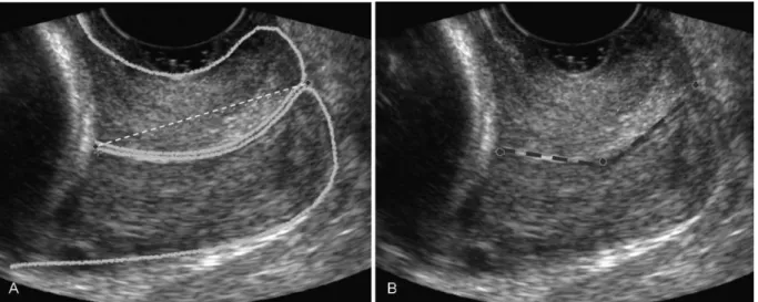
greater than 120°. All the ultrasound examinations were performed with a Logiq P5 apparatus (General Electric Healthcare, Milwaukee, WI, USA). The transducer was cov-ered with a lubricant and gel-free condom and was inserted into the anterior vaginal fornix. This method allowed ob-serving the morphology of the uterine cervix (straight or curved), the internal and external os of the cervix, and the endocervical canal surrounded by the cervical gland area. Continuous biometry was performed for straight uterine cervices, in which the examiner placed one measuring cali-per in the internal os and another in the external os (►Fig. 1A). For curved uterine cervices, the examiner used
two measurement techniques; the continuous technique as described above and the segmented technique, in which the
first measuring caliper was placed in the internal cervical os, the second caliper was placed at the beginning of the cervical
bend, and the last caliper was placed in the external cervical os (►Fig. 1B). The sum of these segments was described as
the length of the uterine cervix. All measurements were performed by only two experienced examiners.
The qualitative variables (gestational age, number of gestations, number of vaginal deliveries, maternal age, eth-nicity, smoking, curved cervix, and straight cervix) were expressed as absolute and relative values, whereas the quantitative variables (cervical length measured by both the continuous and the segmented techniques) were expressed as means, standard deviations (SD), medians, and minimum and maximum values. Polynomial regressions were performed to evaluate the correlation between cervical length and gestational age, and the quality offit was assessed using the coefficient of determination (R2). The
measure-ments of the uterine cervix performed using the continuous and segmented techniques were compared using the paired Studentt-test. The statistical analysis was performed using the SPSS software version 13.0 (SPSS Inc., Chicago, IL, USA), with the level of significance set atp<0.05.
Results
The assessment included 752 pregnant women at gestational ages between 18 and 24 weeks, with the following distribu-tion: 87 (11.5%) at 18 weeks, 91 (12.1%) at 19 weeks, 86 (11.4%) at 20 weeks, 84 (11.2%) at 21 weeks, 83 (11.0%) at 22 weeks, 211 (28.1%) at 23 weeks, and 110 (14.6%) at 24 weeks. The mean maternal age was 30.35.3 years (16–42 years); 68.8% women were white, 51.2% were
primi-gravidas, and 9.6% were smokers.
The results of the continuous cervical biometry varied from 38.77.4 mm (16–60 mm) at the 18th week to 36.3
7.5 mm (12–53 mm) at the 24th week. The results of the
segmented cervical biometry varied from 40.27.8 mm (16–60 mm) at the 18thweek to 38.17.9 mm (12–53 mm)
at the 24th week. Uterine cervical biometry did not vary
significantly with gestational age and was best represented by a linear equation both for the continuous (R2¼0.0075) and
the segmented (R2¼0.0017) techniques (
►Fig. 2).
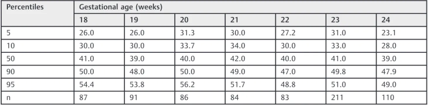
►Tables 1and2show the percentiles 5, 10, 50, 90, and 95
of the cervical length, using the continuous and segmented techniques as a function of gestational age, respectively.
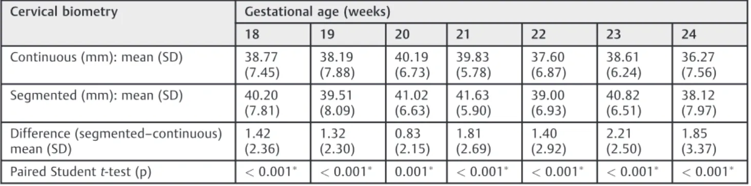
►Table 3shows that there was a statistically significant
difference between cervical lengths measured by both tech-niques at all gestational ages (p<0.001), the mean
differ-ence being 1.7 mm2.6 mm.
Of these 752 pregnant women, 390 (51.9%) exhibited a curved uterine cervix and 362 (48.1%) had a straight cervix.►Table 4shows the distribution of curved and straight
cervical morphology between 18 and 24 weeks of gestation.
According to►Fig. 3, there was a predominance of curved
uterine cervix up to the 21stweek and a predominance of
straight cervix after this gestational age.
Discussion
Until the late 1970s, the methods of assessing the uterine cervix consisted basically of subjective methods, namely direct observation through speculum examination and vagi-nal bidigital palpation. With the advent of the transvagivagi-nal ultrasound, in the early 1980s, the morphology and biometry
Table 2 Percentiles of uterine cervical length (mm) using the segmented technique between 18 and 24 weeks
Percentiles Gestational age (weeks)
18 19 20 21 22 23 24
5 26.0 26.0 31.3 30.0 27.2 31.0 23.1
10 30.0 30.0 33.7 34.0 30.0 33.0 28.0
50 41.0 39.0 40.0 42.0 40.0 41.0 39.0
90 50.0 48.0 50.0 49.0 47.0 49.8 47.9
95 54.4 53.8 56.2 51.7 48.8 51.0 49.0
n 87 91 86 84 83 211 110
Fig. 2 Scatter plots of uterine cervical biometry, using the continuous(A)and the segmented(B)techniques, as a function of gestational age(B).
Table 1 Percentiles of uterine cervical length (mm) using the continuous technique between 18 and 24 weeks of gestation
Percentiles Gestational age (weeks)
18 19 20 21 22 23 24
5 26.0 26.0 31.3 30.0 26.4 27.5 22.0
10 29.0 28.4 33.0 32.0 29.1 31.7 27.0
50 39.0 38.0 39.0 40.0 38.3 38.7 37.3
90 47.2 46.8 49.0 47.5 45.3 46.7 44.9
95 52.6 53.8 56.3 48.7 46.2 47.9 46.8
of the uterine cervix have been increasingly studied, which led to a thorough investigations for the assessment of physi-ological changes during pregnancy.14
In the present study, by means of transvaginal ultrasound, we determined reference values for cervical length between 18 and 24 weeks of gestation; our sample was a Brazilian population at low risk for preterm labor, with a small variation within this gestational interval. Jafari-Dehkordi et al15 determined reference values for cervical length between 8 and 38 weeks of gestation using abdominal ultrasound in Iranian women. The mean cervical length did not show much difference between 18 and 24 weeks of gestation (38.0 mm). A recent study established conditional
intervals of cervical length using transvaginal ultrasound between 11 and 40 weeks in 4,397 Greek women. They observed that the best correlation between uterine cervical length and gestational age was a second-degree equation and the mean length between 18 and 24 weeks changed from 32.1 to 31.3 mm.16In a recent Brazilian study, Peixoto et al5 established a reference curve for cervical length in 996 singleton pregnancies between 20 and 24 weeks of gestation using the continuous technique of transvaginal ultrasound. The values did not vary significantly with gestational age and the mean length was 37 mm in this gestational interval. In the randomized controlled trial, the mean cervical length between 20 and 25 weeks of gestation was 33 mm. This value was lower than the one found in our study (38 mm); however, there are several differences between both studies, such as low versus unselected population different number of cases (752 versus 24,620) and type of pregnancy (single-ton versus single(single-ton and twin).17
With regard to morphology, there was a predominance of curved uterine cervices.10,17In the study by Yost et al,18to determine parameters that could be predictors of preterm labor between 16 and 18 weeks of gestation, the curved cervical morphology predominated over the straight mor-phology (59% versus 41%); however, this type of mormor-phology was shown to be a poor predictor. The results of this study are in line with our findings, namely the results of 51.9% of curved uterine cervix and 48.1% of straight cervix, thus confirming the predominance of the curved morphology up to the 21stweek. This morphology may be explained by
local changes in collagen concentration in the uterine cervix during thefirst trimester of gestation.19
Table 3 Comparison between the continuous and the segmented techniques of cervical biometry between 18 and 24 weeks
Cervical biometry Gestational age (weeks)
18 19 20 21 22 23 24
Continuous (mm): mean (SD) 38.77 (7.45)
38.19 (7.88)
40.19 (6.73)
39.83 (5.78)
37.60 (6.87)
38.61 (6.24)
36.27 (7.56)
Segmented (mm): mean (SD) 40.20 (7.81)
39.51 (8.09)
41.02 (6.63)
41.63 (5.90)
39.00 (6.93)
40.82 (6.51)
38.12 (7.97)
Difference (segmented–continuous) mean (SD)
1.42 (2.36)
1.32 (2.30)
0.83 (2.15)
1.81 (2.69)
1.40 (2.92)
2.21 (2.50)
1.85 (3.37)
Paired Studentt-test (p) <0.001 <0.001 0.001 <0.001 <0.001 <0.001 <0.001
Abbreviation: SD, standard deviation. Chi-square test for trend:p¼0.001 .
Table 4 Distribution of uterine cervical morphology between 18 and 24 weeks of gestation
Variables Gestational age (weeks)
18 19 20 21 22 23 24
n (%) n (%) n (%) n (%) n (%) n (%) n (%)
Curved cervix 58 (66.7) 51 (56.0) 46 (53.5) 50 (59.5) 34 (41.0) 97 (46.0) 54 (49.1)
Straight cervix 29 (33.3) 40 (44.0) 40 (46.5) 34 (40.5) 49 (59.0) 114 (54.0) 56 (50.9)
Total 87 91 86 84 83 211 110
We used two measuring techniques to determine the cervical length: continuous and segmented. In women with a straight uterine cervix the measurements did not differ significantly; however, cervical biometry is hindered when the continuous technique is used in women with a curved cervix because the cervical length is often underestimated. According to the literature, the segmented technique, when performed along the endocervical canal, yields slightly higher yet more accurate values.10 The most adequate method to measure the uterine cervix when there is a bend seems to be the segmented technique; however, to prove this hypothesis it is necessary to compare the measurements to a gold standard or to establish the correlation between measurement by both techniques and the outcome of spontaneous preterm birth. So, in the interval between the 18th and the 24th weeks of
gestation, the ideal gestational age for measuring the curved uterine cervix was the 21st week using transvaginal
ultra-sound. At this stage, the phenomenon of uterine conversion has already occurred and the straight cervical morphology predominates; accordingly, biometric measurements are eas-ier to perform and are more accurate.
As limitation, neither intra- nor inter-observer reproduc-ibility were performed. However, all measurements were taken by only two experienced examiners, which could decrease this reproducibility. Furthermore, all cervical length measurements were performed by transvaginal route. In a previous study, transabdominal ultrasound measure-ment overestimated the mean cervical length by 8 mm among pregnant women with a short cervix and resulted in the underdiagnosis of 57% of cases.20
Conclusion
In conclusion, we determined reference values for uterine cervical length between 18 and 24 weeks of gestation in a Brazilian low-risk population using the continuous and segmented techniques. The values did not vary significantly with gestational age. The ideal gestational age for measuring cervical length was21 weeks.
Contributors
Andrade S. G. A., Andrade F. M., Araujo Júnior E., Pires C. R., Mattar R. and Moron A. F. contributed with the project conception, analysis and interpretation of data, critical review of the intellectual content andfinal approval of the version to be published.
Conflicts to Interest
Authors declare no conflict of interest.
References
1 Tsoi E, Akmal S, Geerts L, Jeffery B, Nicolaides KH. Sonographic measurement of cervical length and fetalfibronectin testing in threatened preterm labor. Ultrasound Obstet Gynecol 2006;27 (04):368–372
2 Hassan S, Romero R, Hendler I, et al. A sonographic short cervix as the only clinical manifestation of intra-amniotic infection. J Perinat Med 2006;34(01):13–19
3 Berghella V, Tolosa JE, Kuhlman K, Weiner S, Bolognese RJ, Wapner RJ. Cervical ultrasonography compared with manual examination as a predictor of preterm delivery. Am J Obstet Gynecol 1997; 177(04):723–730
4 Sonek JD, Iams JD, Blumenfeld M, Johnson F, Landon M, Gabbe S. Measurement of cervical length in pregnancy: comparison between vaginal ultrasonography and digital examination. Obstet Gynecol 1990;76(02):172–175
5 Peixoto AB, da Cunha Caldas TM, Alamy AH, Martins WP, Bruns RF, Araujo Júnior E. Reference values for the cervical length measure-ment in the second trimester of pregnancy using the transvaginal ultrasound in a large Brazilian population. Obstet Gynecol Sci 2016;59(04):303–306
6 Salomon LJ, Diaz-Garcia C, Bernard JP, Ville Y. Reference range for cervical length throughout pregnancy: non-parametric LMS-based model applied to a large sample. Ultrasound Obstet Gynecol 2009;33(04):459–464
7 Guzman ER, Walters C, Ananth CV, et al. A comparison of sono-graphic cervical parameters in predicting spontaneous preterm birth in high-risk singleton gestations. Ultrasound Obstet Gynecol 2001;18(03):204–210
8 Bagga R, Takhtani M, Suri V, Adhikari K, Arora S, Bhardwaj S. Cervical length and cervicovaginal HCG for prediction of pre-term birth in women with signs and symptoms of pre-term labour. J Obstet Gynaecol 2010;30(05):451–455
9 Jwala S, Tran TL, Terenna C, et al. Evaluation of additive effect of quantitative fetalfibronectin to cervical length for prediction of spontaneous preterm birth among asymptomatic low-risk women. Acta Obstet Gynecol Scand 2016;95(08):948–955
10 Gramellini D, Fieni S, Molina E, Berretta R, Vadora E. Transvaginal sonographic cervical length changes during normal pregnancy. J Ultrasound Med 2002;21(03):227–232, quiz 234–235
11 Rozenberg P, Gillet A, Ville Y. Transvaginal sonographic examina-tion of the cervix in asymptomatic pregnant women: review of the literature. Ultrasound Obstet Gynecol 2002;19(03):302–311
12 Khalifeh A, Quist-Nelson J, Berghella V. Universal cervical length screening for preterm birth prevention in the United States. J Matern Fetal Neonatal Med 2017;30(12):1500–1503
13 Pedretti MK, Kazemier BM, Dickinson JE, Mol BW. Implementing universal cervical length screening in asymptomatic women with singleton pregnancies: challenges and opportunities. Aust N Z J Obstet Gynaecol 2017;57(02):221–227
14 Dodson MG, Deter RL. Definition of anatomical planes for use in transvaginal sonography. J Clin Ultrasound 1990;18(04):239–242
15 Jafari-Dehkordi E, Adibi A, Sirus M. Reference range of the weekly uterine cervical length at 8 to 38 weeks of gestation in the center of Iran. Adv Biomed Res 2015;4:115
16 Papastefanou I, Pilalis A, Kappou D, Souka AP. Cervical length at 11-40 weeks: unconditional and conditional longitudinal refer-ence ranges. Acta Obstet Gynecol Scand 2016;95(12):1376–1382
17 Fonseca EB, Celik E, Parra M, Singh M, Nicolaides KH; Fetal Medicine Foundation Second Trimester Screening Group. Proges-terone and the risk of preterm birth among women with a short cervix. N Engl J Med 2007;357(05):462–469
18 Yost NP, Owen J, Berghella V, et al; National Institute of Child Health and Human Development, Maternal-Fetal Medicine Units Network. Second-trimester cervical sonography: features other than cervical length to predict spontaneous preterm birth. Obstet Gynecol 2004;103(03):457–462
19 Gedikbasi A, Yücel B, Arslan O, Giris M, Gedikbasi A, Abbasoglu SD. Dynamic collagen changes in cervix during thefirst trimester and decreased collagen content in cervical insufficiency. J Matern Fetal Neonatal Med 2016;29(18):2968–2972