Stress-related hormone norepinephrine
induces interleukin-6 expression
in GES-1 cells
R. Yang, Q. Lin, H.B. Gao and P. Zhang
Department of Biochemistry and Molecular Cell Biology, School of Medicine, Shanghai Jiao Tong University, Shanghai, China
Abstract
In the current literature, there is evidence that psychological factors can affect the incidence and progression of some cancers. Interleukin 6 (IL-6) is known to be elevated in individuals experiencing chronic stress and is also involved in oncogenesis and cancer progression. However, the precise mechanism of IL-6 induction by the stress-related hormone norepinephrine (NE) is not clear, and, furthermore, there are no reports about the effect of NE on IL-6 expression in gastric epithelial cells. In this study, we examined the effect of NE on IL-6 expression in immortalized human gastric epithelial cells (GES-1 cells). Using real-time PCR and enzyme-linked immunoassay, we demonstrated that NE can induce IL-6 mRNA and protein expression in GES-1 cells. The induction is through theb-adrenergic receptor-cAMP-protein kinase A pathway and mainly at the transcriptional level. Progressive 59-deletions and site-directed mutagenesis of the parental construct show that, although activating-protein-1 (AP-1), cAMP-responsive element binding protein (CREB), CCAAT-enhancer binding protein-b(C/EBP-b), and nuclear factor k-light-chain-enhancer of activated B cells (NF-kB) binding sites are all required in the basal transcription of IL-6, only AP-1 and CREB binding sites in the IL-6 promoter are required in NE-induced IL-6 expression. The results suggest that chronic stress may increase IL-6 secretion of human gastric epithelial cells, at least in part, by the stress-associated hormone norepinephrine, and provides basic data on stress and gastric cancer progression.
Key words: Norepinephrine; GES-1 cells; Interleukin-6
Introduction
The amount of literature reporting the role of psycho-logical stress in cancer onset and development is growing. Regarding the mechanisms connecting stress and cancer progression, most of the early studies concentrated on the indirect effects of stress through immunosuppression in cancer patients (1-3). Recently, it has been reported that the stress-related hormones norepinephrine (NE) and epinephrine, especially NE, can directly induce gene expression and are involved in angiogenesis and metastasis in cancer progression in a variety of cancer cell lines (4-8), stromal cells (9), and immortalized epithelial cell lines (10). Several epidemio-logical studies have demonstrated that chronic stress may accelerate the progression of gastric cancer (11-13). However, the precise mechanism by which chronic stress acts in gastric cancer progression is unclear, and little is known about the effects of stress-related hormones on gastric cancer cells and gastric epithelial cells. We recently performed Bio-Plex analyses (Bio-Rad, USA) to
examine the effect of NE on gastric cancer cells and gastric epithelial cells by detecting several cytokines associated with gastric cancer progression (Figure S1). These results showed the only substantial change by NE was the upregulation of interleukin-6 (IL-6) expression in GES-1 cells, an immortalized human gastric epithelial cell line. Because there are no normal human gastric epithelial cells that are commercially available, we used GES-1 cells (14) to study the effect of NE on gastric epithelial cells.
IL-6 is a pleiotropic cytokine produced by a wide variety of cells including macrophages, T cells, B cells, fibroblasts, and endothelial cells. IL-6 plays important roles in a wide range of biological activities including immune regulation, hematopoiesis, inflammation, and oncogenesis (15-17). IL-6 is also involved in gastric cancer progression, and several reports demonstrated that significant relationships existed between elevated serum IL-6 levels and tumor stages, with unfavorable
Correspondence: P. Zhang, Department of Biochemistry and Molecular Cell Biology, School of Medicine, Shanghai Jiao Tong University, Building 7-203, No. 280, South Chongqing Road, Shanghai 200025, China. E-mail: [email protected]
outcomes in gastric cancer patients (18-20). Interestingly, IL-6 is closely related to chronic stress. Epidemiological studies demonstrated that IL-6 was elevated in a population suffering from chronic stress (21,22). Animal models also showed that stress can lead to elevated IL-6 in rats, and the sources were not immune cells (23). However, the precise mechanism of NE-induced IL-6 expression is not clear, and, furthermore, there are still no data available regarding the effect of NE on IL-6 expression in gastric epithelial cells. The present study, therefore, is aimed at investigating the effect of NE on IL-6 expression in GES-1 cells and exploring the signaling pathway and molecular mechanism involved.
Material and Methods
Cell lines and culture conditions
GES-1 (kindly provided by Dr. Zhu Zhenggang, China) is an immortalized human gastric epithelial cell line, established from fetal gastric epithelial cells after simian virus 40 (SV40) transfection (14). GES-1 cells were maintained in RPMI 1640 medium supplemented with 10% fetal bovine serum (FBS), 2 mM L-glutamine, 100 U/mL penicillin, and 100mg/mL streptomycin at 376C in a humidified atmosphere with 5% CO2. The cells were
allowed to reach 80% confluency before passage. The culture medium was replenished with fresh medium every 2 or 3 days. Although the initial NE-treated experiments (Figure 1) were carried out by maintaining the cells throughout in a medium containing 10% FBS, the other protocol has been used more extensively. Cells were cultured in 10% FBS, and, for isoproterenol treatment, blocking, and the following NE treatments, a medium containing Advanced 1640 (Invitrogen, USA) with 1% FBS was utilized in order to eliminate the possible influence of factors, which can be found in FBS, on the effect of NE in GES-1 cells.
To evaluate the effects of stress hormones on IL-6 secretion, 66104GES-1 cells were seeded into individual
wells of a 24-well plate. Following a 24-h incubation, triplicate cultures (wells) were stimulated by replacing the complete media containing NE or the synthetic
b-adrenergic receptor agonist isoproterenol, at specific concentrations. Culture supernatants were collected at various time points, centrifuged, and stored at ––706C until assayed by enzyme-linked immunoassay (ELISA). Cells were homogenized in TRIzol reagent and stored at ––706C until assayed by real-time PCR.
Reagents
Phentolamine mesylate was purchased from Santa Cruz (USA), forskolin from Calbiochem (USA), KT5720 from Tocris (UK), and actinomycin D (Act D) from Beyotime Institute of Biotechnology Co. (China). Other chemicals were purchased from Sigma-Aldrich (USA).
ELISA
The concentration of IL-6 was measured using a human IL-6 ELISA Kit (Dakewe Biotech Company Limited, China) following the manufacturer’s protocol. The resultant color was read at 450 nm using a Multiskan Spectrum microplate reader (Thermo Fisher Scientific, Finland) with the SkanIt software (version 2.4.2, Thermo Fisher Scientific). The concentration of IL-6 in a sample was determined by interpolation from a standard curve.
Real-time PCR
We utilized real-time RT-PCR on NE-treated cell lines in order to determine the effect of NE on IL-6 gene expression. Total RNA from cultured cells was isolated using TRIzol reagent following the manufacturer’s instruc-tions (Invitrogen). First-strand cDNAs were synthesized using random primers and RevertAidTM M-MuLV reverse
transcriptase (Fermentas, Lithuania). Reactions were performed with SYBR1Premix Ex TaqTMand the
speci-fic primers, following the manufacturer’s instructions (TaKaRa BIO Inc., China). Levels of IL-6 mRNA were measured and amplified using the 7300 real-time PCR system (Applied Biosystems, USA). The cycler conditions were as follows: incubation for 30 s at 956C, followed by 5 s at 956C, and 31 s at 606C for 40 cycles. The levels of expression of IL-6 mRNA in each sample were normalized to the GAPDH mRNA levels. The relative expression of mRNA species was calculated using the 2–DDCt method. All primer sequences span across two adjacent exons of the target genes and are thus specific for mRNAs, as follows: IL-6 forward primer: 59-AACCTGAACCTTCCA AAGATGG-39; IL-6 reverse primer: 59-TCTGGCTTGTTC CTCACTACT-39; GAPDH forward primer: 59-TGTTGC CATCAATGACCCCTT-39; GAPDH reverse primer: 59 -CTCCACGACGTACTCAGCG-39.
In order to elucidate the mechanism in the NE-dependent regulation of IL-6 mRNA levels in GES-1 cells, the effect of Act D, an inhibitor ofde novotranscription, was assessed on mRNA levels. GES-1 cells were grown
in the presence of 5mg/mL Act D and 10mM NE for 1 h. Total RNA was isolated, and the levels of IL-6 mRNA were measured using real-time PCR as described earlier.
Assessment of signaling pathways
In order to examine the signaling pathway involved in NE-induced IL-6 expression, we treated GES-1 cells with a variety of agonists and antagonists. Theb-adrenoreceptor antagonist propranolol (10mM) and the protein kinase A (PKA) inhibitor KT5720 (10mM) were added to the cell cultures 3 h before adding 10mM NE. Thea-adrenoreceptor antagonist phentolamine (10mM) was added to the cell cultures 1 h prior to the addition of 10mM NE. After blocking, the media was replaced with 1% FBS Advanced 1640 containing 10mM NE and the cells continued to incubate for 3 h. GES-1 cells were treated with the b-adrenoreceptor agonist isoproterenol (10mM) and the adenylate cyclase agonist forskolin (10mM). Conditioned medium was col-lected after a 3-h incubation, centrifuged at 300gfor 10 min, and stored at ––706C until tested for the presence of IL-6 by ELISA.
Plasmid construction, 59-deletion constructs, and site-directed mutagenesis
Genomic DNA was isolated from GES-1 cells using a Tianamp Genomic DNA kit (Tiangen, Biotech, China). A 2092-bp PCR fragment corresponding to the promoter region of IL-6 (––2035 to ++53 bp) was generated using the upstream primer: 59-GTGGTACCCCCGTTTTATAGG-39
and the downstream primer: 59-CTGGAGGGGAGA TAGAGCTTC-39 in a PCR using PrimerSTAR HS1
DNA polymerase (TaKaRa BIO Inc.). The 2092-bp fragment was subcloned (59-KpnI, 39-XhoI) into a pGL3 basic luciferase reporter gene vector (Promega, USA) and sequenced for orientation and fidelity. This plasmid served as the source of inserts for further plasmid constructions.
To make the 59-deletion constructs, shorter fragments were generated using new upstream primers, and the
common downstream primer was the same as the reverse primer of the 2092-bp fragment. From this parental construct, pIL-6P-luc1184 and three 59-deletion mutants were generated by using the upstream primer as shown in Table 1. pIL-6P-luc1184 contains the full-length sequence (24) of the IL-6 promoter (from 1173 bp upstream of the transcription start site to ++11 bp). pIL-6P-luc710 contains the four consensus sequences for the transcription factors activating-protein-1 (AP-1), cAMP-responsive element binding protein (CREB), CCAAT-enhancer binding pro-tein-b(C/EBP-b), and nuclear factork -light-chain-enhan-cer of activated B cells (NF-kB) as pIL-6P-luc1184. The other two 59-deletion mutants were deleted fragments containing the consensus sequences for transcription factors C/EBP-b and NF-kB (pIL-6P-luc204) or NF-kB (pIL-6P-luc107). DNA sequence analysis confirmed these sequences.
Within pIL-6P-luc1184, critical nucleotides necessary for transcription factor (AP-1, CREB, C/EBP-b, and NF-kB) binding to the four consensus sites were inactivated by using a QuikChange Lightning site-directed mutagenesis kit (Agilent Technology, USA) and are listed in Table 1. These mutations have previously been shown to inactivate the described consensus sequences (25). All mutant clones designated as pIL-6P-AP1-m, pIL-6P-CREB-m, pIL-6P-C/EBP-m, and pIL-6P-NFkB-m were verified by DNA sequencing.
Cell transfection and luciferase assays
Cell transfection was performed according to the manufacturer’s instruction using the LipofectamineTM
2000 reagent (Invitrogen). Briefly, the cells were seeded onto 24-well plates at a density of 66104cells/well and
incubated at 376C overnight. For each transfection sample, cells were transfected with 0.1mg luciferase plasmid and 1 ng pRL-TK reporter plasmid using 2mL LipofectamineTM2000.
Sixteen hours following transfection, the cells were treated with 10mM NE for 3 h. After treatment, a dual
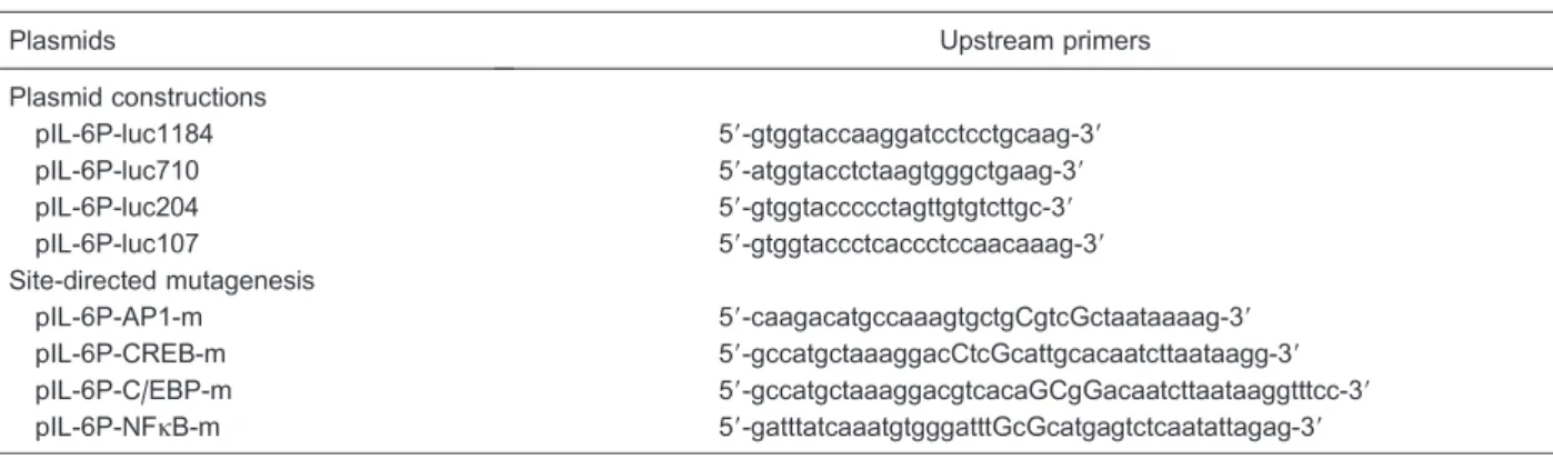
Table 1. Sequences of upstream primers used in the plasmid constructions and site-directed mutagenesis.
Plasmids Upstream primers
Plasmid constructions
pIL-6P-luc1184 59-gtggtaccaaggatcctcctgcaag-39
pIL-6P-luc710 59-atggtacctctaagtgggctgaag-39
pIL-6P-luc204 59-gtggtaccccctagttgtgtcttgc-39
pIL-6P-luc107 59-gtggtaccctcaccctccaacaaag-39
Site-directed mutagenesis
pIL-6P-AP1-m 59-caagacatgccaaagtgctgCgtcGctaataaaag-39
pIL-6P-CREB-m 59-gccatgctaaaggacCtcGcattgcacaatcttaataagg-39
pIL-6P-C/EBP-m 59-gccatgctaaaggacgtcacaGCgGacaatcttaataaggtttcc-39
pIL-6P-NFkB-m 59-gatttatcaaatgtgggatttGcGcatgagtctcaatattagag-39
luciferase assay (Promega) was performed. In brief, cells were lysed with passive lysis buffer, and 20mL aliquots of
each sample were assayed on an amber 96-well plate by a luminometer (NOVOstar, Germany). As an internal control for transfection efficiency, pRL-TK, the expression plasmid encodingRenillaluciferase driven by the thymi-dine kinase promoter, was used (1 ng/well). Firefly and
Renillaluciferase have distinct substrate properties, and thus the activities of both enzymes can be assessed in the same sample using two substrates sequentially. Each transfection was performed in triplicate and in a minimum of three independent experiments.
Statistical analysis
Statistically significant differences between groups were determined by ANOVA using GraphPad Prism v5.0 (GraphPad Software Inc., USA). P,0.05 was considered to indicate a statistically significant difference. When a significant main effect of drug treatment was identified (P,0.05), the Newman-Keulspost hoctest was used to compare groups.
Results
NE stimulation increases IL-6 protein in GES-1 cells
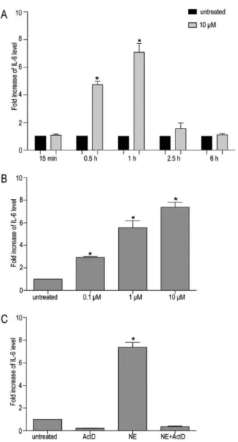
GES-1 cells were stimulated with increasing concen-trations of NE, and the supernatants were assayed for IL-6 by ELISA as shown in Figure 1. Using ANOVA and the Newman-Keulspost hoctest, we demonstrated that an NE dose-dependent and time-dependent increase of IL-6 expression in culture supernatants of GES-1 cells with the 1 h treatment yielded the greatest effect. Treatment of GES-1 cells with 1 and 10mM NE for 1 h produced a 3.15±1.31 and 4.71±2.02 fold increase, respectively. The overall IL-6 induction by 10mM NE in 3 and 6 h differed significantly from the control values, but the fold increase was less than that produced in 1 h by 10mM NE (4.16±0.53 fold in 3 h and 2.21±0.38 fold in 6 h). Because the mean basal production of IL-6 by GES-1 cells at GES-1 h was 9.47±0.87 pg/mL, which was very near the lower detection limit of the ELISA kit, we chose the time point of 3 h and 10mM NE in the following experimental protocols.
NE stimulation increases IL-6 mRNA in GES-1 cells
To further elucidate the mechanism involved in the induction of IL-6 expression in GES-1 cells, we examined the effect of exposure to NE on the transcription of IL-6 expression. GES-1 cells were stimulated with 10mM NE, and IL-6 mRNA levels were quantified using real-time RT-PCR at multiple time points ranging from 15 min to 6 h. The level of IL-6 mRNA was normalized against GAPDH mRNA levels. As shown in Figure 2A, treatment of GES-1 cells with NE resulted in a significant increase of IL-6 mRNA levels, which peaked after 1 h, decreased there-after, and returned to baseline within 6 h. However, for
cells exposed to specified doses of NE, IL-6 mRNA levels showed a dose-dependent increase (Figure 2B).
Co-treatment of GES-1 cells with 5mg/mL Act D and 10mM NE efficiently inhibited the NE-dependent upregu-lation of IL-6 mRNA levels (Figure 2C). This suggests that the NE-dependent upregulation of the IL-6 protein in GES-1 cells is mainly due to the stimulation of de novo
transcriptional activity of the IL-6 gene.
Effect of NE on IL-6 upregulation is mediated by the b-adrenoreceptor-adenylyl cyclase-cAMP-PKA signaling pathway
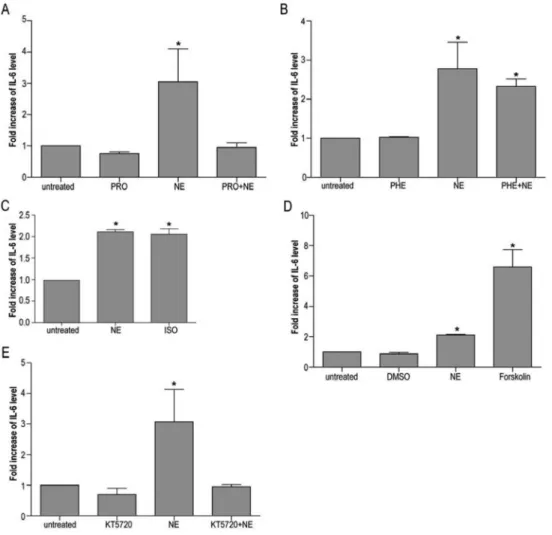
To determine whether the effect of NE on IL-6 expres-sion is transduced by ligation of the b-adrenoreceptor, GES-1 cells were incubated in the presence of the
b-antagonist propranolol (10mM) prior to 10mM NE
stimulation. As shown in Figure 3A, propranolol completely abolished NE-induced IL-6 expression. In contrast, thea -adrenoreceptor antagonist phentolamine (10mM) had no effect on the ability of NE to induce IL-6 production (Figure 3B). Treatment with the b-adrenoreceptor agonist iso-proterenol (10mM) resulted in significant stimulation of IL-6 gene expression, supporting the hypothesis that ligation of b-adrenoreceptors is involved in the observed NE-dependent effect (Figure 3C).
To determine whether activation of the adenylyl cyclase-cAMP-PKA signaling pathway is capable of IL-6 upregulation, GES-1 cells were stimulated in the presence of the adenylyl cyclase activator forskolin (10mM). Forskolin increased IL-6 expression to levels similar to those observed in NE-treated cultures (Figure 3D). To determine whether the effects of NE on IL-6 expression
were mediated by activation of PKA, GES-1 cells were incubated in the presence of the PKA inhibitor KT5720 (10mM) prior to NE stimulation. PKA blockade strongly inhibited the effect of NE on IL-6 expression (Figure 3E), reducing IL-6 expression to levels that were statistically indistinguishable from control cultures. Thus, activation of the PKA signaling cascade via cell surfaceb-adrenoreceptor appears to mediate the effect of NE on IL-6 expression.
IL-6 promoter is inducible by NE in GES-1 cells
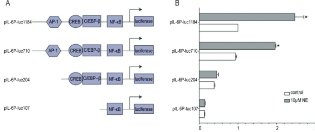
In order to identifycis-regulatory sequences that were responsible for upregulation of IL-6 expression by NE, we generated the full-length IL-6 promoter (24) construct (1184 bp upstream of the transcription start site), desig-nated as pIL-6P-luc1184, and the following three deletion mutants of pIL-6P-luc1184 as follows: i) pIL-6P-luc710, containing four cis-acting elements; ii) pIL-6P-luc204, deficient in a known AP-1 consensus sequence; and iii) pIL-6P-luc107, deficient in both a known AP-1 and a known CREB consensus sequence (Figure 4A).
As demonstrated in Figure 4B, pIL-6P-luc710 has the same basal activity as pIL-6P-luc1184, and both can be induced by NE with a 210±15.6% increase (P,0.05). However, the IL-6 induction by NE in GES-1 cells transfected with pIL-6P-luc204 and pIL-6P-luc107 did not differ significantly from control values. This suggests that the NE-responsive cis-acting elements are located from ––699 to ++11 bp of the IL-6 promoter.
AP-1 and CREB binding sites are required for induction of the IL-6 gene by NE
To examine which regulatory elements in the IL-6
promoter are responsive to NE, we introduced 2- to 3-bp mutations into the core regions of the transcription factor (AP-1, CREB, C/EBP-b, and NF-kB) binding sites within the context of the 1.2-kb IL-6 promoter as outlined in Figure 5A, with the intention to completely abolish the interaction between individual trans-acting factors and their cognate recognition sequences. In contrast to deletion studies, this approach should enable us to identify the contribution of single regulatory elements to gene activation in the context of the full-length promoter. As shown in Figure 5B, when AP-1 and CREB binding sites were mutated, the basal promoter activity was reduced to 31±3.3% and 78.6±10.3% of the wild-type promoter, respectively. Also, there was no statistical difference between GES-1 cells transfected with pIL-6P-CREB-m and pIL-6P-luc1184, while there was a statistical difference between GES-1 cells transfected with pIL-6P-AP1-m and pIL-6P-luc1184. However, mutations of the AP-1 and CREB binding sites can both diminish induci-bility by NE. Therefore, it can be concluded that AP-1 and CREB binding sites were found to be functionally required for inducibility of the IL-6 promoter by NE.
In contrast, as shown in Figure 5B, mutation of the C/EBP-b and NF-kB binding sites did not significantly affect inducibility by NE, and basal promoter activity was reduced to 79.4±16.7% (P.0.05) and 46.1±15% (P,0.05) of the wild-type promoter, respectively. These data indicate that C/EBP-b and NF-kB binding sites are not essential for NE inducibility.
Briefly, AP-1, CREB, C/EBP-b, and NF-kB binding sites in the IL-6 promoter are all involved in the basal transciption of IL-6. AP-1 and CREB binding sites are
involved in NE-induced IL-6 expression, whereas
C/EBP-band NF-kB binding sites are not.
Discussion
In the present study, we found that the stress-related hormone NE can induce IL-6 expression in GES-1 cells and does so via theb-adrenoreceptor-adenylyl cyclase-cAMP-PKA signaling cascade. Moreover, we demon-strated that NE induces IL-6 expression mainly at the transcriptional level. Further evidence demonstrated that NE-induced IL-6 expression needs AP-1 and CREB binding sites in the IL-6 promoter.
We first examined the effect of NE on IL-6 expression of human gastric epithelial cells. Because normal human gastric epithelial cells are not commercially available, we used the immortalized human gastric epithelial cell line GES-1. The results demonstrated that NE induced IL-6 expression in GES-1 cells. Because IL-6 is a pleiotropic cytokine closely related to chronic stress, regulation of IL-6 by stress-related hormones has been suggested previously (7,26). Upregulation of IL-6 is also associated with gastric diseases; therefore, a better understanding of the molecular regulation of IL-6 production by NE in gastric epithelial cells has important clinical implications. However, until now, the precise mechanism of IL-6 induction after exposure to NE in gastric epithelial cells has not been explored. Therefore, we further examined the mechanism involved in NE-induced IL-6 expression in GES-1 cells.
In the classical NE signaling pathway, ligation of the cell surfaceb-adrenoreceptor activates adenylyl cyclase, which triggers increased synthesis of cAMP and thereby activates the cAMP-dependent kinase PKA, leading to the phosphorylation of downstream molecules. In our study, propranolol, the b-adrenoreceptor antagonist, completely abrogated the effect of NE on IL-6 expres-sion, while the a-adrenoreceptor antagonist phento-lamine had no effect. The b-adrenoreceptor agonist isoproterenol mimicked the effect of NE on IL-6 expres-sion. Given that GES-1 cells express both b1- and b2-adrenoreceptors by Western blot (see Figure S2), it is
concluded thatb-adrenoreceptor is involved in NE-induced IL-6 expression whilea-adrenoreceptor is not. Forskolin, an adenylyl cyclase activator, mimicked the effect of NE on IL-6 expression, and the PKA inhibitor KT5720 completely abro-gated the effect of NE on IL-6 expression. These results suggest that the classical cAMP-PKA signaling pathway mediates NE-induced IL-6 expression in GES-1 cells.
IL-6 secretion and mRNA increased after NE stimula-tion of GES-1 cells. The increase of IL-6 transcripts was completely blocked if the cells were co-treated with the transcriptional inhibitor Act D and NE, indicating IL-6 gene regulation at the transcriptional level in response to NE.
In general, transcriptional induction is due to the specific binding of activated transcription factors at functional DNA recognition elements within accessible regulatory regions of the inducible genes. To determine the effect of NE on transcriptional regulation of IL-6 gene expression, we used a transient transfection assay and
reporter constructs with the luciferase gene placed under the transcriptional control of the human IL-6 promoter. The deletion analysis of pIL-6P-luc1184 showed the NE-responsivecis-acting elements are located from ––699 to +11 bp of the IL-6 promoter. This result is in agreement with other reports that this region contains specific sequence motifs bound by multiple cis-activating tran-scription factors including NF-kB, AP-1, C/EBP-b, and CREB (24,25,27,28).
As shown above, NE induced IL-6 expression through the classical cAMP-PKA signaling pathway. The classical example for a cAMP-regulated transcription factor is CREB, which becomes activated upon phosphorylation by cAMP-dependent PKA and could potentially bind to the CREB protein binding site (29,30). However, transcription factors binding to AP-1 (31), C/EBP-b (32), and NF-kB (33) sites are also considered candidates for the transmission of cAMP-mediated signals to the transcrip-tional machinery. In order to further identify NE-induced cAMP-responsive elements in the IL-6 promoter, we therefore studied the effect of NE on the mutated individual AP-1, CREB, C/EBP-b, and NF-kB binding sites in the IL-6 promoter-reporter gene constructs in transiently transfected GES-1 cells. The results indicate that AP-1 and CREB binding sites are essential for NE induction of IL-6 expression in GES-1 cells, while all four binding sites may be involved in basal transcription of the IL-6 gene. Our finding is in concert with a prior study that has implicated AP-1 and CREB activation in the regulation of IL-6 expression induced by NE or isoproterenol in cardiomyocytes (26). Similarly, AP-1 activation via PKA and p38 MAPK (p38 mitogen-activated protein kinase) is reported to contribute to IL-6 induction in osteoblastic cells under b-adrenergic stimulation (34). In contrast, it has been demonstrated that only the C/EBP-b(NF-IL-6) motif
is involved in NE-mediated IL-6 expression (7) in human ovarian carcinoma cells. Although the NF-kB binding site is reported to be involved in many stimuli (28,35,36), it is not essential for NE-mediated IL-6 expression in GES-1 cells. These studies suggest that, although NE can induce IL-6 expression in various cell types, the roles of these transcription factors in NE-induced IL-6 expression are shown to be cell specific.
In conclusion, the present study demonstrated that stress-related hormone NE induced IL-6 expression in GES-1 cells. The induction was via the b-adrenergic receptor-cAMP-PKA pathway and mainly at the transcrip-tional level. Although AP-1, CREB, C/EBP-b, and NF-kB binding sites are all required in the basal transcription of IL-6, only AP-1 and CREB binding sites in the IL-6 promoter were required for NE-induced IL-6 expression. The present study suggests that chronic stress may increase IL-6 secretion of human gastric epithelial cells, at least in part, by the stress-associated hormone NE, and provides basic data on stress and gastric cancer progression.
Supplementary Material
Click here to view [pdf]
Acknowledgments
GES-1 cells were kindly provided by Dr. Zhu Zhenggang, Shanghai Key Laboratory of Gastric Neoplasms, Ruijin Hospital, Shanghai Jiao Tong University School of Medicine. Research supported by grants from the Leading Academic Discipline Project, Shanghai Municipal Education Commission (#J50201) and the Natural Science Foundation of China (grant
#31000662).
References
1. Ben-Eliyahu S, Yirmiya R, Liebeskind JC, Taylor AN, Gale RP. Stress increases metastatic spread of a mammary tumor in rats: evidence for mediation by the immune system. Brain Behav Immun1991; 5: 193-205, doi: 10.1016/0889-1591(91)90016-4.
2. Ben-Eliyahu S, Shakhar G, Page GG, Stefanski V, Shakhar K. Suppression of NK cell activity and of resistance to metastasis by stress: a role for adrenal catecholamines and b-adrenoceptors.Neuroimmunomodulation2000; 8: 154-164, doi: 10.1159/000054276.
3. Reiche EM, Nunes SO, Morimoto HK. Stress, depression, the immune system, and cancer. Lancet Oncol 2004; 5: 617-625, doi: 10.1016/S1470-2045(04)01597-9.
4. Yang EV, Donovan EL, Benson DM, Glaser R. VEGF is differentially regulated in multiple myeloma-derived cell lines by norepinephrine.Brain Behav Immun2008; 22: 318-323, doi: 10.1016/j.bbi.2007.09.010.
5. Lutgendorf SK, Cole S, Costanzo E, Bradley S, Coffin J, Jabbari S, et al. Stress-related mediators stimulate vascular
endothelial growth factor secretion by two ovarian cancer cell lines.Clin Cancer Res2003; 9: 4514-4521.
6. Bernabe DG, Tamae AC, Biasoli ER, Oliveira SH. Stress hormones increase cell proliferation and regulates inter-leukin-6 secretion in human oral squamous cell carcinoma cells.Brain Behav Immun2011; 25: 574-583, doi: 10.1016/ j.bbi.2010.12.012.
7. Nilsson MB, Armaiz-Pena G, Takahashi R, Lin YG, Trevino J, Li Y, et al. Stress hormones regulate interleukin-6 expression by human ovarian carcinoma cells through a Src-dependent mechanism.J Biol Chem2007; 282: 29919-29926, doi: 10.1074/jbc.M611539200.
8. Yang EV, Kim SJ, Donovan EL, Chen M, Gross AC, Webster Marketon JI, et al. Norepinephrine upregulates VEGF, IL-8, and IL-6 expression in human melanoma tumor cell lines: implications for stress-related enhancement of tumor progression.Brain Behav Immun2009; 23: 267-275, doi: 10.1016/j.bbi.2008.10.005.
Penedo F, DeGeest K, et al. Biobehavioral influences on matrix metalloproteinase expression in ovarian carcinoma. Clin Cancer Res2008; 14: 6839-6846, doi: 10.1158/1078-0432.CCR-08-0230.
10. Chan C, Lin HJ, Lin J. Stress-associated hormone, norepinephrine, increases proliferation and IL-6 levels of human pancreatic duct epithelial cells and can be inhibited by the dietary agent, sulforaphane.Int J Oncol2008; 33: 415-419.
11. Watabe K, Nishi M, Miyake H, Hirata K. Lifestyle and gastric cancer: a case-control study. Oncol Rep 1998; 5: 1191-1194.
12. Jansson C, Johansson AL, Jeding K, Dickman PW, Nyren O, Lagergren J. Psychosocial working conditions and the risk of esophageal and gastric cardia cancers.Eur J Epidemiol2004; 19: 631-641, doi: 10.1023/B:EJEP.0000036806.51918.40. 13. Oh DY, Choi KS, Shin HR, Bang YJ. Public awareness of
gastric cancer risk factors and disease screening in a high risk region: a population-based study. Cancer Res Treat 2009; 41: 59-66, doi: 10.4143/crt.2009.41.2.59.
14. Ke Y, Ning T, Wang B. Establishment and characterization of a SV40 transformed human fetal gastric epithelial cell line-GES-1.Zhonghua Zhong Liu Za Zhi1994; 16: 7-10. 15. Hodge DR, Hurt EM, Farrar WL. The role of IL-6 and STAT3
in inflammation and cancer.Eur J Cancer2005; 41: 2502-2512, doi: 10.1016/j.ejca.2005.08.016.
16. Bromberg J, Wang TC. Inflammation and cancer: IL-6 and STAT3 complete the link.Cancer Cell2009; 15: 79-80, doi: 10.1016/j.ccr.2009.01.009.
17. Kishimoto T. IL-6: from its discovery to clinical applications. Int Immunol2010; 22: 347-352, doi: 10.1093/intimm/dxq030. 18. Kai H, Kitadai Y, Kodama M, Cho S, Kuroda T, Ito M, et al. Involvement of proinflammatory cytokines IL-1band IL-6 in progression of human gastric carcinoma. Anticancer Res 2005; 25: 709-713.
19. Heikkila K, Ebrahim S, Lawlor DA. Systematic review of the association between circulating interleukin-6 (IL-6) and cancer. Eur J Cancer 2008; 44: 937-945, doi: 10.1016/ j.ejca.2008.02.047.
20. Tsujimoto H, Ono S, Ichikura T, Matsumoto Y, Yamamoto J, Hase K. Roles of inflammatory cytokines in the progression of gastric cancer: friends or foes?Gastric Cancer2010; 13: 212-221, doi: 10.1007/s10120-010-0568-x.
21. Kiecolt-Glaser JK, Preacher KJ, MacCallum RC, Atkinson C, Malarkey WB, Glaser R. Chronic stress and age-related increases in the proinflammatory cytokine IL-6. Proc Natl Acad Sci U S A2003; 100: 9090-9095, doi: 10.1073/pnas. 1531903100.
22. Costanzo ES, Lutgendorf SK, Sood AK, Anderson B, Sorosky J, Lubaroff DM. Psychosocial factors and inter-leukin-6 among women with advanced ovarian cancer. Cancer2005; 104: 305-313, doi: 10.1002/cncr.21147. 23. LeMay LG, Vander AJ, Kluger MJ. The effects of
psychological stress on plasma interleukin-6 activity in rats.
Physiol Behav 1990; 47: 957-961, doi: 10.1016/0031-9384(90)90024-X.
24. Ray A, Tatter SB, May LT, Sehgal PB. Activation of the human ‘‘b2-interferon/hepatocyte-stimulating
factor/interleu-kin 6’’ promoter by cytofactor/interleu-kines, viruses, and second messen-ger agonists.Proc Natl Acad Sci U S A 1988; 85: 6701-6705, doi: 10.1073/pnas.85.18.6701.
25. Xiao W, Hodge DR, Wang L, Yang X, Zhang X, Farrar WL. Co-operative functions between nuclear factors NFkB and CCAT/enhancer-binding protein-b (C/EBP-b) regulate the IL-6 promoter in autocrine human prostate cancer cells. Prostate2004; 61: 354-370, doi: 10.1002/pros.20113. 26. Rohrbach S, Engelhardt S, Lohse MJ, Werdan K, Holtz J,
Muller-Werdan U. Activation of AP-1 contributes to the b -adrenoceptor-mediated myocardial induction of interleukin-6. Mol Med 2007; 13: 605-614, doi: 10.2119/2007-00071. Rohrbach.
27. Ray A, Sassone-Corsi P, Sehgal PB. A multiple cytokine-and second messenger-responsive element in the enhancer of the human interleukin-6 gene: similarities with c-fos gene regulation.Mol Cell Biol1989; 9: 5537-5547.
28. Libermann TA, Baltimore D. Activation of interleukin-6 gene expression through the NF-kB transcription factor.Mol Cell Biol1990; 10: 2327-2334.
29. Sands WA, Palmer TM. Regulating gene transcription in response to cyclic AMP elevation.Cell Signal2008; 20: 460-466, doi: 10.1016/j.cellsig.2007.10.005.
30. Shaywitz AJ, Greenberg ME. CREB: a stimulus-induced transcription factor activated by a diverse array of extra-cellular signals.Annu Rev Biochem1999; 68: 821-861, doi: 10.1146/annurev.biochem.68.1.821.
31. de Groot RP, Sassone-Corsi P. Activation of Jun/AP-1 by protein kinase A.Oncogene1992; 7: 2281-2286.
32. Metz R, Ziff E. cAMP stimulates the C/EBP-related transcription factor rNFIL-6 to trans-locate to the nucleus and induce c-fos transcription.Genes Dev1991; 5: 1754-1766, doi: 10.1101/gad.5.10.1754.
33. Shirakawa F, Mizel SB. In vitro activation and nuclear translocation of NF-kB catalyzed by cyclic AMP-dependent protein kinase and protein kinase C.Mol Cell Biol1989; 9: 2424-2430.
34. Kondo A, Mogi M, Koshihara Y, Togari A. Signal transduc-tion system for interleukin-6 and interleukin-11 synthesis stimulated by epinephrine in human osteoblasts and human osteogenic sarcoma cells. Biochem Pharmacol2001; 61: 319-326, doi: 10.1016/S0006-2952(00)00544-X.