Breathing pattern, thoracoabdominal
motion and muscular activity during
three breathing exercises
Departamento de Fisioterapia, Escola de Educação Física,
Fisioterapia e Terapia Ocupacional, Universidade Federal de Minas Gerais, Belo Horizonte, MG, Brasil
G.M. Tomich, D.C. França, A.C.M. Diório, R.R. Britto, R.F. Sampaio and V.F. Parreira
Abstract
The objective of the present study was to evaluate breathing pattern, thoracoabdominal motionand muscular activity during three breath-ing exercises: diaphragmatic breathbreath-ing (DB), flow-oriented(Triflo II) incentive spirometry and volume-oriented (Voldyne) incentive spi-rometry. Seventeen healthy subjects (12 females, 5 males) aged 23 ± 5 years (mean ± SD) were studied. Calibrated respiratory inductive plethysmography was used to measure the following variables during rest (baseline) and breathing exercises: tidal volume (Vt), respiratory frequency (f), rib cage contribution to Vt (RC/Vt), inspiratory duty cycle (Ti/Ttot), and phase angle (PhAng). Sternocleidomastoid muscle activity was assessed by surface electromyography. Statistical analy-sis was performed by ANOVA and Tukey or Friedman and Wilcoxon tests, with the level of significance set at P < 0.05. Comparisons between baseline and breathing exercise periods showed a significant increase of Vt and PhAng during all exercises, a significant decrease of f during DB and Voldyne, a significant increase of Ti/Ttot during Voldyne, and no significant difference in RC/Vt. Comparisons among exercises revealed higher f and sternocleidomastoid activity during Triflo II (P < 0.05) with respect to DB and Voldyne, without a significant difference in Vt, Ti/Ttot, PhAng, or RC/Vt. Exercises changed the breathing pattern and increased PhAng, a variable of thoracoabdominal asynchrony, compared to baseline. The only differ-ence between DB and Voldyne was a significant increase of Ti/Ttot compared to baseline. TrifloII was associated with higher f values and electromyographic activity of the sternocleidomastoid. In conclusion, DB and Voldyne showed similar results while Triflo II showed disadvantages compared to the other breathing exercises.
Correspondence V.F. Parreira
Departamento de Fisioterapia Escola de Educação Física, Fisioterapia e Terapia Ocupacional UFMG
Av. Antônio Carlos, 6627 31270-091 Belo Horizonte, MG Brasil
Fax: +55-31-3499-4783 E-mail: [email protected] or [email protected]
Research partially supported by CNPq (No. 477577/2004-0) and CAPES (Master’s fellowship).
Received December 15, 2006 Accepted July 4, 2007
Key words
•Diaphragmatic breathing •Incentive spirometry •Chest physiotherapy •Breathing pattern •Electromyography •Respiratory inductive
plethysmography
Introduction
Deep and slow inspiration is considered to be a therapeutic breathing exercise. Deep inspirations imitating a yawn or a sigh mech-anism promote an increase in
complica-tions, especially atelectasis (1,2).
This maneuver can be done with or with-out auxiliary instruments. When carried with-out without instruments, it is commonly called diaphragmatic breathing (DB) (3). Although DB is present in many chest physiotherapy procedures, little attention has been paid to the analysis of the mechanisms responsible for the specific effects of this type of treat-ment (4).
In contrast, incentive spirometry (IS) in-volves the use of incentive spirometer de-vices (5). IS has been used since the 1970’s when Bartlett et al. (2) developed and de-scribed the first incentive spirometer with the objective of ensuring that the patient would perform slow and sustained deep in-spirations, encouraged by visual feedback. Since then, volume- or flow-oriented incen-tive spirometers have become more popular, with different brands being available (6-8). Deep breathing exercises and IS have been compared in two controlled and ran-domized clinical trials (9,10). Jenkins et al. (10) observed that the addition of deep breath-ing exercises or IS to conventional chest physiotherapy (early mobilization, huffing and coughing) did not alter pulmonary func-tion or the prevenfunc-tion of postoperative pul-monary complications after coronary artery bypass surgery. Celli et al. (9) conducted a study in which three kinds of treatment (in-termittent positive pressure breathing, IS and deep breathing exercises) showed equiva-lent efficacy in the prevention of postopera-tive pulmonary complications after abdomi-nal surgery. Patients treated with IS pre-sented a significantly shorter hospitalization period. Taking into consideration the results of this study and given the economic impli-cations of each type of treatment, Celli et al. (9) pointed out the need for more studies comparing IS and deep breathing exercises. Recently, we conducted two studies re-lated to the evaluation of breathing pattern and thoracoabdominal motion during IS (11,12). Initially, four different incentive
spirometers were analyzed, two volume-ori-ented and two flow-orivolume-ori-ented. The results showed that there was a significantly higher abdominal motion during the use of volume-oriented incentive spirometers compared to flow-oriented devices (12). Later, the influ-ence of body position (inclinations of 30º and 45º) was analyzed during the use of the two spirometers (flow-oriented and volume-oriented). The abdominal motion was sig-nificantly higher at 30ºC during the use of the volume-oriented device (11). A higher abdominal motion can be considered to be an important result since IS is indicated for the prevention of atelectasis, which mainly affects the pulmonary bases (8).
In addition to breathing pattern and tho-racoabdominal motion, muscular activity is another important parameter that can contri-bute to the evaluation of the respiratory sys-tem. Surface electromyography is a non-invasive method for the evaluation of mus-cular activity and has been used in research for the analysis of surface muscles, includ-ing neck muscles that participate in breath-ing (13-15). Costa et al. (13) studied the participation of the sternocleidomastoid muscle (SCM) during different inspiratory maneuvers in healthy subjects and observed that this muscle was active during high lev-els of ventilation. To the best of our knowl-edge, no studies comparing muscular activ-ity during DB and IS have been conducted. The main objective of the present study was to analyze breathing pattern, thoracoab-dominal motion and SCM muscular activity during DB and IS using a volume-oriented device (Voldyne) and a flow-oriented de-vice (Triflo II).
Subjects and Methods
Subjects
being 18 to 44 years old, having a normal body mass index, being a non-smoker, not knowing the DB and the IS techniques, and reporting the absence of respiratory diseases. The exclusion criteria were: presenting al-teration in respiratory function detected by functional analysis of lung volume and ca-pacity and/or inability to understand or per-form the procedure. The Ethics Research Committee of the Universidade Federal de Minas Gerais approved the protocol used, and all subjects gave informed written con-sent.
Signals and measurements
Initial interview and functional analysis of lung volume and capacity. Evaluation was carried out using questions about physi-cal activities, exposure to a risky environ-ment for respiratory illnesses, and previous and/or current diseases, based on a guideline for pulmonary function tests (16). Func-tional analysis of lung volume and capacity was carried out using a portable spirometer (Vitalograph 2120®, Vitalograph, Bucking-ham, England) to ensure a normal lung func-tion. Criteria of acceptance and reproduc-ibility were observed (16). The values of the spirometric variables were compared to pre-dicted values according to Knudson et al. (17).
Breathing pattern and thoracoabdomi-nal motion. Inductive respiratory plethys-mography (Respitrace® 204, Nims, Miami, FL, USA) was used to assess breathing pat-tern and thoracoabdominal motion. The ac-curacy of plethysmography in the evalua-tion of breathing pattern has been deter-mined at rest and during physical activity in both adults and children (18). Tidal volume (Vt) measurements are satisfactory as long as the body position remained constant after the calibration procedure (18). The system consisted of two bands (Teflon®-coated in-ductance) measuring changes in cross-sec-tional area of the rib cage (RC) and abdomen
(AB). Bands of appropriate size were placed around the RC and AB; the upper edge of the RC band was placed at the level of the axilla and the abdominal band at the level of the umbilicus. Signals were calibrated using qualitative diagnostic calibration (19). A detailed description of the calibration proce-dure has been recently published (12).
The following variables were measured via a digital acquisition system on a breath-by-breath basis (RespiEvents, Nims): Vt, respiratory frequency (f), minute ventilation (VE), inspiratory time (Ti), inspiratory duty cycle (Ti/Ttot), mean inspiratory flow (Vt/ Ti), rib cage motion contribution to Vt (RC/ Vt), and phase angle (PhAng). The contribu-tion of abdominal mocontribu-tion to Vt (AB/Vt) was calculated as AB/Vt = 100 - RC/Vt. PhAng is a variable that reflects thoracoabdominal asynchrony.
Electromyographic activity of the ster-nocleidomastoid muscle. Surface electromy-ography was used to record electromyo-graphic muscular activity of the right SCM muscle during maximal inspiratory pressure (MIP) measurements and during the breath-ing exercises.
For data collection, a pair of bipolar active surface electrodes (silver/silver chloride, round-shaped, measuring 1 cm2 and equipped with an internal amplifier) were used. After cleaning the skin with 70% isopropyl alcohol the electrodes were positioned at the mid-point of the belly of the right SCM. Initial positioning and orientation of the electrodes were based on palpation of the muscle belly during manually resisted neck flexion con-tractions (20). Next, the distance between the mastoid process and the sternoclavicular joint was measured in order to locate the midpoint of the sternal portion of the muscle belly. The electrodes were positioned in this area sepa-rated by a distance of approximately 2.5 cm. For protection of the volunteers and to avoid electromagnetic interference during data col-lection, a reference electrode was positioned on the ulnar styloid process of the right fore-arm. Subjects were required to perform a small neck flexion isometric contraction to ensure that the electrodes were in the appropriate position (20).
After confirming the position of the elec-trodes, MIP measurements were made in or-der to normalize the electromyographic data. Measurements were made with a manovacu-ometer (GeRar®, São Paulo, SP, Brazil) using a specific protocol (21). For an adequate com-parison of the electromyographic activity of the right SCM during DB, Triflo II and Voldyne, data collected during MIP measure-ments were used to calculate the percentage of muscle activation during each respiratory ex-ercise related to the electromyographic activ-ity recorded during MIP measurements.
Intervention
Two modalities of respiratory exercises based on deep and slow inspirations were used: DB and IS. During DB, the researcher placed one hand slightly below the lower ribs in the abdominal region of the subject and the subject was instructed to perform inspirations up to the maximum level of volume avoiding
rib cage displacement (3); this recommenda-tion was also valid for exercises using incen-tive spirometers. IS was executed using two different devices: Triflo II (Hudson RCI, Temecula, CA, USA) and Voldyne (Hudson RCI). During exercise with Triflo II, subjects were instructed to execute deep inspirations raising the first two balls of the device, which corresponded to a mean Vt/Ti of 900 mL/s. This criterion was based on manufacturer rec-ommendations. During exercise with Voldyne, subjects were instructed to perform maximum inspirations regardless of the level observed in the cylinder.
Procedure
After an initial interview and functional analysis of lung volume and capacity, MIP was measured. The subjects comfortably lay in bed in the supine position with 30º of inclination (11,22) and were instructed about how to perform the breathing exercises. Sub-jects repeated each exercise about three times and had the chance to express their doubts before the beginning of data collection.
Breathing pattern and thoracoabdominal motion, as well as electromyographic activity of the right SCM muscle were recorded at rest (baseline) and during the respiratory exer-cises. A 5-min baseline period was first re-corded, followed by recording of the first breathing exercise. Eight to 10 respiratory cycles were required during each exercise (6). After each period of exercise, a baseline pe-riod was recorded. A total of three baseline periods and three exercise periods were re-corded. The order of execution of the three exercises was randomized by a specific com-puter program (MatLab®, Natick, MA, USA). Transcutaneous oxygen saturation (SaO2) and heart rate were measured by pulse oximetry (Datex-Ohmeda Inc., Louisville, CO, USA).
Statistical analysis
Distri-bution analysis was performed using the Kolmogorov-Smirnov test. When distribu-tion was normal, comparisons between base-line and breathing exercises or between ex-ercises were performed with ANOVA for repeated measures followed by the post hoc
Tukey test; when distribution was not nor-mal, the Friedman and Wilcoxon tests were used for these comparisons, respectively. The Student t-test for paired samples was used to compare pre- and post-trial SaO2 and pulse rate when distribution was considered to be normal; for data not normally distrib-uted, the Wilcoxon test was used. The level of significance (α) was set at 0.05
(two-tailed) for all tests. Bonferroni correction was used, modifying the level of signifi-cance to 0.017 or 0.008 according to the number of contrasts performed (23). Data were analyzed with the Statistical Package for the Social Sciences (SPSS 10.0, Chi-cago, IL, USA).
Results
Nine of the 26 volunteers were excluded (6 for presenting alteration in respiratory func-tion and 3 for being unable to complete the procedures). Thus, 17 subjects (5 men and 12 women) were studied, with the following char-acteristics (mean ± SD): age, 22.71 ± 4.74 years (range 19 to 38 years); weight, 59.82 ± 9.90 kg; height, 1.67 ± 0.10 m; body mass index, 21.33 ± 1.65 kg/m2. The mean values of the spirometric variables, shown as a percent-age of the values predicted by Knudson et al. (17), were: forced vital capacity (FVC) = 92.54 ± 6.08%, forced expiratory volume in 1 s (FEV1) = 96.07 ± 5.12%, FEV1/FVC = 104.32 ± 7.31%, peak expiratory flow (PEF) = 99.21 ± 11.14%, and forced expiratory flow at 25-75% of maximum lung volume = 96.43 ± 17.23%, characterizing subjects with normal respiratory function (16). SaO2 and heart rate were within normal values, ranging from 96.24 ± 1.39 to 98.00 ± 1.00% and from 71.13 ± 9.23 to 80.65 ± 9.80 bpm, respectively (24).
Among the 17 subjects, 4 repeated one of the exercise periods (1 with DB, 2 with Triflo II, and 1 with Voldyne) because of artifacts on the plethysmographic or electro-myographic tracing. Twelve of the 17 sub-jects practiced a regular physical activity, and 5 were sedentary at the time of data collection.
Breathing pattern and thoracoabdominal motion
Data regarding breathing pattern and tho-racoabdominal motion were obtained dur-ing the three baseline periods and the three exercise periods for all subjects except one, whose Voldyne data could not be analyzed due to excessive artifacts. A minimum of 1 min of steady state during the baseline peri-ods and a minimum of five breath cycles during the exercises were analyzed. A total of 1397 breath cycles were analyzed: 957 during the baseline periods (average of 17.72 ± 3.92 breathing cycles per subject), 141 cycles during DB (7.83 ± 2.01), 160 during the use of Triflo II (8.89 ± 2.47), and 139 during the use of Voldyne (8.18 ± 2.04). There were no significant differences among the three baseline periods considering the variables Vt, f, RC/Vt, and PhAng (P = 0.958, 0.945, 0.990, and 0.398, respective-ly). Thus, only the first baseline period was considered in the subsequent comparisons.
II, or in f during Triflo II.
Comparisons between the three types of exercise showed no significant differences in Vt, Ti/Ttot, RC/Vt, AB/Vt, or PhAng. A higher f value was observed and, conse-quently, a higher VE during Triflo II com-pared to DB and Voldyne. Ti was cantly lower and associated with a signifi-cantly higher Vt/Ti during Triflo II com-pared to DB and Voldyne.
Electromyographic activity of the sternocleidomastoid muscle
The electromyographic activity of the right SCM was determined in all 17 subjects
during measurements of MIP and breathing exercises. A minimum of 1 s of recording during MIP measurements and 30 s of re-cording during each exercise were analyzed. Figure 1 presents percent electromyographic activity of the SCM during breathing exer-cises in relation to MIP measurements. Dur-ing the use of Triflo II, there was a signifi-cantly higher SCM activity compared to both Voldyne and DB, without a significant dif-ference between DB and Voldyne.
Discussion
The main results of this study were: 1) comparisons between baseline and exercise periods showed a significant increase in Vt during all exercises and a significant de-crease in f during the use of DB and Voldyne; there was a significant increase in Ti/Ttot only during the use of Voldyne. 2) Compari-sons between exercises showed no signifi-cant differences between DB and Voldyne in any variable. During exercise with Triflo II, f, VE, and Vt/Ti values were significantly higher, and Ti values were significantly lower. 3) Regarding thoracoabdominal
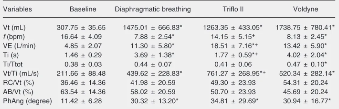
mo-Table 1. Respiratory variables at rest (baseline), and during diaphragmatic breathing, flow-oriented incentive spirometry (Triflo II) and volume-oriented incentive spirometry (Voldyne) for 17 subjects.
Variables Baseline Diaphragmatic breathing Triflo II Voldyne
Vt (mL) 307.75 ± 35.65 1475.01 ± 666.83* 1263.35 ± 433.05* 1738.75 ± 780.41*
f (bpm) 16.64 ± 4.09 7.88 ± 2.54* 14.15 ± 5.15+ 8.13 ± 2.45*
VE (L/min) 4.85 ± 2.07 11.30 ± 5.80* 18.51 ± 7.16*+ 13.42 ± 5.90*
Ti (s) 1.46 ± 0.29 3.69 ± 1.38* 1.77 ± 0.59*+ 4.02 ± 2.04*
Ti/Ttot 0.38 ± 0.03 0.44 ± 0.07 0.41 ± 0.06 0.47 ± 0.10*
Vt/Ti (mL/s) 211.66 ± 88.48 439.62 ± 228.83* 761.27 ± 268.95*+ 520.34 ± 282.14*
RC/Vt (%) 36.46 ± 14.36 41.98 ± 20.59 49.30 ± 23.93 54.31 ± 20.24
AB/Vt (%) 63.54 ± 14.36 58.02 ± 20.59 50.70 ± 23.93 45.69 ± 20.24
PhAng (degree) 11.42 ± 6.28 30.32 ± 13.20* 34.81 ± 29.69* 30.94 ± 16.77*
Data are reported as means ± SD. Vt = tidal volume, f = respiratory frequency, VE = minute ventilation, Ti = inspiratory time, Ti/Ttot = inspiratory duty cycle, Vt/Ti = mean inspiratory flow, RC/Vt = rib cage motion contribution to Vt, AB/Vt = abdominal motion contribution to Vt, PhAng = phase angle.
Statistical comparisons were performed with ANOVA for repeated measures and the post hoc Tukey test (data normally distributed: Ti/Ttot, RC/Vt, and AB/Vt), or the Friedman and Wilcoxon tests (data not normally distributed: Vt, f, VE, Ti, Vt/Ti, and PhAng). The level of significance (α) was set at 0.05 and adjusted for 0.008 with Bonferroni correction (0.05/6 = 0.008). *P < 0.05 for comparisons between baseline and breathing exercises; +P < 0.05 for comparisons between breathing exercises.
tion, comparisons between baseline and ex-ercises or between exex-ercises showed no sig-nificant changes in RC/Vt or AB/Vt. During all exercises, there was an increase in PhAng compared to baseline, reflecting an increase in thoracoabdominal asynchrony. 4) A higher electromyographic activity of the SCM oc-curred during exercise with Triflo II com-pared to the other exercises.
A question may be raised about the fact that only the first baseline period was con-sidered in the comparisons between baseline and exercises. This choice was made be-cause there were no significant differences between the three baselines. The periods of baseline between exercises were necessary to prevent carryover effects. A period of 5 min was sufficient for the recovery of basal values, as also observed in previous studies (11,12). In order to eliminate cumulative effects, exercises were randomized.
Regarding the physiology of deep and slow inspiration, the basis of DB and IS, an increase in Vt is expected, associated with a decrease in f (1,6) and an increase in Ti/Ttot (25), which can contribute to a laminar in-spiratory flow, resulting in a more uniform distribution of air through the pulmonary parenchyma. The variables analyzed showed the same results during DB and Voldyne, except for Ti/Ttot, which only increased sig-nificantly during exercise with Voldyne com-pared to baseline. Thus, the changes ex-pected as a consequence of deep and slow inspirations occurred in a significant and concomitant way compared to baseline only during Voldyne. Comparisons between deep breathing exercise and IS were performed in previous studies (9,10) in terms of different aspects of pulmonary function, but not in terms of breathing pattern variables as was done in the present study.
Minute ventilation was significantly higher during Triflo II and this can be associated with a decreased risk of hypoxemia (26). However, this higher VE seems to be a consequence of high values of f, since Vt was similar between
exercises, an important objective of deep breathing exercises (25). Vt/Ti was signifi-cantly higher during exercise with Triflo II. It should be pointed out that during exercise with Triflo II, Vt/Ti was limited to the level corresponding to the elevation of the first two balls. However, f was not pre-determined for any exercise and was also significantly higher during exercise with Triflo II in relation to the other exercises.
Another result that can be considered to be a disadvantage of Triflo II was a higher electromyographic activity of the SCM dur-ing the use of this device compared to the other exercises. Previous studies have shown a higher electromyographic activity of the SCM associated with an increased load in the respiratory system. Costa et al. (13) as-sessed the participation of the SCM during deep inspiration in healthy adults and ob-served a higher electromyographic activity of the SCM when inspiratory movements were performed in a fast and abrupt way compared to slow inspirations. De Andrade et al. (27) reported an increase of 7% in the electromyographic activity of the SCM dur-ing muscular traindur-ing with Threshold® (Res-pironics, Cedar Grove, NJ, USA).
volume-oriented device. During the postop-erative period of abdominal surgery, when IS is classically indicated, breathing work can increase significantly as a consequence of atelectasis, weakness of inspiratory muscles and excessive airway mucus secretion (30). On this basis, we suggest that a device associ-ated with a lower additional imposed work should be chosen.
With respect to thoracoabdominal mo-tion, there were no significant differences in RC/Vt or AB/Vt between baseline and exer-cise or between exerexer-cises. These results are different from those reported in previous studies, probably because of differences be-tween healthy subjects and patients. Chuter et al. (31) observed an increase in RC/Vt during IS compared to baseline after chole-cystectomy. We may speculate that this re-sult was consequent to an increased abdomi-nal pressure related to surgery (32).
Sharp et al. (33), in a study with healthy subjects, observed that 61 and 50% of sub-jects, respectively, showed an increasing par-ticipation of rib cage and abdomen with increasing tidal volume during deep inspira-tions performed in the supine position. In the upright position, nearly all the volume change was due to rib cage displacement, with much less abdominal participation compared to the supine position. In this case, results dif-ferent from ours may be related to difdif-ferent methods used to instruct subjects and differ-ent devices used to assess thoracoabdominal motion, since the cited investigators used magnetometer, whereas we used respiratory inductive plethysmography.
Parreira et al. (12), in 2005, observed an increase in the contribution of abdominal motion to Vt during volume-oriented IS com-pared to flow-oriented IS, with subjects in the supine position with 45º of inclination
and with parameter limitation during IS. In the present study, subjects were positioned at 30º of inclination and instructed to per-form deep inspirations without limitations. Thus, different study designs probably con-tributed to differences in the results.
Asynchrony between the thoracic and ab-dominal compartments was assessed by PhAng (34,35). When rib cage and abdomen move in perfect synchrony, PhAng is 0º, and with in-creasing thoracoabdominal asynchrony its value approaches 180º (34). We did not find any literature reports about PhAng values for healthy adults during breathing exercises. The baseline PhAng values observed here during baseline were similar to those observed in previous studies with healthy subjects during breathing at rest (35-37). When compared to baseline, there was a significant increase in PhAng during all exercises, without differ-ences between them. A priori, these results can be related to an increased respiratory load imposed on the respiratory system (37).
Our results suggest that the execution of the three breathing exercises caused changes in the volume and time components of the breathing pattern and increased asynchrony when compared to baseline. Diaphragmatic breathing and the volume-oriented incentive spirometer Voldyne showed similar perfor-mance. Considering breathing pattern vari-ables, SCM electromyographic activity and the physiology of techniques based on deep and slow inspirations, our results suggest that the flow-oriented incentive spirometer Triflo II presented important disadvantages compared to the other exercises studied.
Acknowledgments
References
1. Bakow ED. Sustained maximal inspiration - a rationale for its use.
Respir Care 1977; 22: 379-382.
2. Bartlett RH, Gazzaniga AB, Geraghty TR. Respiratory maneuvers to prevent postoperative pulmonary complications. A critical review.
JAMA 1973; 224: 1017-1021.
3. Gosselink HAAM. Breathing exercises in patients with chronic ob-structive pulmonary disease - An experimental study on the effi-ciency and coordination of breathing. Amsterdam: Vrije Universiteit te Amsterdam; 1991.
4. Gosselink R, Schrever K, Cops P, Witvrouwen H, De Leyn P, Troosters T, et al. Incentive spirometry does not enhance recovery after thoracic surgery. Crit Care Med 2000; 28: 679-683.
5. Grant-Paterson L, Moodie NB. Incentive spirometry: an adjunct to chest physiotherapy. Physiother Can 1985; 37: 388-393.
6. AARC (American Association for Respiratory Care) clinical practice guideline. Incentive spirometry. Respir Care 1991; 36: 1402-1405. 7. Douce FH. Incentive spirometry and other aids to lung inflation. In:
Barnes G (Editor), Core text book. 1st edn. New York: McGraw Hill; 1994. p 231-241.
8. Wojciechowski WV. Incentive spirometers, secretion evacuation devices, and inspiratory muscle training devices. In: Barnes G (Edi-tor), Core text book. 1st edn. New York: McGraw Hill; 1994. p 499-522.
9. Celli BR, Rodriguez KS, Snider GL. A controlled trial of intermittent positive pressure breathing, incentive spirometry, and deep breath-ing exercises in preventbreath-ing pulmonary complications after abdomi-nal surgery. Am Rev Respir Dis 1984; 130: 12-15.
10. Jenkins SC, Soutar SA, Loukota JM, Johnson LC, Moxham J. Phys-iotherapy after coronary artery surgery: are breathing exercises necessary? Thorax 1989; 44: 634-639.
11. Parreira VF, Coelho EM, Tomich GM, Alvim AMA, Sampaio RF, Britto RR. Avaliação do volume corrente e da configuração toraco-abdominal durante o uso de espirômetros de incentivo a volume e a fluxo, em sujeitos saudáveis: influência da posição corporal. Rev Bras Fisioter 2004; 8: 45-51.
12. Parreira VF, Tomich GM, Britto RR, Sampaio RF. Assessment of tidal volume and thoracoabdominal motion using volume and flow-oriented incentive spirometers in healthy subjects. Braz J Med Biol Res 2005; 38: 1105-1112.
13. Costa D, Vitti M, de Oliveira TD, Costa RP. Participation of the sternocleidomastoid muscle on deep inspiration in man. An electro-myographic study. Electromyogr Clin Neurophysiol 1994; 34: 315-320.
14. Maarsingh EJ, van Eykern LA, Sprikkelman AB, Hoekstra MO, van Aalderen WM. Respiratory muscle activity measured with a nonin-vasive EMG technique: technical aspects and reproducibility. J Appl Physiol 2000; 88: 1955-1961.
15. Winter DA, Fuglevand AJ, Archer SE. Crosstalk in surface electro-myography: theoretical and practical estimates. J Electromyogr Kinesiol 1994; 4: 15-26.
16. Pereira CAC. Espirometria. J Bras Pneumol 2002; 28 (Suppl 3): S1-S82.
17. Knudson RJ, Lebowitz MD, Holberg CJ, Burrows B. Changes in the normal maximal expiratory flow-volume curve with growth and ag-ing. Am Rev Respir Dis 1983; 127: 725-734.
18. Chadha TS, Watson H, Birch S, Jenouri GA, Schneider AW, Cohn MA, et al. Validation of respiratory inductive plethysmography using different calibration procedures. Am Rev Respir Dis 1982; 125: 644-649.
19. Sackner MA, Watson H, Belsito AS, Feinerman D, Suarez M, Gon-zalez G, et al. Calibration of respiratory inductive plethysmograph during natural breathing. J Appl Physiol 1989; 66: 410-420. 20. Falla D, Dall’Alba P, Rainoldi A, Merletti R, Jull G. Repeatability of
surface EMG variables in the sternocleidomastoid and anterior scalene muscles. Eur J Appl Physiol 2002; 87: 542-549.
21. Souza RB. Pressões respiratórias estáticas máximas. J Bras Pneumol 2002; 28 (Suppl 3): S155-S165.
22. Melendez JA, Alagesan R, Reinsel R, Weissman C, Burt M. Postthoracotomy respiratory muscle mechanics during incentive spirometry using respiratory inductance plethysmography. Chest
1992; 101: 432-436.
23. Munro BH. Repeated measures analysis of variance. In: Munro BH (Editor), Statistical methods for health and care research. 4th edn. Philadelphia: Lippincott Williams & Wilkins; 2000. p 201-221. 24. Webber BA, Pryor JA. Physiotherapy for respiratory and cardiac
problems. New York: Churchill Livingstone; 1998.
25. Grassi C, Brambilla C, Fishman AP. Pulmonary diseases. London: McGraw-Hill International; 1999.
26. West JB. Mecânica da respiração. Fisiologia respiratória moderna. São Paulo: Manole Ltda.; 1996.
27. De Andrade AD, Silva TN, Vasconcelos H, Marcelino M, Rodrigues-Machado MG, Galindo VC, et al. Inspiratory muscular activation during threshold therapy in elderly healthy and patients with COPD.
J Electromyogr Kinesiol 2005; 15: 631-639.
28. Weindler J, Kiefer RT. The efficacy of postoperative incentive spi-rometry is influenced by the device-specific imposed work of breath-ing. Chest 2001; 119: 1858-1864.
29. Mang H, Obermayer A. Imposed work of breathing during sustained maximal inspiration: comparison of six different incentive spirom-eters. Respir Care 1989; 34: 1122-1128.
30. Marini JJ. Postoperative atelectasis: pathophysiology, clinical im-portance, and principles of management. Respir Care 1984; 29: 516-522.
31. Chuter TAM, Weisman C, Starker P, Gump FE. Effect of incentive spirometry on diaphragmatic function after surgery. Surgery 1989; 105: 488-493.
32. Simonneau G, Vivien A, Sartene R, Kunstlinger F, Samii K, Noviant Y, et al. Diaphragm dysfunction induced by upper abdominal sur-gery. Role of postoperative pain. Am Rev Respir Dis 1983; 128: 899-903.
33. Sharp JT, Goldberg NB, Druz WS, Danon J. Relative contributions of rib cage and abdomen to breathing in normal subjects. J Appl Physiol 1975; 39: 608-618.
34. Mayer OH, Clayton RG Sr, Jawad AF, McDonough JM, Allen JL. Respiratory inductance plethysmography in healthy 3- to 5-year-old children. Chest 2003; 124: 1812-1819.
35. Sackner MA, Gonzalez H, Rodriguez M, Belsito A, Sackner DR, Grenvik S. Assessment of asynchronous and paradoxic motion between rib cage and abdomen in normal subjects and in patients with chronic obstructive pulmonary disease. Am Rev Respir Dis
1984; 130: 588-593.
36. Bloch KE, Li Y, Zhang J, Bingisser R, Kaplan V, Weder W, et al. Effect of surgical lung volume reduction on breathing patterns in severe pulmonary emphysema. Am J Respir Crit Care Med 1997; 156: 553-560.