BREATHING CHARACTERISTICS OF INDIVIDUALS
WITH DENTOFACIAL DEFORMITY
Características respiratórias de indivíduos
com deformidade dentofacial
Daniela Galvão de Almeida Prado(1), Hugo Nary Filho(2),
Giédre Berretin-Felix (3), Alcione Ghedini Brasolotto(4)
(1) Post Graduation Program in Dentistry at Piracicaba School
of Dentistry, Campinas State University – FOP-UNICAMP, Piracicaba-SP, Brazil.
(2) University of Sagrado Coração, Bauru-SP, Brazil.
(3) Speech-language pathologist; PhD Professor of the
Spe-ech-Language Pathology Department at Bauru School of Dentistry, University of São Paulo FOB-USP, Bauru-SP, Brazil; PhD in Physiopathology in Medical Clinics/UNESP; Postdoctoral research in Swallowing Disorders at the Uni-versity of Florida; MSc in Dentistry/UNICAMP; Specialist in Orofacial Motricity.
(4) Speech-Language Pathology Department at Bauru School
of Dentistry, University of São Paulo FOB-USP, Bauru-SP, Brazil.
This study was conducted at the Speech-Language Pathology Department at Bauru School of Dentistry – University of São Paulo, Bauru-SP, Brazil.
Grant: São Paulo State Research Foundation (FAPESP)
Conlict of interest: non-existent
soft and hard tissues. When this happens, dento-facial deformities may occur, which interfere with the functional aspects, facial esthetics, personality, attitudes and behavior of individuals1.
The anatomical characteristics in dentofacial deformities (DFD) may be related to breathing manifestations of individuals with these deformities2,3.
There has been report of smaller volume of
the oropharynx airway in individuals with Class II
malocclusion compared to individuals with Class I and III malocclusions, reporting that mandibular
positioning in relation to the cranial base inluences the oropharynx volume. Also, individuals with Class
II malocclusion present smaller nasal air volume compared to Class I individuals4. Functional
abnor-malities that affect nasal breathing, including septal deviation, nasal valve constriction and turbinate hypertrophy, are observed in individuals with skeletal
maxillary deformity5.
In cases of maxillary constriction, which produce a narrow nasal valve, a study revealed that maxillary expansion increased the nasal permeability in the
INTRODUCTION
The balance of the stomatognathic system may be disrupted by factors that alter the structure of
ABSTRACT
Purpose: comprehend the respiratory characteristics of individuals with dentofacial deformities and verify if there are differences comparatively to individuals with dentofacial balance. Methods:
participated 60 individuals (18 to 40 years old), 30 with a dentofacial deformities and 30 of a control
group. The assessment of the Maximum Phonation Time for the emissions /a/, /i/, /u/, /s/, /z/ and
the number counting was evaluated using the program Sound Forge (Sony); the vital capacity and
pneumophonic coordination by the PonyFx spirometer. The results were compared by using the “t”
Student test. Results: the individuals with dentofacial deformities presented lower Maximum Phonation
Time values than individuals with dentofacial balance in the emissions: “s” for those with skeletal Class II malocclusion and men; “z” for individuals with Class II malocclusion; number counting for men. The measures extracted by the spirometry were similar between the individuals with and without dentofacial
deformities. Conclusion: there were no differences regarding the vital capacity and pneumophonic
coordination, but the dentofacial deformities group presented lower values of Maximum Phonation
Time in the emissions that contain consonant phonemes.
be triggered by allergic pulmonary disorders16.
Evaluation of spirometry measurements including vital capacity, pulmonary volume and pulmonary
low, besides evaluation of the maximum phonation
time (MPT), may contribute to the understanding on breathing disorders in these individuals.
Therefore, this study aimed to analyze the respi-ratory characteristics of individuals with dentofacial deformities submitted to preoperative orthodontic treatment compared to individuals with dentofacial
balance, as to the following aspects: maximum
phonation times, vital capacity and pneumophonic coordination.
METHODS
This study was approved by the Institutional Review Board of Bauru School of Dentistry, University of São Paulo, process n. 049/2009. All participants signed the Informed Consent Form.
Young adults with dentofacial deformities in orthodontic treatment and candidates for orthog-nathic surgery, with the agreement of directors of orthodontic clinics and institutes, were invited to participate in this study. Young adults without dento-facial deformities from the community were also invited to participate.
The inclusion criteria for the study were age between 18 and 40 years, regardless of gender, presenting Class II or Class III dentofacial deformity, having completed preoperative orthodontic treatment for orthognathic surgery, and presenting oral or oronasal breathing in accordance with the Orofacial Myofunctional Evaluation, MBGR protocol 19.
The experimental group (EG) consisted of 22
young adults aged 18 to 40 years with dentofacial deformities, being 14 with Class III skeletal maloc-clusion and 8 with Class II malocmaloc-clusion, among whom 13 were females and nine were males. All individuals showed oral or oronasal breathing. The control group (CG) consisted of 22 individuals with dentofacial balance, adjusted by gender and age according to the study group, with relationship between dental arches with horizontal and vertical overlap between 1 and 3 mm, natural teeth at least up to the second premolar, and average facial type and nasal breathing, in accordance with the Orofacial Myofunctional Evaluation, MBGR protocol 19.
Exclusion criteria for the experimental and control
group, obtained by reports from the participants, included neurological and/or psychiatric syndromes, chronic pulmonary obstruction, smoking, voice changes and previous laryngeal surgery, facial trauma, or prior orthognathic surgery.
short term; however, the effect did not persist over time and was unable to change the breathing pattern for most individuals6.
In cases of individuals with Class III
maloc-clusion submitted to bimaxillary osteotomy, involving mandibular setback and maxillary advancement,
there are reports of inferior repositioning of the hyoid bone, posterior displacement of the tongue
and soft palate, narrowing of the oropharynx and hypopharynx and widening of the nasopharynx and velopharynx ive months after surgery7. Other
authors reported signiicant increase in the low limitation index and reduced oxygen saturation
in the ventilation during sleep in individuals with Class III malocclusion, eight and a half months after osteotomy8. In case of surgery involving only
mandibular setback, studies did not report signif-icant changes in the upper airway9,10. Conversely,
in surgeries involving only maxillary advancement,
there are reports of reduced nasal obstruction11.
The anatomical changes present in individuals with dentofacial deformities are related with mouth breathing, since several authors describe that this breathing pattern promotes elevation and greater
head extension related to the cervical spine, inlu
-encing the hyoid bone and different intermaxillary
positions12. The mandibular positioning and tongue
posture are inluenced by the breathing needs,
altering the balance of mandibular and teeth pressures, affecting both the cranial morphology and tooth positioning13.
The relationship between breathing and maloc-clusion has usually been described in studies on children, yet it is necessary to consider the possibility of maintenance of mouth breathing up to adulthood. Some authors report a close relationship between mouth breathing and malocclusion, since studies on mouth-breathing children revealed that most of them presented Angle Class II malocclusion14,15.
Also, some studies demonstrate that allergies
inluence the occlusal development, with greater inluence from rhinitis, both allergic and vasomotor, and atopic asthma to a lesser extent. The changes
in nasal function induced by rhinitis may lead to the development of bronchial asthma due to loss of the
natural mechanism of nasal iltering, because of
the development of edema. This change in nasal function may trigger the antigen presentation in
the airway, and the inlammatory reaction initiated on the nose may lead to airway inlammation by a
systemic pathway16.
Therefore, several studies have addressed aspects of breathing in individuals with DFD, especially focusing on upper airway disorders4,5,7,8,11,
vowel /a/. The examiner also verbally instructed that
the emission should be as long as possible, and this procedure was repeated three times to calculate a mean value. The phonation volume, measured in milliliters, refers to the quantity of air used for phonation of the prolonged vowel, with normality parameters relative to vital capacity mean values of 67% for men and 59% for women18. The phonation
medium low, calculated in milliliters per second,
indicates the air outlet control for speech and was calculated as the ratio between the phonation volume and the MPT.
Besides these parameters, the study also calcu-lated the simple phonic quotients (SPQ) obtained by evaluating the ratio between vital capacity and MPT of the vowel /a/.
The results were compared between experi -mental and control groups, which were subdivided by gender and malocclusion type. The statistical
Student t test was used, at a signiicance level of
5%.
RESULTS
Analysis of the relative results for MPT showed
statistically signiicant difference in “s” and “z”
emissions and numbers for individuals in the Class
II malocclusion subgroup and in “s” emissions in the male subgroup, in which the values of the experi -mental group were smaller than the control group (Tables 1 and 2).
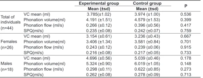
Concerning the relative results for the spirometry evaluation, differences were not found between the
experimental and control groups, for all individuals,
men and women subgroups and Class II and III malocclusions concerning the vital capacity,
phonation volume, phonation low and simple phonic coeficient (Tables 3 and 4).
Characterization of the control group revealed mean height of 1.70 cm and weight 69.07 kg; for the
experimental group, the values were 1.70 cm and
67.75 kg.
To evaluate the maximum phonation time (MPT),
three prolonged emissions of the vowels /a/, /i/ and /u/; the fricatives /s/ and /z/; and the counting of numbers were recorded, and the means of the three productions of each emission were considered. The time for such measures was counted using the auditory aid and visual timescale of the software Sound Forge 9.0
Spirometry was performed using the spirometer PonyFX of 12 L. For that purpose, the individual was comfortably seated in a chair with the arms
supported. A mouthpiece coupled to a ilter and
the spirometer were positioned in the vestibule of the individual’s mouth, and the individual was asked to breathe normally to get accustomed to the system. To obtain the vital capacity, the individual was instructed to practice some breaths cycles and, when he or she felt comfortable, to perform
maximum inspiration followed by a pause of few seconds and then maximum expiration in the form of forced blowing. During expiration, the examiner
verbally asked the individual to perform the longest
possible expiration, and this procedure was
repeated three times to calculate the mean in liters of the three values obtained.
For quantitative evaluation of pneumophonic coordination, the same equipment was used, which graphically recorded the equivalent curve of each
emission. The phonation volume and mean low
were obtained from the vowel emissions produced with the same spirometer. The individual was instructed to take some breaths cycles and, when
he or she felt comfortable, to perform maximum
Table 1- Values of maximum phonation time (MPT) emissions /a/, /i, /u/,/s/,/z/; and counting of numbers from individuals in the Experimental Group and Control Group for the total of individuals and gender subgroup
Experimental group Control group
p
Mean (±sd) Mean (±sd)
Total of individuals (n=44)
“a” 17.00(±5.45) 17.20(±5.19) 0.901
“i” 17.75(±6.27) 17.27(±5.56) 0.852
“u” 15.60(±6.52) 14.74(±3.77) 0.595
“s” 13.67(±6.00) 16.25(±4.97) 0.122
“z” 14.48(±6.59) 17.39(±6.82) 0.157
“numbers” 18.41(±5.26) 20.32(±4.19) 0.190
females (n=26)
“a” 15.42(±4.98) 15.63(±3.58) 0.902
“i” 16.49 (±6.39) 15.60 (±4.15) 0.677
“u” 15.02(±7.05) 13.49(±3.73) 0.496
“s” 14.27(±7.28) 15.30(±4.30) 0.664
“z” 13.98 (±7.16) 14.77 (±3.79) 0.728
“numbers” 18.21 (±4.77) 18.54 (±3.51) 0.842
males (n=18)
“a” 19.28(±5.55) 19.49(±6.46) 0.941
“i” 19.58 (±5.96) 19.69 (±6.66) 0.971
“u” 16.44 (±5.96) 16.54 (±3.20) 0.965
“s” 12.80 (±3.68) 17.62 (±5.78) 0.049*
“z” 15.21 (±6.00) 21.17 (±8.57) 0.106
“numbers” 18.70 (±6.18) 22.90 (±3.87) 0.103
*p<0.05 – statistically signiicant
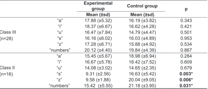
Table 2 – Values of maximum phonation time (MPT) emissions /a/, /i, /u/,/s/,/z/; and counting of numbers from individuals in the Experimental Group and Control Group subdivided by malocclusion type
Experimental
group Control group p
Mean (±sd) Mean (±sd)
Class III (n=28)
“a” 17.88 (±5.32) 16.19 (±3.82) 0.343
“i” 18.37 (±6.67) 16.62 (±4.28) 0.421
“u” 16.47 (±7.84) 14.79 (±4.47) 0.501
“s” 16.16 (±6.02) 16.03 (±4.89) 0.953
“z” 17.28 (±6.71) 15.88 (±4.92) 0.534
“numbers” 20.12 (±4.40) 19.84 (±4.39) 0.867
Class II (n=16)
“a” 15.45 (±5.67) 18.98 (±6.94) 0.284
“i” 16.67 (±5.78) 18.42 (±7.52) 0.609
“u” 14.08 (±3.02) 14.65 (±2.35) 0.679
“s” 9.31 (±2.56) 16.63 (±5.42) 0.003*
“z” 9.58 (±1.88) 20.04 (±9.05) 0.006*
“numbers” 15.42 (±5.55) 21.18 (±3.95) 0.031*
was altered respiratory airlow control in individuals
with DFD, because MPT was altered only for frica-tives production and chained speech (Tables 1 and
2), which suggests that the individuals’ dificulty is
related to articulation.
The production of vowels “a”, “i”, “u” involves
opening the mouth wider and increasing the distance
between the teeth. Conversely, the production of “s” and “z” involves control of the airlow so that the teeth
stay close, at which point adjustments may occur to the muscle that brings the mandible to a position at which premature dental and occlusal interference can result. Therefore, individuals with malocclusion
have dificulty in producing these phonemes 21.
DISCUSSION
Considering that previous studies demonstrated relationship between malocclusion and breathing disorders, this study investigated whether there is difference in the breathing pattern of individuals with dentofacial deformities (DFD) compared to individuals with normal occlusion, by measuring the vital capacity and pneumophonic coordination, besides analysis of MPT.
Because MPTs are related to sustaining
phonation, they experience interference from
several factors, such as breathing function control,
glottal eficiency, vital capacity, and laryngeal
control 20. However, this study cannot state that there
Table 3 – Values of spirometry measures including mean vital capacity, phonation volume and low, simple phonic quotients, from individuals in the Experimental Group and Control Group for the total of individuals and gender subgroup
Experimental group Control group
P
Mean (±sd) Mean (±sd)
Total of individuals (n=44)
VC mean (ml) 3.785(±1.02) 3.974 (±1.00) 0.536 Phonation volume(ml) 4.191 (±1.51) 4.579 (±1.53) 0.399
Phonation low (ml/s) 0.266 (±0.12) 0.396 (±0.56) 0.417 SPQ(ml/s) 0.235 (±0.08) 0.242 (±0.07) 0.759
Females (n=26)
VC mean (ml) 3.154 (±0.61) 3.236 (±0.43) 0.667 Phonation volume(ml) 3.406 (±1.34) 3.581 (±0.84) 0.701
Phonation low (ml/s) 0.243 (±0.12) 0.239 (±0.06) 0.915 SPQ(ml/s) 0.216 (±0.08) 0.217 (±0.05) 0.733
Males (n=18)
VC mean (ml) 4.696 (±0.56) 5.039 (±0.46) 0.178 Phonation volume(ml) 5.324 (±0.90) 6.019 (±1.05) 0.148
Phonation low (ml/s) 0.298 (±0.11) 0.622 (±0.85) 0.273 SPQ(ml/s) 0.262 (±0.08) 0.278 (±0.09) 0.713
Legend: VC: Vital Capacity and SPQ: Simple Phonic Quotients
Table 4 – Values of spirometry measures including vital capacity, phonation volume and low, simple phonic quotients, from the Experimental Group and the Control Group subdivided by malocclusion type.
Experimental group Control group
p
Mean (±sd) Mean (±sd)
Class III (n=28)
VC mean (ml) 3.859 (±0.91) 3.979 (±1.07) 0.750 Phonation volume (ml) 4.128 (±1.56) 4.675 (±1.74) 0.386
Phonation low (ml/s) 0.250 (±0.12) 0.292 (±0.09) 0.902 SPQ(ml/s) 0.227 (±0.08) 0.244 (±0.07) 0.274
Class II (n=16)
VC mean (ml) 3.654 (±1.10) 3.965 (±0.95) 0.543 Phonation volume (ml) 4.30 (±1.51) 4.41 (±1.14) 0.871
Phonation low (ml/s) 0.29 (±0.10) 0.58 (±0.93) 0.395 SPQ(ml/s) 0.25 (0.07) 0.24 (±0.07) 0.779
Several studies addressed the relationship between DFD and upper airway changes 4,5,7,8,11;
however, not all studies observed changes in these aspects in individuals with DFD7,8, which agrees with
the outcomes of absence of breathing alterations in individuals in this study. Therefore, further studies like this are warranted, analyzing groups with larger number of balanced individuals as to gender and facial pattern.
The contribution of the present study is the obser-vation that, during vocal evaluation of MPT in the clinical practice, a reduction in time of the fricative
emission of “s” can occur due to the presence of
dentofacial deformities, unrelated to alterations in
the respiratory control of airlow.
CONCLUSION
The group of patients with DFD did not show different vital capacity measures and pneumophonic coordination from those of the control group, yet it showed reduced MPT values in emissions of conso-nantal phoneme.
ACKNOWLEDGMENT
The authors thank the São Paulo State Research Foundation (FAPESP) for their support to conduct this study (process n. 2009/04621-4).
In relation to the MPT consonants /s/ and /z/, the emission of /s/ allows evaluation of an individual’s capacity to control the pulmonary air support. Since in its production there is no vibration of the vocal folds, it is possible to evaluate the frictional sound origin. With /z/, there is an additional possibility to
evaluate the glottal origin, because larynx vibration
occurs in its production 22.
In general, the MPTs of individuals in the control group were in accordance with the normal standards established by authors, which preconize values in the 20s for men and 14s for women. The same
cannot be stated for the experimental group, whose
values were below the normal 23.
The results showed that the values related to vital capacity for both study groups are in agreement with normality, which, according to a study, would be 4.64 ± 0.77 liters for males and 3.14 ± 0.65 liters for women 23.
Some studies 16 have shown that mouth breathing
can be triggered by allergic pulmonary alteration, while other authors described a relationship between malocclusion and breathing alteration 14,15.
Considering that the majority of individuals with DFD in this study showed oral or oronasal breathing, the
breathing alteration was expected, yet differences in
spirometry measures were not noted (Tables 3 and 4), which can indicate that the respiratory character-istics of EG did not interfere with pulmonary volumes
or the ability to control the air low during phonation.
RESUMO
Objetivo: compreender as características respiratórias em indivíduos com deformidades dentofaciais
e veriicar se há diferenças comparativamente a indivíduos com equilíbrio dentofacial. Métodos: par-ticiparam 60 indivíduos (18 a 40 anos), 30 portadores de deformidade dentofacial e 30 de um grupo
controle. Foi realizada avaliação do Tempo Maximo de Fonação das emissões /a/, /i/, /u/, /s/, /z/ e
contagem de números pelo programa Sound Forge (Sony); avaliação da capacidade vital e coor-denação pneumofonoarticulatória, pelo espirômetro PonyFx. Os resultados foram comparados pelo
teste “t” de Student. Resultados: os indivíduos com deformidade dentofacial apresentaram valores
de Tempo Maximo de Fonação inferiores aos indivíduos com equilíbrio dentofacial nas emissões: “s” para aqueles com má oclusão esquelética classe II e homens; “z” para indivíduos com má oclusão classe II; contagem de números para os homens. As medidas extraídas pela espirometria foram
semelhantes entre os indivíduos com e sem deformidade dentofacial. Conclusão: não houve dife-renças em relação à capacidade vital e coordenação pneumofonoarticulatória, mas o grupo com
deformidade dentofacial apresentou valores reduzidos de Tempo Maximo de Fonação em emissões
que contêm fonemas consonantais.
11. Williams BJD;Isom A; Laureano-ilho JR; O’Ryan FS. Nasal airway function after maxillary surgery: a
prospective cohort study using the nasal obstruction
symptom evaluation scale. J oral Maxillofac
Surg.2013;71:343-50.
12. Cuccia AM, Lotti M, Caradonna D. Oral breathing and head posture. Angle Orthod. 2008;78(1):77-82. 13. Karacay AKIN, Ortakoglu K, Bengi AO. Dynamic MRI evaluation of tongue posture and deglutitive movements in a surgically corrected open bite. Angle Orthod. 2006;76(6):1057-65.
14. Motta RJ. Relação da postura cervical e oclusão
dentária em crianças respiradoras orais. Rev
CEFAC. 2009;11(3):298-304.
15. Almeida FL, Silva AMT, Serpa E. O. Relação
entre má oclusão e hábitos orais em respiradores
orais. Rev CEFAC. 2009;11(1):86-93.
16. Lampasso JD, lampasso JF. Allergy Nasal Obstruction, and Occlusion Seminars in Orthodontics. 2004;10(1):39-44
17. Almeida FL, Silva AMT, Serpa EO. Relação
entre má oclusão e hábitos orais em respiradores
orais. Rev CEFAC. 2009;11(1):86-93.
18. Sies ML, Faria SR, Vieira MM. Respiração oral:
relação entre o tipo facial e a oclusão dentária
em adolescentes. Rev Soc Bras Fonoaudiol. 2007;12(3):191-8.
19.Genaro KF, Berretin-Felix G, Rehder MIBC,
Marchesan IQ. Avaliação miofuncional orofacial – protocolo MBGR. Rev CEFAC. 2009;11(2):237-55. 20.Mendes A, Castro E. Analise acústica da avaliação vocal I: tarefas fonatória e medidads acústicas. Rev Port ORL. 2005;43(2):127-36.
21.Vallino LD, Tompson AB. Perceptual Characteristics of Consonan errors associated with
malocclusion. J Oral Maxillofac Surg. 1993;51:850-6.
22.Behlau M, Azevedo R, Pontes P. Avaliação de voz. In: Behlau MS. (org.) Voz – O livro do especialista. Rio de Janeiro: Revinter; 2001, vol. 1. P. 85-246.
23.Pereira C, Sato T, Rodrigues SC. New reference values for forced spirometry in white adults in Brazil. J Bras Pneumol. 2007;33(4):397-406.
REFERENCES
1. Ribas MO, Reis LFG, França BHS, Lima AAS.
Cirurgia ortognática: orientações legais aos ortodontistas e cirurgiões bucofaciais. R Dental
Press Ortodon Ortop Facial. 2005;10(6):75-83. 2. Hwang S, Chung CJ, Choi YJ Huh JK, Kim KH. Changes of hyoid, tongue and pharyngeal airway after mandibular set-back surgery by intraoral vertical ramus osteotomy. Angle Orthod. 2010;80:302-8. 3. Kim JS, Kim JK, Hong S-C, Cho JH. Pharyngeal airway changes after sagittal split ramus osteotomy of the mandible: a comparison between genders. J
Oral Maxillofac Surg. 2010;68:1802-6.
4. El Hakan, Palomo JM. Airway volume for different dentofacial skeletal patterns. Am J Orthod Dentofacial Orthop. 2011;139:511-21.
5. Moche JA; Palmer O. Surgical management of
nasal obstruction. Oral Maxillofac Surg Clin North
Am. 2012 ;24:229.
6. Berretin-Felix G, Yamashita RP, Nary-Filho
H, Gonçales ES, Trindade Jr AS, Trindade IEK. Short- and Long-Term Effect of Surgically Assisted
Maxillary Expansion on Nasal Airway Size. Journal
of Craniofacial Surgery. 2006;17(6):1045-97.
7. Gokce SM, S Gorgulu, Gokce HS, Bengi O, Sabuncuoglu F, Ozgen F et al. Changes in posterior airway space, pulmonary fuction and sleep
quality following bimaxillary orthognathic surgery.
2012;41:820-9.
8. Foltan R Hoffmannová J, Donev F, Vlk M, Sedy ,
Kufa R, Bulik O. The impact of Le Fort I advancement and bilateral sagital split osteotomy setback on
ventilation during sleep. Int J Oral Maxillofac Surg.
2009;38:1036-40.
9. Gu GM, Nagata J, Suto M, Anraku Y, Nakamura K, Kuroe K, Ito G. Hyoid position, pharyngeal airway and head posture in relation to relapse after the mandibular setback in skeletal class III. Clin Orthod Res. 2000;3:67-77.
10. Kiatagawara K, Kobayashi T, Goto H, Yokobayashi T, Kitamura N, Saito C. Effects of mandibular setback surgery on oropharyngeal
airway and arterial oxygen saturation. Int J Oral Maxillofac Surg. 2008;37:328-33.
Received on: May 28, 2013 Accepted on: September 03, 2013 Mailing address:
Daniela Galvão de Almeida Prado
Rua Pio Sbrissa 1001 – Condominio Reserva do Engenho – Reserva do Engenho
Piracicaba – SP – Brasil CEP: 13402-330