O
RIGINALA
RTICLE ©Revista Brasileira de FisioterapiaVentilatory and muscular assessment in
healthy subjects during an activity of daily living
with unsupported arm elevation
Avaliação ventilatória e muscular de indivíduos saudáveis durante atividade de
vida diária com os braços elevados e sem apoio
Giselle F. L. Panka1, Marina M. Oliveira2, Danielle C. França1, Verônica F. Parreira3, Raquel R. Britto3, Marcelo Velloso3.
Abstract
Introduction: Patients with chronic obstructive pulmonary disease (COPD) report dyspnea when performing activities of daily living (ADLs) with elevated upper limbs. To elucidate the determinants of dyspnea, it is important to study the changes in the respiratory pattern of these patients and the electromyographic activity of their accessory muscles of respiration during ADLs. In the literature, there are no reports of a normative parameter, therefore it is necessary to study how these variables behave in healthy subjects. Objectives: To verify, in healthy subjects, the existence of changes in the respiratory pattern and activation of the sternocleidomastoid (SCM) muscle during an ADL with unsupported arm elevation. Methods:Thirteen male subjects, 60.57 (±6.42) years old, with normal spirometry values for age and sex, were evaluated using surface electromyography (EMG) and respiratory inductive plethysmography (RIP) collected at rest and during the activity of combing their hair with elevated and unsupported upper limbs. The data distribution was assessed using Shapiro-Wilk’s test. ANOVA was used to compare the phases, and when the difference was significant (p<0.05), Tukey’s test was applied. Results:TheRIP during the ADL showed a significant increase in tidal volume, minute ventilation, respiratory frequency and mean inspiratory flow. Thoracoabdominal asynchrony was identified in percentage of asynchrony in inspiration, expiration, and Phase Angle (p<0.05). The EMG showed an increase in SMC muscle recruitment (p<0.05). Conclusions: Healthy subjects increased their SCM muscle activation and changed their breathing pattern when performing the ADL with unsupported arm elevation, resulting in thoracoabdominal asynchrony.
Key Words: Physical therapy; upper limbs; activities of daily living.
Resumo
Introdução: Pacientes com doença pulmonar obstrutiva crônica (DPOC) relatam dispneia quando realizam atividades da vida diária (AVD) com membros superiores (MMSS) elevados. Estudar as alterações do padrão respiratório e a atividade eletromiográfica dos músculos acessórios da respiração desses pacientes nas AVD pode contribuir para esclarecer os determinantes de dispneia. Entretanto, não se encontrou, na literatura, um padrão de normalidade, fazendo-se necessário estudar, primeiramente, o comportamento dessas variáveis em saudáveis. Objetivos: Verificar, em indivíduos saudáveis, a existência de mudanças no padrão respiratório e na ativação do músculo esternocleidomastoideo (ECM) durante a realização de uma AVD com os MMSS elevados e sem apoio. Métodos:
Treze voluntários masculinos, com média de idade 60,57 (6,42) anos, com valores espirométricos normais para idade e sexo foram avaliados com eletromiografia (EMG) de superfície e pletismografia respiratória por indutância (PRI), coletadas durante o repouso e na atividade de pentear cabelos com MMSS elevados sem apoio. A distribuição dos dados foi analisada pelo teste Shapiro-Wilk. Para a comparação entre as fases avaliadas, utilizou-se ANOVA e, quando a diferença foi considerada significativa (p<0,05), aplicou-se o teste de Tuckey. Resultados:A PRI, durante a AVD, mostrou aumentos significativos do volume corrente, do volume minuto, da frequência respiratória e do fluxo inspiratório médio. Observou-se assincronia toracoabdominal tanto em porcentagem de assincronia na inspiração e na expiração quanto na análise do Ângulo de Fase (p<0,05). A EMG mostrou aumento no recrutamento do ECM (p<0,05). Conclusões: Indivíduos saudáveis apresentam maior ativação do músculo ECM e modificam o padrão respiratório durante AVD com MMSS elevados sem apoio, gerando assincronia toracoabdominal.
Palavras-chaves: Fisioterapia; membro superior; atividades cotidianas.
Received: 22/04/2009 – Revised: 05/08/2009 – Accepted: 21/10/2009
1 Graduate Program in Rehabilitation Sciences, Universidade Federal de Minas Gerais (UFMG), Belo Horizonte (MG), Brazil 2 Physical Therapy Course, UFMG
3 Physical Therapy Department, UFMG
Correspondence to: Marcelo Velloso, Departamento de Fisioterapia, UFMG, Avenida Presidente Antônio Carlos, 6627, CEP 31270-901, Belo Horizonte (MG), Brazil, e-mail: [email protected]
Introduction
In healthy subjects1 and in patients with chronic
obstruc-tive pulmonary disease (COPD), arm elevation results in chan-ges in the pattern of respiratory muscle recruitment and in an increase in metabolic demand, the latter being more marked in subjects with airlow obstruction2-9. his could be explained by
an eiciency reduction in the respiratory mechanics, by a dual activity of shoulder girdle muscles in COPD and by an increase in dead space2.
Patients with COPD report fatigue and dyspnea when performing activities of daily living (ADLs) with unsupported arm elevation9, such as combing their hair4 and brushing their
teeth. In contrast, they ind it less diicult to perform more de-manding activities involving their legs1,10. It is likely that these
patients’ ability to sustain unsupported arm elevation is not de-termined by the endurance and strength of the shoulder girdle muscles alone. A number of studies suggest that this limitation in respiratory mechanics is inluenced by arm positioning, with changes being observed in the respiratory breathing pattern and thoracoabdominal asynchrony during unsupported arm elevation8,10,11.
According to Epstein et al.6, when COPD patients sustain
arm elevation, two factors become determinants of the altered respiratory pattern: lung hyperinlation and, to a small degree, diaphragm reserve strength. During inspiration due to dyna-mic hyperinlation, the respiratory muscles are placed in an unfavorable position of their length-tension curve, reducing the strength generation capacity12. herefore, unsupported
arm elevation may force the shoulder girdle muscles and the upper torso to participate simultaneously in the ventilatory and non-ventilatory activities1,4,6. Simultaneous aferent inputs
and outputs of the central nervous system (CNS), responsible for the respiratory and tonic functions of these muscles, can result in a signiicant lack of coordination of respiratory muscle action and can result in an increase in dyspnea, with thoracoa-bdominal asynchrony being observed during unsupported arm exercise6.
Based on the literature on the possible reasons for dyspnea and changes in the respiratory pattern and respiratory muscle recruitment in COPD patients during ADL with unsupported arm elevation, it is necessary to establish parameters for he-althy subjects for further comparison studies involving COPD patients. he indings of these studies will contribute to the de-velopment of future interventions and/or improvements in the existing ones, providing more functional independence and, consequently, better quality of life for these individuals. Also, the indings of the present study will be used in another study, cur-rently underway by our group, comparing the results of healthy subjects with COPD patients using the same protocol during
an ADL with unsupported upper limbs. herefore, the current data will also be part of a more comprehensive research. he aim of the present study was to assess, in healthy subjects, the breathing pattern, thoracoabdominal motion and electromyo-graphic activity of the sternocleidomastoid (SCM) muscle at rest and during an ADL with unsupported arm elevation.
Methods
he Ethics Research Committee of Universidade Federal de Minas Gerais, Belo Horizonte (MG), Brazil, approved the protocol (ETIC 551/06), and all subjects gave their informed consent. he sample calculation was accomplished after a pilot study with the irst ten subjects, using all respiratory pattern variables, thoracoabdominal motion and electromyography (EMG) to a power of 80% and a signiicance level of 0.05 (non-directional). he calculation resulted in a value of 13.8 healthy subjects. he sample consisted of 13 healthy subjects selected from the com-munity. he inclusion criteria were: male subjects, age between 50 and 80 years, absence of bone and joint impairment in the shoulder girdle and normal lung function tests according to Brazilian standards of normality13. he exclusion criteria were:
inability to maintain overhead arm elevation for the ive minu-tes of data collection and inability to perform any of the steps determined in the protocol. A signed informed consent form was obtained from all subjects.
he subjects underwent a functional analysis of lung vo-lume and capacity. A portable spirometer (Vitalograph 2120® , Vitalograph, Buckingham, England) was used to ensure normal lung function according to the recommendations of the Bra-zilian Society of Pneumology13. he values of the spirometric
variables were compared to predicted values according to Pereira et al.14. he breathing pattern was accessed by
respira-tory inductive plethysmography (RIP) based on the principle irst described by Konno and Mead15, and the
electromyogra-phic activity of the SCM muscle was accessed by surface EMG. Respiratory variables were obtained by RIP (Respitrace® , Nims, Miami, FL, USA). Telon-coated inductance coils of ap-propriate size were placed around the rib cage (RC) and abdo-men (AB); the upper edge of the RC band was placed at the level of the axilla and the AB band at the level of the umbilicus. he signals were calibrated using qualitative diagnostic calibration (QDC)16 during ive minutes of natural breathing; this is a
two-step procedure whereby the RC and AB electrical gains of the RIP ampliiers are correctly partitioned during tidal breathing and subsequently compared to the output of a spirometer to attain equivalence. he subject then breathed into a spirome-ter via a mouthpiece (Vitatrace, Pro Médico, Rio de Janeiro, RJ, Brazil) with the nose clipped, for 30 to 60 s, and the electrical
spirometer output was recorded with a computer and used to calibrate the RIP sum signal for absolute volume in mL17.
he spirometer was calibrated with a one-liter syringe (Vita-lograph, Ennis, Ireland). he entire procedure was performed using a computer software (RespiPanel 4.0, Nims Miami, FL, USA)18,19 that allows a tidal volume (V
T) variation ≤5% during
calibration process.
he following variables were measured by a digital acquisi-tion system on a breath-by-breath basis (RespiEvents®
, Nims, Miami, FL, USA): tidal volume (VT), respiratory frequency (f), minute ventilation (VE), inspiratory time (Ti), inspiratory duty cycle (Ti/Ttot), mean inspiratory low (VT/Ti), RC motion
contribution to VT (RC/VT), phase angle (PhAng; 0° for
com-plete RC-AB synchrony and 180° for comcom-plete asynchrony)20,
inspiratory RC-AB synchrony (PhRIB), expiratory RC-AB synchrony (PhREB), inspiratory and expiratory synchrony (PhRTB), variables that represent the time percentage during a respiratory cycle in which the RC and AB move in oppo-site directions. When the RC and AB are moving in perfect synchrony, the values are equivalent to 0%, and when there is paradoxical movement, they are equivalent to 100%20. he
contribution of abdominal motion to VT (AB/VT) was
calcula-ted as AB/VT = 100 - RC/VT.
he surface EMG device (EMG System do Brasil LTDA, São Paulo, SP, Brazil) was used to record the electromyographic ac-tivity of the SCM muscle. After the skin was cleansed, ECG bi-polar surface electrodes (Mini Medi Trace 100, Kendall – LTP/ Tyco, Canada) were placed on the lower third of the left sternal portion of the SCM muscle belly. he electrodes were posi-tioned 2.5 cm apart over this area21,22. he EMG signals were
recorded using one of the eight channels of the biological sig-nal acquisition system consisting of a sigsig-nal conditioner with a gain of 1000, a high-pass ilter of 20 Hz, and a low-pass ilter of 500 Hz, a speciic software for data acquisition and analysis (Aqdados, São José dos Campos, SP, Brazil) and a 12-bit analog-to-digital converter, with a sample frequency of 2000 Hz for the channel and an entry band of 5 mV. he mean amplitude of the signal was estimated by its root mean square (RMS) value. In the present study, the data were normalized by rest, given that the objective was to compare the analyzed variables at rest and at diferent moments of an ADL with unsupported arm eleva-tion. he subject was his own control group.
here was a simultaneous collection for the surface EMG and for the RIP data, there were two diferent collections. In the irst collection, the data were collected with the subject in a seated position and arms resting along the body during ive minutes. For the statistical analyses, the last regular minute of the RIP was considered. Next, the second data collection was conducted with the subject in a seated position and combing their hair with unsupported arm elevation for ive minutes. he
subjects were instructed to perform the combing movement on the top of the head, alternating both arms with the domi-nant hand holding the comb while the other followed the mo-vement. he activity had to be constant and uninterrupted for the entire period and at a minimum of 90° arm elevation. he last 30 seconds of the 1st, 3rd and 5th minute were considered for
analyses.
Data are reported as mean and standard deviation (SD). The data distribution for each variable was assessed using Shapiro-Wilk’s test. The comparisons between the four analyzed phases (rest, 1st, 3rd and 5th minute of exercise) were
performed with ANOVA for repeated measures. When the difference was considered significant, Tukey’s test was per-formed to identify the difference between pairs. The level of significance (α) was set at 0.05 for all tests. Data were analyzed with the Statistical Package for the Social Sciences software (SPSS 15.0, Chicago, IL, USA).
Results
Demographic data of the 13 subjects are presented in Table 1. The values for the variables during rest, 1st, 3rd, and
5th minutes of exercise with unsupported arm elevation and
the respective analysis are presented in Table 2.
Breathing pattern
he VT increased signiicantly from the 3rd minute; its value
was 31% higher than the rest value. At the 5th minute, a slight
increase in VT was observed when compared with the 3rd
mi-nute corresponding to a value 33% higher than rest. here was a statistically signiicant diference between rest and the 3rd
mi-nute (P=0.008), rest and the 5th minute (P=0.033) and between
the 1st and the 3rd minute (P=0.048) of activity (Table 2).
he f increased between rest and the 1st minute of activity,
which was 21% higher than the baseline value, and then remai-ned stable during the course of the exercise. here was a sta-tistically signiicant diference between rest and the 1st minute
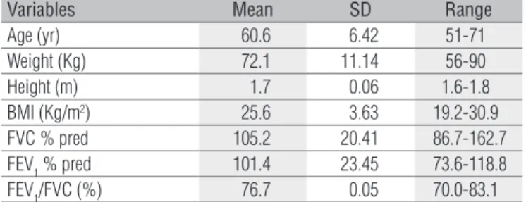
Table 1. Characteristics of the 13 subjects.
Variables Mean SD Range
Age (yr) 60.6 6.42 51-71
Weight (Kg) 72.1 11.14 56-90
Height (m) 1.7 0.06 1.6-1.8
BMI (Kg/m2) 25.6 3.63 19.2-30.9
FVC % pred 105.2 20.41 86.7-162.7
FEV1 % pred 101.4 23.45 73.6-118.8
FEV1/FVC (%) 76.7 0.05 70.0-83.1
SD=standard deviation; Kg=kilogram; m=meters; BMI=body mass index; FVC=forced vital
capacity; VEF1=forced expiratory volume in one second.
(P=0.013), rest and the 3rd minute (P=0.003) and between rest
and the 5th minute (P=0.01) of activity (Table 2). Consequently,
the changes in f in the 1st minute, in addition to the changes in
VT, led to a progressive increase in VE until the 3rd minute, with
a 67% higher value than rest that remained unchanged through the 5th minute. here was a signiicant diference between rest
and the 1st minute (p=0.001), rest and the 3rd minute (p<0.001),
rest and the 5th minute (p<0.001), and between the 1st and the
3rd minute (p=0.013) and the 1st and the 5th minute of exercise
(p=0.02; Table 2).
he VT/Ti presented a 36% increase in the 1
st minute when
compared with rest, a 56% increase in the 3rd minute, stabilizing
in the 5th minute of activity. here was a signiicant diference
when comparing rest and the 1st minute (p<0.001), rest and
the 3rd minute (p<0.001) and rest and the 5th minute of activity
(p=0.003; Table 2).
Thoracoabdominal motion
No statistically signiicant diference was found for %RC/VT and for %AB/VT during the activity period, however
it was observed that at rest %RC/VT was 4.54% higher than
%AB/VT, and that during the exercise there was a shift to AB
in which the %AB/VC increased its participation by 6.54% compared with %RC/VT (Table 2). Considering the PhAng,
we observed that the subjects presented a PhAng of 23.82o at
the end of the activity, which corresponds to a 129% increase from rest. here was a signiicant diference when conside-ring rest and the 1st minute (p=0.038), rest and the 5th minute
(p=0.003), the 1st and the 5th minute (p=0.045) and the 3rd and
the 5th minute of exercise (p=0.026; Figure 1 - A).
A progressive increase in PhRIB was observed during the activity, corresponding to 6.36% at rest, 14.77% in the 1st minute,
14.59% in the 3rd minute and 17.73% in the 5th minute, which
represents a 79% increase between rest and the end of the exer-cise. here was a signiicant diference when considering rest and the 1st minute (p<0.001), rest and the 3rd minute (p<0.001),
rest and the 5th minute (p<0.001) and the 3rd and the 5th minute
of exercise (p=0.034; Figure 1 - B). Similarly, a progressive incre-ase in PhREB was observed during the activity, corresponding to 11.63% at rest, 17.18% in the 1st minute, 18.07% in the 3rd
minute and 19.05% in the 5th minute, which represents a 64%
increase between rest and the end of the exercise. here was a signiicant diference when considering rest and the 1st minute
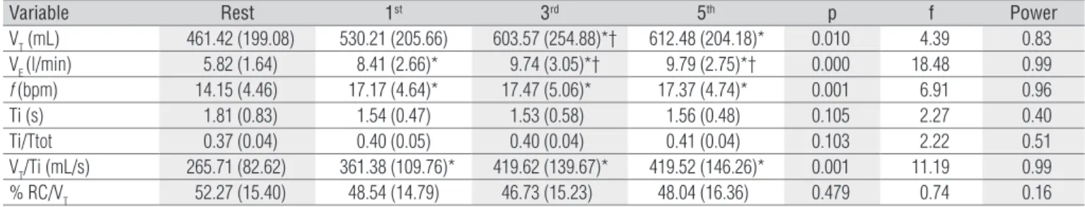
(p=0.042), rest and the 3rd minute (0.021), and rest and the 5th Table 2. The analyzed variables during rest, 1st, 3rd, and 5th minutes of exercise with unsupported arm elevation. The values of p, f and Power of ANOVA for repeated measures are also presented.
Variable Rest 1st 3rd 5th p f Power
VT (mL) 461.42 (199.08) 530.21 (205.66) 603.57 (254.88)*† 612.48 (204.18)* 0.010 4.39 0.83
VE (l/min) 5.82 (1.64) 8.41 (2.66)* 9.74 (3.05)*† 9.79 (2.75)*† 0.000 18.48 0.99
f (bpm) 14.15 (4.46) 17.17 (4.64)* 17.47 (5.06)* 17.37 (4.74)* 0.001 6.91 0.96
Ti (s) 1.81 (0.83) 1.54 (0.47) 1.53 (0.58) 1.56 (0.48) 0.105 2.27 0.40
Ti/Ttot 0.37 (0.04) 0.40 (0.05) 0.40 (0.04) 0.41 (0.04) 0.103 2.22 0.51
VT/Ti (mL/s) 265.71 (82.62) 361.38 (109.76)* 419.62 (139.67)* 419.52 (146.26)* 0.001 11.19 0.99
% RC/VT 52.27 (15.40) 48.54 (14.79) 46.73 (15.23) 48.04 (16.36) 0.479 0.74 0.16
Data expressed as mean (standard deviation). Significant difference was considered when p<0.05; * Statistically different in comparison to rest; † Statistically different in comparison to the 1st minute.
Significant difference was considered when p<0.05;
* Statistically significant for rest; †Statistically significant for the 1 minute; #Statistically
SCM=sternocleidomastoid muscle; EMG=electromyography.
significant for the 3 minute; PhAng=Phase Angle; PhRIB=inspiratory RC-AB synchrony; PhREB=expiratory RC-
st rd
AB synchrony; PhRTB=inspiratory and expiratory synchrony;
*
B A
C
30 25 20 15 10
0 5
PhAng
PhRIB PhREB
SCM EMG
PhRTB
D e g r e e
28 24 20 16 8 12
0 4 % R M S 25
20 15 10
0 5 %
1st min.
Rest 3rd min. 5rd min.
*
*
*
*
##
†
Figure 1. A) Comparison between PhAng during rest and during the analyzed moments of the hair-combing activity. B) Comparison between PhRIB, PhREB and PhRTB during rest and during the analyzed moments of the hair-combing activity. C) Comparison between SCM EMG during rest and during the analyzed moments of the hair-combing activity.
minute of exercise (p=0.001; Figure 1- B). When we consider the PhRTB, a progressive increase in synchrony between the two compartments was also observed during the activity, cor-responding to 9.93% at rest, 16.61% in the 1st minute, 17.09% in
the 3rd minute and 19.12% in the 5th minute, which represents
a 92% increase between rest and the end of the exercise. here was a statistically signiicant diference when considering rest and the 1st minute (p=0.005), rest and the 3rd minute (p=0.002)
and rest and the 5th minute of exercise (p<0.001; Figure 1 - B).
Electromyography activity
he subjects’ SCM muscle activity between rest and the ac-tivity of combing hair with unsupported arm elevation ranged in the following magnitude compared with rest: 168% RMS in the 1st minute, 196% RMS in the 3rd minute and 224% RMS in
the 5th minute. here was a statistically signiicant diference
when comparing rest with the 1st (p=0.005), the 3rd (p=0.001)
and the 5th minute of exercise (p=0.001; Figure 1 - C). No
corre-lations were found between the respiratory pattern variables, thoracoabdominal motion and electromyographic activity of the SCM muscle.
Discussion
he main results of this study were: increased f and VT, follo-wed by increased VE, increased VT/Ti and thoracoabdominal asynchrony demonstrated by PhAng, PhRIB, PhREB, PhRTB, and increase in SCM muscle activity during the unsupported arm elevation. In addition, a change was observed, though not statistically signiicant, in the predominance of the contribu-tion of the RC and AB to VT. here was a signiicant increase in VT, followed by a slightly increased f, resulting in a signiicant increase in VE during the activity time with unsupported arm elevation. he VT and VE absolute values during rest were com-parable with those reported by Tobin et al.23 using RIP in
heal-thy subjects in the supine position. However, when comparing breathing patterns from diferent studies, body posture must also be taken into account23,24.
In the sitting position, Couser, Martinez and Celli3 and
Baarends et al.2 measured V
E and VT using other methods, and
the results were markedly higher than those recorded for healthy subjects during rest in the present study. his diference probably relates to direct measuring techniques which employ breathing through a mouthpiece with the nose clipped23. As a consequence
of the small variation in f (21%) and especially in VT in the 3rd
minute (31%), there was a progressive increase in VE until the 3rd minute, achieving 67% of the rest value, remaining stable
until the 5th minute. hese results are partially comparable with
those reported by Couser, Martinez and Celli3 and by Baarends
et al.2, who analyzed healthy subjects with static elevated arms
for 2 minutes and observed a VE increase due to an increase in VT while the f remained unchanged. he diference in f variation may be due to the type of exercise performed by our subjects, i.e. combing hair with alternating unsupported arm elevation, which is more diicult than static arm elevation. Younes and Kivinen25 also reported that, at low levels of exercise, ventilation
increases primarily due to increases in VT and, at higher levels of exercise, the changes occur through changes in f. Tobin et al.23
reported that mean inspiratory low relects respiratory center drive input, which explains the increase in the VT/Ti ratio during unsupported arm elevation and its stabilization by the 3rd
mi-nute with 58% of rest value.
When we analyzed the PhAng, we observed that the sub-ject presented a value of 23.82° at the end of the activity that corresponds to a 129% increase when compared with rest. his demonstrates an increase in RC-AB asynchrony. To our know-ledge, there are no reports in the literature of PhAng values for healthy adults during unsupported arm elevation. With regard to rest values, our results were comparable with those found by Tobin et al.26 and Bloch et al.27 with healthy subjects during
quiet breathing.
he variables PhRIB, PhREB and PhRTB represent the time percentage during a respiratory cycle in which the RC and the AB move in opposite directions. If both compartments move in the same direction along a respiratory phase (inspiratory, expi-ratory or both), a 0% value is computed. If the compartments move in opposite directions, a 100% value is computed20. In
the present study, there was a progressive increase in PhRIB, PhREB and PhRTB, representing an increase of approximately 18% when comparing rest and the last minute of activity.
In healthy subjects, arm elevation leads to increased ven-tilatory and metabolic demands similar to mild exercise. At rest, the diaphragm is the dominant active inspiratory muscle, displacing the abdomen. During exercise, the inspiratory ac-cessories are progressively recruited to assist the diaphragm and elevate the RC. During arm elevation, some of the upper torso muscles become involved in arm positioning, their par-ticipation in ventilation is decreased, and there is a shift of ventilatory work to the diaphragm. his disproportionate in-crease in diaphragmatic work contributes to the generation of ventilatory pressures3. hese changes in RC and/or AB
mecha-nics may be the cause of the increase in asynchrony in COPD patients and, to a small degree, in healthy subjects. However, COPD patients have insuicient diaphragmatic function, lea-ding to considerable thoracoabdominal asynchrony.
In a study that assessed healthy subjects, Tobin et al.26
concluded that thoracoabdominal asynchrony and paradox in compartmental contribution to VT are predominantly due to
increases in respiratory load rather than muscle fatigue. In the present study, the RC and AB movements at rest were similar to those reported by Tobin et al.23. It appears that the slight
increase in the AB contribution in activity in exchange for RC contribution at rest is related to the arm elevation and to a gre-ater participation of the diaphragm in ventilation. According to Goldman et al.28, abdominal wall displacement is intimately
related to diaphragm displacement, however the same cannot be said for RC displacement.
he changes in the respiratory pattern mentioned above seem to be related to the hypothesis raised by many authors1,3,4,6
when referring to an increase in diaphragm activity in tasks in-volving unsupported arms, which may suggest an increase in diaphragmatic work to generate a VT due to a decrease in partic-ipation of the accessory muscles needed to stabilize the shoul-der girdle during unsupported arm movements. Tobin et al.23
reported that, although there was a slight predominance of AB contribution to VT compared to RC contribution in healthy subjects in the supine position, there was a large individual variation in the respiratory pattern at rest. Sharp et al.29, in
their study on the sitting position at rest, reported that the RC was responsible for about 70% of VT whereas, in the supine position, its contribution was only about 25%. Grimby, Bunn and Mead30 also reported a 25% contribution of AB to V
T in
individuals sitting on a cycle ergometer without exercising. hese indings were attributed to the position of the legs with a tonic contraction of the muscles in the torso and abdominal wall that could efectively reduce abdominal compliance.
Grimby, Bunn and Mead30 noted the variability of V T
dur-ing exercise and the need for caution when analyzdur-ing the esti-mated changes to RC and AB contribution to VT. However, they reported that, although there is a large individual variation, there appears to be a consistent pattern of changes in relative RC and AB movements with an increase in VT between rest and moderate exercise with a tendency toward a greater AB con-tribution to VT. In contrast, when exercise intensity increases, there is an increase in RC contribution.
In the present study, for the calibration of RIP, the QDC was used during natural breathing, a procedure irst described by Sackner et al.16 in 1989. his method computes the calibration
factor (K) by considering breaths of constant VT31. In 2001, De
Groote, Paiva and Verbandt31 criticized the QDC calibration
method for the possibility of error when there is a variation in VT during the procedure. It should be noted that, in the pre-sent study, there was variation in VT, but this occurred mainly during the exercise. During the calibration performed at rest, there was a slight variation in VT in 13 evaluated subjects, with maximum standard deviation of 12.2 mL (1.98% VT variation).
Furthermore, this is the current method employed by various studies that used RIP to assess the respiratory pattern18,19,32-35.
Regarding surface EMG, we observed a steady increase in the amplitude of SCM muscle recruitment between rest and activity. hese changes suggest an increase in SCM muscle participation in upper limb support during activities with unsupported arm elevation, reducing its respiratory function. his would lead to an increase in AB contribution to VT during exercise.
Our indings are comparable with those reported by Martinez, Couser and Celli8, who observed changes in
respira-tory muscle recruitment pattern indicating a more efective contraction of the diaphragm and an increase in SCM muscle amplitude of activation in EMG during activities with unsup-ported arm elevation. his suggests that some RC muscles are recruited during arm elevation. he present study also shows that there was an increase in VT and f with a subsequent increase in VE during the activity with unsupported arm elevation, and there was an increase in VT/Ti as the activity progressed, which shows a greater neural activation during exercise. We also ob-served an increase in thoracoabdominal asynchrony during un-supported arm elevation as well as an abrupt increase in SCM muscle amplitude of activation in the beginning of the exercise, showing a steady increase until the 5th minute of activity.
he changes detected in the respiratory pattern and the EMG activity of the respiratory accessory muscle of the healthy subjects during the studied ADL can contribute to elucidate the determinants of dyspnea in patients with COPD. However, no reports of a normative parameter were found in the litera-ture. herefore, it was necessary to study how these variables behave in healthy subjects. Considering the indings of the present study, it is possible to use this data as normality pa-rameters for clinical application to assess upper limb ADLs in patients with COPD.
Study limitations
Although the sample number was calculated in the pilot study to ind signiicant diferences between the moments (rest and activity), the number of subjects was insuicient for the correlations between respiratory pattern variables, thora-coabdominal motion and SCM muscle EMG. For the variables RC/VT andAB/VT, although the sample calculated to a power of 80% was small (eight), after the statistical analysis, the power found by ANOVA was low.
Partially funded by Conselho Nacional de Desenvolvimento Cientíico e Tecnológico (CNPq) and Fundação de Amparo à Pesquisa do Estado de Minas Gerais (FAPEMIG), Brazil.
1. Celli B, Criner G, Rassulo J. Ventilatory muscle recruitment during unsupported arm exercise in normal subjects. J Appl Physiol. 1988;64(5):1936-41.
2. Baarends EM, Schols AMWJ, Slebos DJ, Mostert R, Janssen PP, Wouters EF. Metabolic and ventilatory response pattern to arm elevation in patients with COPD and healthy age-matched subjects. Eur Respir J. 1995;8(8):1345-51.
3. Couser JI Jr, Martinez FJ, Celli BR. Respiratory response and ventilatory muscle recruitment during arm elevation in normal subjects. Chest. 1992;101(2):336-40.
4. Criner GJ, Celli BR. Effect of unsupported arm exercise on ventilatory muscle recruitment in patients with severe chronic airflow obstruction. Am Rev Respir Dis. 1988;138(4):856-61.
5. Dodd DS, Brancatisano T, Engel LA. Chest wall mechanics during exercise in patients with severe chronic air-flow obstruction. Am Rev Respir Dis. 1984;129(1):33-8.
6. Epstein SK, Celli BR, Williams J, Tarpy S, Roa J, Shannon T. Ventilatory response to arm elevation. Its determinants and use in patients with chronic obstructive pulmonary disease. Am J Respir Crit Care Med. 1995;152(1):211-6.
7. Martinez FJ, Couser JI, Celli BR. Factors influencing ventilatory muscle recruitment in patients with chronic airflow obstruction. Am Rev Respir Dis. 1990;142(2):276-82.
8. Martinez FJ, Couser JI, Celli BR. Respiratory response to arm elevation in patients with chronic airflow obstruction. Am Rev Respir Dis. 1991;43(3):76-80.
9. Velloso M, Stella SG, Cendon S, Silva AC, Jardim JR. Metabolic and ventilatory parameters of four activities of daily living accomplished with arms in COPD patients. Chest. 2003;123(4): 1047-53.
10. Celli BR, Rassulo J, Make BJ. Dyssynchronous breathing during arm but not leg exercise in patients with chronic airflow obstruction. N Engl J Med. 1986;314(23):1485-90.
11. Dolmage TE, Maestro L, Avendano MA, Goldstein RS. The Ventilatory response to arm elevation of patients with chronic obstructive pulmonary disease. Chest. 1993;104(4):1097-100.
12. Gigliotti F, Coli C, Bianchi R, Grazzini M, Stendardi L, Castellani C, et al. Arm exercise and hyperinsuflation in patients with COPD. Chest. 2005;128(3):1225-32.
13. Pereira CAC. Espirometria. J Pneumol. 2002;28 (Suppl 3 ):1-82.
14. Pereira CAC, Barreto SP, Simões JG, Pereira FQL, Gerstler JG, Nakatani J. Valores de referência para espirometria em uma amostra da população brasileira adulta. J Pneumol. 1992;18(1): 10-22.
15. Konno K, Mead J. Measurement of separate volume changes of the rib cage and abdomen during breathing. J Appl Physiol. 1967;22(3):407-22.
16. Sackner MA, Watson H, Belsito AS, Feinerman D, Suarez M, Gonzalez G, et al. Calibration of respiratory inductive plethysmograph during natural breathing. J Appl Physiol. 1989;66(1): 410-20.
17. Bloch KE, Barandun J, Sackner MA. Effect of mouthpiece breathing on cardiorespiratory response to intense exercise. Am J Respir Crit Care Med. 1995;151(4):1087-92.
18. Parreira VF, Tomich GM, Britto RR, Sampaio RF. Assessment of tidal volume and thoracoabdominal motion using volume and flow-oriented incentive spirometers in healthy subjects. Braz J Med Biol Res. 2005;38(7):1105-12.
19. Parreira VF, Coelho EM, Tomich GM, Alvim AMA, Sampaio RF, Britto RR. Avaliação do volume corrente e da configuração toracoabdominal durante o uso de espirômetros de incentivo a volume e a fluxo, em sujeitos saudáveis: influência da posição corporal. Rev Bras Fisioter. 2004;8(1):45-51.
20. Reber A, Geiduschek JM, Bobbia SA, Bruppacher HR, Frei FJ. Effect of continuous positive airway pressure on the measurement of thoracoabdominal asynchrony and minute ventilation in children anesthetized with sevoflurane and nitrous oxide. Chest. 2002;122(2):473-8.
21. Falla D, Dall’Alba P, Rainoldi A, Merletti R, Jull G. Location of innervation zones of sternocleidomastoid and scalene muscles-a basis for clinical and research electromyography applications. Clin Neurophysiol. 2002;113(1):57-63.
22. Falla D, Dall’Alba P, Rainoldi A, Merletti R, Jull G. Repeatability of surface EMG variables in the sternocleidomastoid and anterior scalene muscles. Eur J Appl Physiol. 2002;87(6):542-9.
23. Tobin MJ, Chadha TS, Jenouri G, Birch SJ, Gazeroglu HB, Sackner MA. Breathing patterns. 1. Normal subjects. Chest. 1983;84(2):202-5.
24. Feltrin M. Estudo do padrão respiratório e da congiguração tóraco-abdominal em indivíduos normais, nas posições sentada, dorsal e laterais, com o uso de pletismografia respiratória por indutância [dissertação]. São Paulo: Unifesp; 1994.
25. Younes M, Kivinen G. Respiratory mechanics and breathing pattern during and following maximal exercise. J Appl Physiol. 1984;57(6):1773-82.
26. Tobin MJ, Perez W, Guenther SM, Lodato RF, Dantzker DR. Does rib cage-abdominal paradox signify respiratory muscle fatigue? J Appl Physiol. 1987;63(2):851-60.
27. Bloch KE, Li Y, Zhang J, Bingisser R, Kaplan V, Weder W, et al. Effect of surgical lung volume reduction on breathing patterns in severe pulmonary emphysema. Am J Respir Crit Care Med. 1997;156(2 Pt 1):553-60.
28. Goldman MD, Grassino A, Mead J, Sears TA. Mechanics of the human diaphragm during voluntary contraction: dynamics. J Appl Physiol. 1978;44(6):840-8.
29. Sharp JT, Goldberg NB, Druz WS, Danon J. Relative contributions of rib cage and abdomen to breathing in normal subjects. J Appl Physiol. 1975;39(4):608-18.
30. Grimby G, Bunn J, Mead J. Relative contribution of rib cage and abdomen to ventilation during exercise. J Appl Physiol. 1968;24(2):159-66.
31. De Groote A, Paiva M, Verbandt Y. Mathematical assessment of qualitative diagnostic calibration for respiratory inductive plethysmography. J Appl Physiol. 2001;90(3):1025-30.
32. Tomich GM, Franca DC, Diorio AC, Britto RR, Sampaio RF, Parreira VF. Breathing pattern, thoracoabdominal motion and muscular activity during three breathing exercises. Braz J Med Biol Res. 2007;40(10):1409-17.
33. Brant TCS, Parreira VF, Mancini MC, Becker HMG, Reis AFC, Britto RR. Padrão respiratório e movimento toracoabdominal de crianças respiradoras orais. Rev Bras Fisioter. 2008;12(6):495-501.
34. Teramoto S, Fukuchi Y, Nagase T, Matsuse T, Orimo H. A comparison of ventilation components in young and elderly men during exercise. J Gerontol A Biol Sci Med Sci. 1995;50(1):B34-9.
35. Brown K, Aun C, Jackon E, Mackersie A, Hatch D, Stocks J. Validation of respiratory inductive plethysmography using the Qualitative Diagnostic Calibration method in anaesthetized infants. Eur Respir J. 1998;12(4):935-43.