Chronic HIV-1 Infection Frequently Fails
to Protect against Superinfection
Anne Piantadosi1,2[, Bhavna Chohan1,2[, Vrasha Chohan3, R. Scott McClelland3,4,5, Julie Overbaugh1,2*
1Division of Human Biology, Fred Hutchinson Cancer Research Center, Seattle, Washington, United States of America,2Department of Pathobiology, University of Washington, Seattle, Washington, United States of America,3Department of Medical Microbiology, University of Nairobi, Nairobi, Kenya,4Department of Medicine, University of Washington, Seattle, Washington, United States of America,5Department of Epidemiology, University of Washington, Seattle, Washington, United States of America
Reports of HIV-1 superinfection (re-infection) have demonstrated that the immune response generated against one strain of HIV-1 does not always protect against other strains. However, studies to determine the incidence of HIV-1 superinfection have yielded conflicting results. Furthermore, few studies have attempted to identify superinfection cases occurring more than a year after initial infection, a time when HIV-1-specific immune responses would be most likely to have developed. We screened a cohort of high-risk Kenyan women for HIV-1 superinfection by comparing partial gag and envelope sequences over a 5-y period beginning at primary infection. Among 36 individuals, we detected seven cases of superinfection, including cases in which both viruses belonged to the same HIV-1 subtype, subtype A. In five of these cases, the superinfecting strain was detected in only one of the two genome regions examined, suggesting that recombination frequently occurs following HIV-1 superinfection. In addition, we found that superinfection occurred throughout the course of the first infection: during acute infection in two cases, between 1–2 y after infection in three cases, and as late as 5 y after infection in two cases. Our results indicate that superinfection commonly occurs after the immune response against the initial infection has had time to develop and mature. Implications from HIV-1 superinfection cases, in which natural re-exposure leads to re-infection, will need to be considered in developing strategies for eliciting protective immunity to HIV-1.
Citation: Piantadosi A, Chohan B, Chohan V, McClelland RS, Overbaugh J (2007) Chronic HIV-1 infection frequently fails to protect against superinfection. PLoS Pathog 3(11): e177. doi:10.1371/journal.ppat.0030177
Introduction
HIV-1 superinfection (also referred to in the literature as re-infection) has been described in more than 20 cases [1–5], demonstrating that natural infection with HIV-1 does not always generate a protective immune response. Evidence of superinfection presents a potential challenge to HIV-1 vaccine design because most of the other viruses for which vaccines exist do elicit protective immunity in natural infection. However, the implications of HIV-1 superinfection for vaccine design remain controversial, in part because it is unclear how often superinfection occurs and whether it is restricted to times in infection when an HIV-specific immune response has not yet developed, such as soon after the first infection, or has become impaired, such as during advanced AIDS.
To date, several population-based studies have measured the incidence of HIV-1 superinfection, with conflicting results. At one extreme, two studies found that superinfection occurred at a rate close to that of initial infection: 4% per year among women in Kenya [1], compared to an initial infection rate of 8% per year in the same cohort [6]; and 5% per year among men in southern California [2], compared to an initial infection rate of 5% per year in a comparable cohort [7]. At the other extreme, two studies did not identify any cases of HIV-1 superinfection despite extensive follow-up [8,9]. Differences in study design and methodology may account for some variability in the detection of super-infection. It is also likely that the incidence of HIV-1 superinfection depends on the frequency of re-exposure and potentially on characteristics of the viruses being studied. One factor that may influence the incidence of HIV-1
superinfection is the relatedness of the virus strains. In approximately half of the published cases of superinfection, the initial and superinfecting viruses belonged to different subtypes, which can differ by ;30% in theenvelope gene. In contrast to these cases of intersubtype superinfection, in cases of intrasubtype superinfection viruses differ by;10% or less inenv[10] and might be expected to share antigenic properties. Intrasubtype superinfection is more difficult to detect and has primarily been documented for subtype B viruses [2,11–16]. It is not known how often intrasubtype superinfection occurs in regions where non-subtype B viruses predominate, and these are the regions where HIV-1 is most prevalent.
The rate of HIV-1 superinfection may also depend on whether an HIV-specific immune response has been gener-ated at the time of exposure to the second virus. It is possible that superinfection occurs more frequently in the period soon after the initial infection, before the development of an HIV-1-specific immune response, although there are reports that indicate this is not always the case [12,17,18]. The
Editor:Richard A. Koup, National Institutes of Health, United States of America ReceivedAugust 3, 2007;AcceptedOctober 3, 2007;PublishedNovember 16, 2007
Copyright:Ó2007 Piantadosi et al. This is an open-access article distributed under the terms of the Creative Commons Attribution License, which permits unrestricted use, distribution, and reproduction in any medium, provided the original author and source are credited.
Abbreviations:CTL, cytotoxic T lymphocyte; DPI, days post-infection; MRCA, most recent common ancestor; ssPCR, strain-specific polymerase chain reaction * To whom correspondence should be addressed. E-mail: joverbau@fhcrc.org
cytotoxic T lymphocyte (CTL) response generally arises within the first month after infection [19], presenting the first potential barrier to HIV-1 superinfection. Experimental superinfection of macaques with HIV-2 demonstrated that superinfection was limited to the first month after infection in that model [20]. However, HIV-1 superinfection in humans has been documented to occur after the development of a CTL response [12,16,18,21], including one case in which CTLs recognized the superinfecting strain [12].
The other arm of the adaptive immune response to HIV-1, neutralizing antibodies, does not arise until later in infection but may play a stronger role in preventing superinfection. Neutralizing antibodies have been shown to block primary infection in an experimental animal setting [22–25]. Prelimi-nary evidence suggests that they may also block super-infection; Smith et al. recently found that three individuals who became superinfected had weaker neutralizing antibody responses to their initial infections than did control individuals [26]. However, neutralizing antibodies generally do not arise until 2 mo after infection, and antibodies do not broaden to recognize heterologous strains until approxi-mately 1 y after infection, if at all [27]. Because most of the documented cases of superinfection occurred within the first year after infection [11], it is tempting to speculate that antibodies could protect against later superinfections. How-ever, in many studies, individuals were not followed for longer than a year. Studies with long follow-up are needed to determine whether later superinfections occur despite the opportunity for the host to develop a broad immune response.
Finally, most of the previous studies are likely to have underestimated the incidence of superinfection because they examined only one region of the HIV-1 genome [1,2,9] or two neighboring regions [8]. This approach may have missed cases of superinfection in which the initial and superinfecting viruses recombined, a frequent event during HIV-1 repli-cation [28]. Examining more than one region of the HIV-1 genome may provide a better estimate of the incidence of HIV-1 superinfection.
To address these issues, we screened a prospective cohort of Kenyan women for HIV-1 superinfection. We compared sequences from two non-neighboring regions of the genome (gagp17 andenvV1-V5) at time points soon after the initial infection and approximately five years later. We identified seven cases of HIV-1 superinfection among 36 women. Three of these cases were subtype A intrasubtype superinfections. In the other four cases, the initial and superinfecting viruses belonged to different subtypes in at least one of the genome regions examined. In five out of seven cases, the super-infecting strain was only detected in one region of the genome (gagorenv), indicating that recombination frequently occurred following superinfection. We also defined the timing of each superinfection case, and these results indicate that superinfection can occur as late as five years after the initial infection.
Results
To identify potential HIV-1 superinfection cases, we analyzed HIV-1 proviral sequences from 36 individuals at two time points. The first (‘‘initial’’) time point was a median of 111 days post-infection (DPI) (range 17–338), and the second (‘‘chronic’’) was a median of 1,901 DPI (range 1,309– 2,631, which corresponds to 3.6–7.2 y post-infection). For each individual, we studied a median of sevengagsequences (range 3–19) and eight envsequences (range 3–13) from the initial time point and a median of sevengagsequences (range 5–13) and sevenenvsequences (range 4–12) from the chronic time point.
In examining sequences from the initial time point, sequences from each individual formed a monophyletic cluster in both gagand env phylogenetic trees (unpublished data), suggesting that all individuals were initially infected from a single source partner (i.e., individuals were not coinfected). The initialenvsequences had a median pairwise diversity of 2.6% (range 0%–6.4%) and a median divergence from their most recent common ancestor (MRCA) of 1.6%
(range 0%–11.5%). The initial median gag diversity and
divergence were 0.3% (0%–1.3%) and 0.6% (0%–3.3%), respectively. In nine individuals, the initial gag sequences
and env sequences belonged to different HIV-1 subtypes,
indicating initial infection with intersubtype recombinant
viruses. Figure 1 shows env (A) and gag (B)
maximum-likelihood phylogenetic trees containing four representative sequences (two initial and two chronic) from each of the 36 individuals. All initial env V1–V5 sequences clustered with subtype A reference sequences (including both sub-subtypes, A1 and A2), which was a selection criterion for this study. However, initialgagp17 sequences clustered with subtype C in one individual and subtype D in three individuals and were A1/D recombinant in two individuals and A2/D recombinant in three individuals.
Through analysis of sequences from both the initial and chronic time points, we identified seven potential cases of superinfection, in which some or all of the chronic sequences clustered separately from the initial sequences on phyloge-netic trees ofenv,gag, or both. These cases are highlighted in color on the phylogenetic trees in Figure 1. Separate
clustering was observed in both env and gag trees for one
case (Case 2), in onlyenvfor four cases (Cases 1, 3, 4, and 5), and in onlygagfor two cases (Cases 6 and 7).
Author Summary
In three of these cases, the chronic sequences belonged to a different subtype than the initial sequences, indicating intersubtype superinfection. Specifically, as shown in Figure 1A, the initialenvsequences from Case 3 are subtype A while
the chronic sequences are subtype D. The initial env
sequences from Case 4 are subtype A, while the chronic sequences are subtype C. As shown in Figure 1B, the initial gagsequences from Case 7 are subtype C, while the chronic sequences cluster between subtypes A and C and are an A/C recombinant (Figure S1). One of the remaining cases was an inter-sub-subtype superinfection: the initial sequences from
Case 2 were sub-subtype A2 inenvand D/A2 recombinant in
gag (Figure S2A), while the chronic sequences were
sub-subtype A1 in env and D/A1 in gag (Figure S2B). In the
remaining three cases (Cases 1, 5, and 6), both the initial and chronic sequences clustered with subtype A reference sequences, indicating intrasubtype superinfection.
Quantitative analysis of virus divergence supported super-infection in these cases. For each individual, we calculated divergence as the maximum genetic distance between any chronic sequence and the MRCA of the initial sequences. To determine what level of divergence would be expected in an intrasubtype superinfection, we also measured the divergence between each pair of individuals who had a subtype A virus (including sub-subtypes A1 and A2). Figure 2 shows plots of env(A) andgag(B) divergence within and between individuals; the potential superinfection cases from Figure 1 are labeled. Excluding these presumed superinfection cases, the diver-Figure 1.Maximum-Likelihood Phylogenetic Trees ofenv(A) andgag(B) Sequences from 36 Individuals
Each tree contains two initial and two chronic sequences from each individual (unlabeled branches) that were selected to represent both strains in presumed superinfection cases and were selected randomly for other cases. Reference sequences representing different subtypes from the LANL database are also included (labeled branches). Cases that demonstrate separate clustering on either or both trees are highlighted in color. In most cases, separate clustering is evident in onlyenvor onlygag. Sequences from each of these presumed superinfection cases are labeled with the case number
and either‘‘initial’’or‘‘chronic’’on the tree(s) that demonstrates separate clustering. Green¼Case 1, orange¼Case 2, blue¼Case 3, purple¼Case 4, red¼Case 5, yellow¼Case 6, light blue¼Case 7. Bootstrap values were omitted for clarity; however, except for the superinfection cases, sequences from each individual form a monophyletic cluster with82% bootstrap support (most have 100% bootstrap support).
gences within each individual ranged from 2.1% to 10.7% in env(median¼4.5%) and from 1.2% to 3.9% ingag(median¼ 2.3%). The between-individual divergence ranged from
10.4% to 38.2% (median¼17.9%) inenvand from 5.4% to
17.1% (median¼10.1%) ingag. These ranges are somewhat
higher than what is typically reported [10,29,30] because we included comparisons between subtype A2 and sub-subtype A1 sequences. Furthermore, we calculated maximum divergence rather than average diversity in order to identify potential superinfection cases in which just one sequence belonged to the superinfecting strain. As shown in Figure 2, the levels of divergence observed in the three potential intrasubtype superinfection cases (Cases 1, 5, and 6) are in the range that would be expected for a superinfection. Consistent with the results of phylogenetic analysis (Figure 1), Cases 1 and 5 demonstrate high divergence inenvbut notgag, while Case 6 demonstrates high divergence ingagbut notenv. Case 2, the inter-sub-subtype superinfection that was detected in
bothenv andgag shows high divergence on both plots. The
divergence in the intersubtype superinfection cases (Cases 3
and 4 in env and Case 7 in gag) is higher than in the
intrasubtype cases, as would be predicted.
For all seven potential cases of HIV-1 superinfection identified by phylogenetic analysis, we examined samples from intervening time points to estimate when superinfec-tion occurred. As described below, we examined samples using both single-copy sequencing and strain-specific PCR (ssPCR), which allowed us to sample a greater number of genome copies. For cases of superinfection in which the second strain was initially detected in only env or gag, this approach also allowed us to carefully screen the other region at intervening time points. In one case (Case 1), we detected a second strain ingagat an intervening time point, whereas it was not found in our initial screen. The timing and characterization of superinfection cases are summarized in Table 1.
Figure 2.Maximum Divergence from the MRCA for Chronicenv(A) andgag(B) Sequences
Comparisons within individuals (black circles) and between subtype A–infected individuals (gray triangles) are arbitrarily distributed along thex-axis.
Superinfection cases are labeled in both plots. For most cases, the high divergence indicative of superinfection is apparent in one region of the genome but not the other, consistent with Figure 1.
doi:10.1371/journal.ppat.0030177.g002
Table 1.Summary and Estimated Timing of HIV-1 Superinfection Cases
Case Subject ID Estimated
Timing (DPI)
Viral Load Changea
CD4b Subtypec (env)
Subtypec (gag)
Inter/Intra Subtyped
Initial Chronic Initial Chronic
1 QB726 749–1,031 4.1–4.0 309 A AþA* D DþA* Inter
2 QB045 1,680–2,048 4.5–4.3 553 A2 A2þA1 D/A2 D/A2þD/A1 Intra
3 QB850 52–73 5.2–5.5 NA A AþA/D A A Inter
4 QD022 1,832–1,957 4.3–5.8 788 A C A A Inter
5 QA413 714–1,007 4.9–5.0 296 A AþA* A A Intra
6 QB685 303–1,453 2.6–4.0 628 A A A A* Intra
7 QC885 58–152 5.6–5.5 NA A A C CþA/C Inter
aViral load (log 10 copies RNA/mL plasma) at the time points immediately before and immediately after superinfection. bCD4 cells/uL at the time point immediately after superinfection. NA, not available.
cFor each time point, the virus subtype(s) detected are indicated. A*, a subtype A virus different from the initial subtype A virus. d
Cases were classified as intersubtype if the strains belonged to different subtypes in at least one genome region (gagorenv).
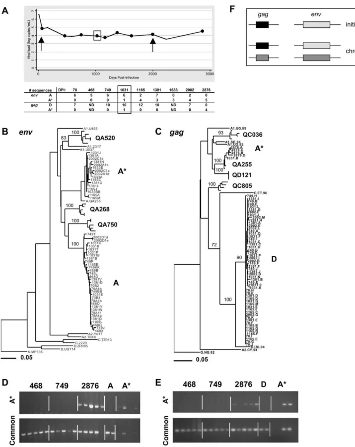
Figure 3.Case 1 (QB726): Intrasubtype Superinfection (Subtype A) between 749 and 1,031 DPI Detected inenvandgag
(A) Plot of viral load (log RNA copies/mL plasma) versus DPI. Sequences were obtained by single-copy PCR from time points marked by circles. Arrows indicate the initial and chronic time points used in the analysis in Figure 1, and the box indicates the first time point at which the superinfecting strain was detected. The number of sequences of each strain at each time point is indicated in a table below the graph. ND, not done.
(B) Maximum-likelihood tree ofenvsequences from all time points. Sequence names from QB726 are comprised of the sample DPI and a unique
identifier. The cluster of strain A contains sequences from all time points except 1,633 and 2,876 DPI. The cluster of strain A* contains sequences from 1,031, 1,165, 1,381, and 2,002 DPI. Sequences from three other individuals (QA520, QA268, and QA750), as well as subtype reference sequences from the LANL database, are also included. Bootstrap values greater than 70% are shown.
Case 1 (QB726): Intrasubtype Superinfection (Subtype A)
between 749 and 1,031 DPI Detected inenvandgag
For Case 1, intrasubtype superinfection was first suggested by separate clustering of initial and chronicenvelope sequen-ces on a phylogenetic tree (Figure 1).Envsequences from 70 DPI formed a monophyletic cluster (strain A), while some sequences from 2,002 DPI clustered separately (strain A*). To determine when strain A* first appeared, we examined multiple sequences from six intervening time points. These time points are highlighted in Figure 3A, a plot of this individual’s viral load throughout infection. A phylogenetic tree of allenvsequences is shown in Figure 3B.Envsequences from 468 DPI and 749 DPI clustered with strain A sequences. Among the sequences from 1,031 DPI, one clustered with strain A*, suggesting that superinfection occurred between 749 and 1,031 DPI. We detected both strain A and strain A* at 1,165, 1,381, and 2,002 DPI, and we detected only strain A* in sequences from a later sample (2,876 DPI). The number of sequences of each strain at each time point is listed in Figure 3A.
We used ssPCR ofenvsequences to assess the presence or absence of strain A* at time points before it was detected by single-copy sequencing. This method allowed us to sample a greater number of genome copies (50 copies per time point versus;6). Using primers specific for strain A*, we did not detect the new strain in 50 genome copies from 468 or 749 DPI but did detect it at 2,876 DPI, consistent with results from the sequence analysis (Figure 3). The 70-DPI sample was not included in this analysis because we did not have enough DNA available.
In our initial analysis ofgagsequences from Case 1, we did not detect a superinfecting strain at 2,002 DPI. As shown in Figure 1, sequences from 70 and 2,002 DPI formed a monophyletic cluster within subtype D. To determine whether a superinfecting strain could be detected in thegag region at intervening time points, we examined a total of 47 single-copy sequences from five samples (listed in Figure 3A). At 1,031 DPI, we found one sequence that clustered with subtype A sequences (A*) (Figure 3C). The subtype A strain was not detected by single-copy sequencing at 1,165 or 1,381 DPI but was the only strain present at 2,876 DPI. Using strain-specificgagprimers, we did not detect the subtype A strain in 50 genome copies from 468 or 749 DPI but did detect it at 2,876 DPI (Figure 3E).
As summarized in Figure 3F and Table 1, these results suggest that this individual was initially infected with an intersubtype recombinant strain, containing subtype A inenv
and subtype D in gag. Between 749 and 1,031 DPI, this
individual acquired a second strain that belonged to subtype A, which was detected in bothenvandgagsequences, although less consistently ingag.
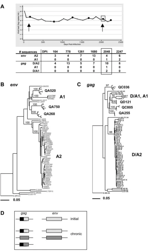
Case 2 (QB045): Intrasubtype Superinfection (Sub-Subtype
A2–A1) between 1,680 and 2,048 DPI Detected inenvand
gag
For Case 2, intrasubtype superinfection was suggested by separate clustering of initial and chronic sequences in both envandgagphylogenetic trees (Figure 1).Envsequences from 164 DPI formed a monophyletic cluster within sub-subtype
A2, while at 2,048 DPI, one env sequence (out of eight)
clustered with sub-subtype A1. We examinedenv sequences
from three available intervening time points (Figure 4A) and found only the initial sub-subtype A2 strain in a total of 24 sequences (Figure 4B). To confirm the presence of the superinfecting strain, we also examined sequences from a
later sample, 2,247 DPI. At this time point, we found env
sequences from both the sub-subtype A2 strain (n¼6) and the sub-subtype A1 strain (n¼2) (Figure 4B).
Gag sequences from Case 2 at 164 DPI clustered most
closely with subtype A2; however, Simplot analysis indicated that thegagsequences were a D/A2 recombinant at the initial time point (Figure S2A). At 2,048 DPI, we detected one subtype A1 strain and two D/A1 recombinants (Figures 1 and S2B). To determine whether the recombinant strain was derived from the initial subtype D/A2 strain, we separately
analyzed the 59 subtype D portion of these sequences.
Sequences from both time points clustered together on a phylogenetic tree (unpublished data), suggesting that the D/ A1 strain present at 2,048 DPI was generated by recombina-tion between the initial subtype D/A2 strain and the novel subtype A1 strain. We did not detect the subtype A1 strain or D/A1 recombinant ingagat any of the three intervening time points (25 total sequences) or at the later time point (eight sequences) (Figure 4C). For this individual, we did not have enough HIV-1 DNA available to confirm the timing of superinfection using ssPCR.
As shown in Figure 4D, our results suggest that this individual was initially infected with a subtype D/A2
recombinant (D/A2 in gag and A2 in env). Between 1,680
and 2,048 DPI, this individual was superinfected with a subtype A1 strain, which was detected in both env and gag.
Within the gag region, we also detected a recombinant
between the initial and superinfecting strains.
Case 3 (QB850): Intersubtype Superinfection (Subtype A–
D) between 52 and 73 DPI Detected inenv
For Case 3,envsequences from 45 DPI belonged to subtype A, while at 1,768 DPI we detected both subtype A and an A/D recombinant (Figures 1 and S3). In analysis of only the subtype A portion (C2–V3), all sequences formed a mono-phyletic cluster (unpublished data), suggesting that the new A/ D sequence arose by recombination between the initial subtype A strain and a novel subtype D strain.
identifier. The cluster of strain D contains sequences from all time points except 2,876 DPI. The cluster of strain A* contains sequences from 1,031 and 2,876 DPI. Sequences from four other individuals (QC036, QA255, QD121, and QC805), as well as subtype reference sequences from the LANL database, are also included. Bootstrap values greater than 70% are shown.
(D) Amplification of 50 HIV-1 genome copies from the indicated time points usingenvstrain–specific primers (top panel) orenvcommon primers
(bottom panel). For each time point, each lane contains an estimated ten HIV-1 genome copies of template input. As a control, we also amplified 103 copies of a cloned A sequence (two lanes) and one copy of a cloned A* sequence (four lanes).
(E) Amplification of 50 HIV-1 genome copies from the indicated time points usinggagstrain–specific primers (top panel) orgagcommon primers
(bottom panel). For each time point, each lane contains an estimated ten HIV-1 genome copies of template input. We also amplified 103copies of a cloned D sequence (two lanes) and one copy of a cloned A* sequence (four lanes).
(F) Summary of the strains detected in each genome region at the initial time point and during chronic infection (all other time points). Light gray¼ subtype A; dark gray¼A*; black¼subtype D.
Figure 4.Case 2 (QB045): Intrasubtype Superinfection (Sub-Subtype A2–A1) between 1,680 and 2,048 DPI Detected inenvandgag
The layout of this figure is similar to that of Figure 3, as described in the legend for Figure 3.
(A) Plot of viral load (log RNA copies/mL plasma) versus DPI with the number of sequences of each strain indicated.
(B) Maximum-likelihood tree ofenvsequences from all time points. The A2 cluster contains sequences from all time points, while the A1 cluster contains
sequences from 2,048 and 2,247 DPI.
(C) Maximum-likelihood tree ofgagsequences from all time points. The cluster of strain D/A2 contains sequences from all time points. The cluster of
strains D/A1 and A1 contains sequences from 2,048 DPI.
In analysis of sequences from intervening time points (Figure 5A), we detected only subtype A at 52 DPI, but both A and A/ D at 73 DPI (Figure 5B). Forty-four sequences obtained from eight more intervening time points between 73 and 1,768 DPI all belonged to subtype A and clustered with the 45 DPI sequences. Using ssPCR to amplify 50 genome copies, the second strain was not detected at 45 or 52 DPI but was detected at 73 DPI (Figure 5).
Gagsequences from 45 and 1,768 DPI belonged to subtype
A and formed a monophyletic cluster (Figure 1). The divergence at 1,768 DPI was unusually high: 6.1%, which is more than 3 standard deviations higher than the average divergence for all individuals. This high divergence could be the result of transmission of a highly diverse virus population from the source partner, rapid de novo evolution of the initial strain in this individual, or superinfection by a strain that is closely related to the initial strain. In examination of sequences from intervening time points, we did not detect
any sequences that formed a separate cluster on a gag
phylogenetic tree (Figure 5C).
Together, these results suggest that this individual was initially infected with a subtype A strain and was super-infected between 52 and 73 DPI with a subtype D strain,
which recombined with the initial strain within env V1–V5
(Figure 5E). After it was first detected at 73 DPI, the A/D recombinant strain was not detected in a total of 44 sequences from eight time points sampled over four and a half years, however it was detected (in three out of 12 sequences) at the last time point examined, 1,768 DPI.
Case 4 (QD022): Intersubtype Superinfection (Subtype A–
C) between 1,832 and 1,957 DPI Detected inenv
For Case 4,envsequences from 51 DPI belonged to subtype A, while those from 1,957 DPI belonged to subtype C, suggesting intersubtype superinfection (Figure 1). Because this individual had limited follow-up, we were only able to study sequences from two intervening time points (Figure 6A). Sequences from 522 and 1,832 DPI belonged to subtype A and clustered with the 51 DPI sequences (Figure 6B). To confirm presence of the subtype C strain, we also examined sequences from a sample obtained later in infection (2,752 DPI). At this time, allenv sequences belonged to subtype C and clustered with the 1,957 DPI sequences (Figure 6B).
Results from env ssPCR supported those from single-copy
sequencing; the subtype C strain was not detected at 51, 522, and 1,832 DPI but was detected at 1,957 DPI (Figure 6C and unpublished data).
In the analysis of initial and chronic gag sequences, all sequences formed a monophyletic cluster within subtype A (Figure 1). We examinedgagsequences from intervening time points and did not observe evidence of superinfection (unpublished data). Overall, these results suggest that this individual was initially infected with a subtype A strain and
was superinfected with a subtype C strain between 1,832 and 1,957 DPI (Figure 6D). Because we did not detect the novel strain in the gag region, it is likely that the two strains recombined, and the strain that predominated contained the initial subtype A ingagand the novel subtype C inenv.
Case 5 (QA413): Intrasubtype Superinfection (Subtype A)
between 714 and 1,007 DPI Detected inenv
In this case, env sequences from 45 DPI formed a
mono-phyletic cluster, while some sequences from 1,346 DPI formed a separate cluster within subtype A, suggesting intrasubtype
superinfection (Figure 1). We examinedenvsequences from
six intervening time points (Figure 7A) and first detected the superinfecting strain (A*) at 1,007 DPI (Figure 7B). Using ssPCR, we did not detect strain A* inenvin 50 genome copies from 57, 275, 411, 607, or 714 DPI but did detect it at 1,007 DPI (Figure 7C and unpublished data).
In examination of gag sequences, we did not identify a
novel strain in 23 total sequences from 714, 1,007, 1,146, or 1,346 DPI (unpublished data). Based on these results, it appears that this individual was initially infected with a subtype A strain and was superinfected with another subtype A strain between 714 and 1,007 DPI. As in Case 4, our failure to detect the superinfecting strain in thegagregion suggests that the strains recombined, generating a variant that contained the initial subtype A strain in gagand the novel subtype A strain inenv(Figure 7D).
Case 6 (QB685): Intrasubtype Superinfection (Subtype A)
between 303 and 1,453 DPI Detected ingag
Intrasubtype superinfection in this case was suggested by separate clustering of initial (86 DPI) and chronic (1,800 DPI) gagsequences (Figure 1). Only two intervening samples were available for estimating the timing of superinfection in this case (Figure 8A). The superinfecting strain (A*) was detected at 1,453 but not 303 DPI by single-copy sequencing (Figure 8B). Using ssPCR to amplify 50 genome copies, we also detected A* at 1,453 but not 303 or 86 DPI (Figure 8C).
In the analysis of env sequences from Case 6, initial and chronic sequences clustered together on a phylogenetic tree (Figure 1), although the divergence at 1,800 DPI was relatively high (8.2%). Examination of sequences from intervening time points did not reveal the presence of a second strain. These results suggest that this individual acquired a subtype A intrasubtype superinfection between 303 and 1,453 DPI (Figure 8D). Because we did not detect the second strain in the env region, it is likely that the two strains recombined (Figure 8D).
Case 7 (QC885): Intersubtype Superinfection (Subtype C–
A) between 58 and 152 DPI Detected ingag
Gagsequences sampled from Case 7 at 58 DPI belonged to
subtype C, while those sampled at 1,405 DPI clustered Figure 5.Case 3 (QB850): Intersubtype Superinfection (Subtype A–D) between 52 and 73 DPI Detected inenv
The layout of this figure is similar to that of Figure 3, as described in the legend for Figure 3.
(A) Plot of viral load (log RNA copies/mL plasma) versus DPI with the number of sequences of each strain indicated.
(B) Maximum-likelihood tree ofenvsequences from all time points. Cluster A contains sequences from all time points, while cluster A/D contains
sequences from 73 and 1,768 DPI.
(C) Maximum-likelihood tree ofgagsequences from all time points, illustrating unusually high divergence.
(D) Amplification of 50 HIV-1envgenome copies from the indicated time points using strain-specific primers (top panel) and common primers (bottom
panel).
between subtypes A and C (Figure 1). Simplot analysis of the chronic sequences revealed A/C recombinant sequences (Figure S1). Phylogenetic analysis of the 39subtype C portion of these sequences indicated that the novel subtype C was derived from the original subtype C (unpublished data).
To determine when the A/C strain first appeared, we examined sequences from five intervening time points (Figure 9A). The A/C recombinant was detected at the earliest available intervening time point (152 DPI) and several other time points (Figure 9B). Using ssPCR, we did not detect the A/ C strain in 50 genome copies from the 58 DPI sample but did detect it at 152 DPI (Figure 9C). In addition, the super-infecting strain was not detected in analysis ofenvsequences from any time point (unpublished data).
As shown in Figure 9D, these results suggest that this individual was initially infected with a C/A recombinant (C in gag, A in env). This individual acquired a novel subtype A strain between 58 and 152 DPI, which recombined with the
initial subtype C strain within thegagregion studied and was not detected in theenvregion.
Summary of Superinfection Cases
Overall, we observed seven cases of HIV-1 superinfection among 36 individuals with 188.7 total person-years of follow-up, corresponding to an incidence of 3.7% per person-year. Superinfection occurred throughout the course of infection, with the earliest between 52–73 DPI and the latest between 1,832–1,957 DPI (Table 1). In most cases, the viral load at the time of superinfection was at the expected level for this cohort. The median viral load at the time point before superinfection was 4.5 log copies RNA/mL plasma (range 2.6– 5.6), compared to a median viral load set point of 4.5 log copies RNA/mL plasma in this cohort [31]. The median log viral load at the time point after superinfection was 5.1 log copies RNA/mL plasma (range 4.0–5.8). Our sample size was too small for a meaningful statistical analysis of the change in Figure 6.Case 4 (QD022): Intersubtype Superinfection (Subtype A–C) between 1,832 and 1,957 DPI Detected inenv
The layout of this figure is similar to that of Figure 3, as described in the legend for Figure 3.
(A) Plot of viral load (log RNA copies/mL plasma) versus DPI with the number of sequences of each strain indicated.
B) Maximum-likelihood tree ofenvsequences from all time points. Cluster A contains sequences from 51, 522, and 1,832 DPI, while cluster C contains
sequences from 1,957 and 2,752 DPI.
(C) Amplification of 50envHIV-1 genome copies from the indicated time points using strain-specific primers (top panel) and common primers (bottom
panel).
viral load between these time points; however, the change was negligible for five of the seven individuals (median 0.0 log copies RNA/mL plasma, range:0.2 to 0.2). For the other two individuals (Cases 4 and 6), the viral load increased substantially (by 1.5 log copies RNA/mL plasma) between the time points immediately before and immediately after superinfection (Figures 6A and 8A). CD4 counts were available for five of the seven individuals at the time point after superinfection and in all five cases were greater than 200 cells/uL (Table 1).
Superinfection was detected in analysis of bothenvandgag sequences in only two of the seven cases, and in one of these cases the superinfecting strain was detected in both regions only at some time points. In the other five cases, the superinfecting and initial strains recombined. In five cases, both the initial and superinfecting strains persisted
through-out infection in at least one genome region, while in the remaining two cases (Cases 4 and 6), the initial strain was not present to our level of detection after superinfection, suggesting that it was substantially replaced by the super-infecting strain.
Discussion
This study represents the most in-depth population-level assessment of the incidence and timing of HIV-1 super-infection to date. Although the number of individuals included in the study was relatively small, they had extensive long-term follow-up. Importantly, because individuals were enrolled before their first infection and followed for approximately five years after infection, we were able to assess the incidence of superinfection throughout the course of HIV-1 infection. By contrast, many of the previously Figure 7.Case 5 (QA413): Intrasubtype Superinfection (Subtype A) between 714 and 1,007 DPI Detected inenv
The layout of this figure is similar to that of Figure 3, as described in the legend for Figure 3.
(A) Plot of viral load (log RNA copies/mL plasma) versus DPI with the number of sequences of each strain indicated.
(B) Maximum-likelihood tree ofenvsequences from all time points. The cluster of strain A contains sequences from all time points, while the cluster of
strain A* contains sequences from 1,007, 1,146, and 1,346 DPI.
(C) Amplification of 50envHIV-1 genome copies from the indicated time points using strain-specific primers (top panel) and common primers (bottom
panel).
published cases of superinfection were identified as clinical case reports [3,12,15,16,18,32,33] or in studies with short follow-up time [2,17,21]. In addition, the design of this cohort included frequent longitudinal sampling, and as a result, in most cases we were able to pinpoint the timing of super-infection to within months. These studies indicate that HIV-1 superinfection occurs both during acute infection and during chronic infection, as late as 5 y post-infection. Five out of seven of the superinfections we detected occurred more than a year after infection and while CD4 counts were above 200 cells/uL (before the onset of AIDS and initiation of treat-ment). In these cases, the immune response to natural infection was not sufficient to protect against re-infection.
We found that superinfection, even during chronic infection, was not limited to viruses that are distantly related. We detected three cases of subtype A intrasubtype super-infection. To our knowledge, these are the first documented cases of intrasubtype superinfection with subtype A. Subtype A is highly prevalent in Africa and is the most common
subtype in this cohort, representing ;80% of initial
infections [34]. Therefore, we expected most superinfections to be with subtype A viruses. To have the best chance of detecting intrasubtype superinfections in this study, we selected individuals whose initial infection was with subtype
A viruses, based onenv V1–V3 sequences [34]. However, by
examining virus sequences from a second region of the genome (gag) during primary infection, we found that nine of the 36 individuals (25%) were initially infected with inter-subtype recombinant viruses. This is consistent with prior studies that reported a high prevalence of intersubtype recombinant viruses in Kenya [35–37]. We found that two of the individuals initially infected with intersubtype recombi-nant viruses acquired superinfections with subtype A viruses. Another two individuals who were initially infected with subtype A viruses became superinfected with subtype C or D viruses. Thus, there were four intersubtype superinfections, which we defined as cases in which the two strains belonged to different subtypes in at least one genome region (gagor Figure 8.Case 6 (QB685): Intrasubtype Superinfection (Subtype A) between 303 and 1,453 DPI Detected ingag
The layout of this figure is similar to that of Figure 3, as described in the legend for Figure 3.
(A) Plot of viral load (log RNA copies/mL plasma) versus DPI with the number of sequences of each strain indicated.
(B) Maximum-likelihood tree ofgagsequences from all time points. The cluster of strain A contains sequences from all time points except 1,800 DPI. The
cluster of strain A* contains sequences from 1,453 and 1,800 DPI.
(C) Amplification of 50gagHIV-1 genome copies from the indicated time points using strain-specific primers (top panel) and common primers (bottom
panel).
env). Although we detected both intersubtype and intra-subtype superinfection, we did not have sufficient power to compare their rates.
Because we screened two regions of the HIV-1 genome, we were able to identify more cases of superinfection than we would have detected by examining only one region. Specif-ically, had we only examinedenv, we would have detected five cases, and had we only examinedgag, we would have detected four cases. We interpret these results to indicate that recombination frequently occurred following superinfection (in five out of seven cases). This is concerning because it suggests that superinfection could provide a mechanism for HIV-1 to rapidly gain fitness. For example, superinfection followed by recombination could allow the rapid acquisition of antiretroviral drug resistance or contribute to immune escape. Moreover, because of the high frequency of recombi-nation that we observed, it is likely that we missed some superinfections that we would have detected by examining a greater proportion of the HIV-1 genome. Specifically, we would have missed cases in which recombination occurred
and the superinfecting strain was no longer represented in either gag p17 or env V1-V5. Similarly, other studies that examined a small proportion of the genome may have underestimated the incidence of HIV-1 superinfection.
It is also possible that we missed some cases of super-infection that occurred transiently, as seen by Yerly et al. [17]. In fact, in one case described here, we detected the super-infecting strain at only two of 12 time points studied. We also could have missed cases in which the superinfecting strain was maintained at a low level. Using a binomial distribution, we calculated that in our initial examination of;7 sequences per time point, we had a 79% probability of detecting a strain present at 20% prevalence, a 52% probability of detecting a strain present at 10% prevalence, and only a 30% probability of detecting a strain present at 5% prevalence. Because our initial screen could have missed a strain present at a low level, our estimated incidence of 3.7% per person-year is likely to be an underestimate of the true incidence of HIV-1 super-infection.
It is important to note that, as in all studies of HIV-1 Figure 9.Case 7 (QC885): Intersubtype Superinfection (Subtype C–A) between 58 and 152 DPI Detected ingag
The layout of this figure is similar to that of Figure 3, as described in the legend for Figure 3.
(A) Plot of viral load (log RNA copies/mL plasma) versus DPI with the number of sequences of each strain indicated.
(B) Maximum-likelihood tree ofgagsequences from all time points. The cluster of strain C contains sequences from all time points except 339 and 1,405
DPI. The cluster of strain A/C contains sequences from 152, 339, 635, and 1,405 DPI.
(C) Amplification of 50gagHIV-1 genome copies from the indicated time points using strain-specific primers (top panel) and common primers (bottom
panel).
superinfection, it is possible that we have included cases in which the two strains were acquired simultaneously or nearly simultaneously (co-infections) and one strain was maintained in a different compartment or at a low level. However, the results from ssPCR, in which we sampled 50 HIV-1 genomes per time point, argue against this. We calculated that by using
ssPCR we had a .99% probability of detecting a strain
present at 10% prevalence, a 92% probability of detecting a strain present at 5% prevalence, and a 40% probability of detecting a strain present at 1% prevalence. It is noteworthy that in all cases examined here, as well as three cases of superinfection described previously [1], the results of ssPCR and sequence analyses were absolutely concordant, and both indicated the same timing of superinfection.
The frequency of HIV-1 superinfection that we detected, 3.7% per year, is approximately half the incidence of primary infection in this cohort,;8% per year [6]. It is likely that the women in this study, who are sex workers, were exposed to HIV-1 more frequently than women in the general popula-tion. However, their level of exposure is lower than in other populations where superinfection and co-infection have been reported [21,38,39]. In the original study from which the group examined here was derived, HIV-negative women reported 1–2 sex partners per week and an 83% frequency of condom use [31]. It is notable that, in the original study cohort, the frequency of unprotected intercourse decreased after HIV-1 infection for women who became HIV-1-infected [40]. Therefore, although the overall incidence of super-infection is lower than that of initial super-infection, the rate per exposure may be quite similar. Because of the relatively small sample size of this study, we were unable to determine whether either the overall incidence or the rate per exposure was statistically significantly different for superinfection versus initial infection. Large population-based studies, controlling for behavioral and other risk factors, are needed to determine whether there is any role of initial HIV-1 infection in preventing subsequent infections.
Our study clearly demonstrates that HIV-positive individ-uals are at continued risk of acquiring a second HIV infection. However, it does not provide conclusive informa-tion on the clinical consequences of HIV-1 superinfecinforma-tion. Many of the previously published studies have shown an increase in viral load and faster disease progression in superinfected individuals [2,3,12,15–18,21,33,39,41] and du-ally infected individuals [13]. In two of the HIV-1 super-infection cases described here, viral load increased substantially between the time points before and after superinfection. However, in the other five cases, we did not observe a change in viral load. The fact that we observed no change in viral load in the majority of superinfection cases may be a consequence of our study design; we did not select individuals with viral load change or any other specific characteristics other than adequate follow-up. In addition, we noticed that our ability to consistently detect both viral strains was different for cases with and without viral load change. Specifically, in the two cases in which viral load changed substantially, the superinfecting strains appeared to replace the initial strains (to our limit of detection). In the five cases with no viral load change, both viruses persisted after superinfection. Among the latter cases, it is unclear whether there were some cases in which the superinfecting strain was maintained at a lower level than the initial strain
because we could not discern what fraction of the overall virus burden was due to each strain. Quantitative analyses of viral RNA using primers specific for each virus will be needed to better address and define the replication dynamics of each virus over time.
The observed association between viral replacement and change in viral load may suggest that virus fitness plays a role in determining the clinical consequences of superinfection. For example, if an individual is superinfected by a highly fit virus, it may out-compete the initial virus and lead to increased viral load. On the other hand, if both viruses are of approximately equal fitness, both might persist without dramatically changing disease progression. Upon inspection of previously published cases of HIV-1 superinfection, increased viral load was associated with apparent replace-ment of the initial strain in many cases [1,2,12,14–18,33,41]. Further studies comparing virus fitness in cases of increased viral load versus stable viral load may reveal virus character-istics that contribute to the occurrence and outcome of HIV-1 superinfection.
Materials and Methods
Study population. The individuals in this study were part of a previously described prospective cohort of high-risk HIV-1 sero-negative women from Mombasa, Kenya [42]. Blood samples were obtained approximately every month and tested for infection by HIV-1 serology. Over the period when samples were collected for this study, 1993–2004, the average seroincidence in the cohort was 8% per year [6]. For individuals who seroconverted, the estimated date of infection was determined by testing stored plasma samples from prior to seroconversion for HIV-1 RNA [31]. HIV-1-infected individuals continued to be followed with blood samples collected approximately every 3 mo. Beginning in 1998, absolute CD4 counts were determined for all infected individuals [6]. All infected individuals acquired HIV-1 through heterosexual contact [31]. The individuals in this cohort had one to two sex partners per week; they engaged in sex work to supplement income, but typically not as a primary source of support [43]. None of the women reported using antiretroviral therapy during follow-up for the present study, although some have since started therapy due to CD4 counts,200/ uL. Thirty-six individuals were included in this study based on the following criteria: infection with a subtype A virus based onenvelope
V1–V3 sequences [34], availability of a blood sample from within the first year of infection, and available samples for approximately 5 y post-infection. Informed consent was obtained from all participants. The study was approved by the ethical review committees of the University of Nairobi, the University of Washington, and the Fred Hutchinson Cancer Research Center.
Molecular analyses.HIV-1 proviral DNA sequences were obtained from two time points for each of the 36 individuals: the first visit after the detection of seroconversion and a visit during chronic infection (approximately 5 y later). For individuals identified as potential superinfection cases, samples were also analyzed from intervening follow-up visits. DNA was extracted from;5 million viable frozen PBMCs using the QIAamp DNA Blood Mini kit (Qiagen).
For each sample, the HIV-1 proviral copy number was estimated using real-time quantitative PCR to amplify a 165-bp region ofpol
[44,45]. From an estimated one copy of proviral template, separate nested PCRs were used to amplify two regions of the HIV-1 genome:
envelopeV1–V5 (;1.2 kb) andgagp17 (;700 bp). PCR primers and conditions are described in Table S1 (env) and Table S2 (gag). For each sample, at least 24 independent PCRs were performed. The PCR products were treated with Exo-SAP (Amersham Biosciences) and directly sequenced.
Phylogenetic analysis.Sequences were assembled using Sequench-er (Gene Codes) and automatically aligned using Clustal X [46]. Alignments were manually edited using MacClade 4.01 [47], and regions that could not be unambiguously aligned (mainly regions of
envV1/V2 and V4) were removed from analysis. We also removed virus sequences that were identified as hypermutated, using a modified version of the method used by Pace et al. [48].
Phylogenetic trees were constructed by two methods: neighbor-joining using an HKY85 model in PAUP* [49] and maximum-likelihood (ML) using a general time reversible model in GARLI 0.95 [50]. Both methods yielded comparable results, and only the ML trees are shown. Trees were prepared for publication, including coloring branches and removing branch names using TreeDyn [51]. For each individual, the MRCA of the sequences from the earliest available sample was reconstructed using maximum likelihood in GARLI. Genetic distances were calculated using a general time reversible model in PAUP*. Genetic diversity was defined as the average pairwise distance between sequences. Divergence was defined as the maximum pairwise distance between a sequence and the MRCA of the same individual. For each pair of individuals, the between-individual divergence was calculated as the maximum pairwise distance between a sequence from one individual and the MRCA of the other individual.
Virus subtypes were determined using the NCBI genotyping tool [52] and by clustering on phylogenetic trees with reference sequences from the Los Alamos National Laboratory HIV database (http://www. hiv.lanl.gov/). Intersubtype recombinant sequences were character-ized using Simplot [53]; sequences were compared to reference sequences of known subtype obtained from the LANL database.
ssPCR. For each case of superinfection, primers were designed to specifically amplify the second (superinfecting) strain. For each region (gagandenv), common primers were also designed to amplify all strains in the superinfection cases. Primers and PCR conditions are listed in Tables S1 (env) and S2 (gag). PCR products representing the first and second strains in each superinfection case were cloned into a TOPO 2.1 vector and amplified with the common and specific primers. In each case, the specific primers could amplify one copy of the superinfecting strain, but not 103 copies of the initial strain. Common primers could amplify all strains using one copy of template. As expected, amplifications from an estimated single copy of template were successful approximately 1/3–1/2 of the time. DNA from PBMCs was amplified using a nested PCR with common primers in the first round and either common or strain-specific primers in the second round.
Supporting Information
Figure S1.Simplot Analysis ofgagSequences from Case 7
A sequence from 1,405 DPI was compared to reference sequences of known subtype from the LANL database.
Found at doi:10.1371/journal.ppat.0030177.sg001 (43 KB PDF).
Figure S2.Simplot Analysis ofgagSequences from Case 2
Sequences from 164 DPI (A) and 2,048 DPI (B) were compared to reference sequences of known subtype from the LANL database. Found at doi:10.1371/journal.ppat.0030177.sg002 (81 KB PDF).
Figure S3.Simplot Analysis ofenvSequences from Case 3
A sequence from 1,768 DPI was compared to reference sequences of known subtype from the LANL database.
Found at doi:10.1371/journal.ppat.0030177.sg003 (45 KB PDF).
Table S1.EnvelopePCR Primers
Cycling conditions for all PCRs were: 948 35 min; 353(948 31 min, annealing temp31 min, 728 33 min); 728 38 min.
Found at doi:10.1371/journal.ppat.0030177.st001 (43 KB DOC).
Table S2.GagPCR Primers
Cycling conditions for all PCRs were: 948 35 min; 253(948 31 min, annealing temp330 s, 728 31 min); 728 36 min.
Found at doi:10.1371/journal.ppat.0030177.st002 (41 KB DOC).
Accession Numbers
Sequences from this study available on GenBank (http// www.ncbi.nlm.nih.gov/Genbank/) are gag sequences (EU158368-EU158771) andenvsequences (EU163983-EU164399).
Acknowledgments
We thank the women who participated in this study. We also thank Kishorchandra Mandaliya, Walter Jaoko, Jeckoniah Ndinya-Achola, and the Mombasa clinic and lab staff for technical assistance; Barbra Richardson, Jared Baeten, and Rachel Brem for helpful discussions; and members of the lab for comments on the manuscript.
Author contributions.JO conceived and designed the experiments. AP and BC performed the experiments and analyzed the data. AP, BC, and JO wrote the paper. BC and VC oversaw the laboratory aspects of this study related to the cohort, including serving as liaison between clinical and laboratory staff. RSM oversaw clinical aspects of the cohort, including directing clinical monitoring and follow-up. JO oversaw all aspects of this research project.
Funding.This work was supported by National Institutes of Health grant A138518. AP was supported by NIH training grants T32 A107140 and T32 GM07266, and BC by D43–00007 through the University of Washington International AIDS Research and Training Program.
Competing interests.The authors have declared that no competing interests exist.
References
1. Chohan B, Lavreys L, Rainwater SMJ, Overbaugh J (2005) Evidence for frequent reinfection with human immunodeficiency virus type 1 of a different subtype. J Virol 79: 10701–10708.
2. Smith DM, Wong JK, Hightower GK, Ignacio CC, Koelsch KK, et al. (2004)
Incidence of HIV superinfection following primary infection. JAMA 292: 1177–1178.
E, et al. (2005) In-depth analysis of a heterosexually acquired human immunodeficiency virus type 1 superinfection: evolution, temporal fluctuation, and intercompartment dynamics from the seronegative window period through 30 months postinfection. J Virol 79: 11693–11704. 5. Pernas M, Casado C, Fuentes R, Perez-Elias MJ, Lopez-Galindez C (2006) A dual superinfection and recombination within HIV-1 subtype B 12 years after primoinfection. J Acquir Immune Defic Syndr 42: 12–18.
6. Lavreys L, Baeten JM, Chohan V, McClelland RS, Hassan WM, et al. (2006) Higher set point plasma viral load and more-severe acute HIV type 1 (HIV-1) illness predict mortality among high-risk HIV-1-infected African women. Clin Infect Dis 42: 1333–1339.
7. Harro CD, Judson FN, Gorse GJ, Mayer KH, Kostman JR, et al. (2004) Recruitment and baseline epidemiologic profile of participants in the first phase 3 HIV vaccine efficacy trial. J Acquir Immune Defic Syndr 37: 1385– 1392.
8. Gonzales MJ, Delwart E, Rhee SY, Tsui R, Zolopa AR, et al. (2003) Lack of detectable human immunodeficiency virus type 1 superinfection during 1072 person-years of observation. J Infect Dis 188: 397–405.
9. Tsui R, Herring BL, Barbour JD, Grant RM, Bacchetti P, et al. (2004) Human immunodeficiency virus type 1 superinfection was not detected following 215 years of injection drug user exposure. J Virol 78: 94–103.
10. Robertson DL, Anderson JP, Bradac JA, Carr JK, Foley B, et al. (2000) HIV-1 nomenclature proposal [letter]. Science 288: 55–56.
11. Smith DM, Richman DD, Little SJ (2005) HIV superinfection. J Infect Dis 192: 438–444.
12. Altfeld M, Allen TM, Yu XG, Johnston MN, Agrawal D, et al. (2002) HIV-1 superinfection despite broad CD8þT-cell responses containing replication of the primary virus. Nature 420: 434–439.
13. Gottlieb GS, Nickle DC, Jensen MA, Wong KG, Grobler J, et al. (2004) Dual HIV-1 infection associated with rapid disease progression. Lancet 363: 619– 622.
14. Brenner B, Routy JP, Quan Y, Moisi D, Oliveira M, et al. (2004) Persistence of multidrug-resistant HIV-1 in primary infection leading to super-infection. AIDS 18: 1653–1660.
15. Koelsch KK, Smith DM, Little SJ, Ignacio CC, Macaranas TR, et al. (2003) Clade B HIV-1 superinfection with wild-type virus after primary infection with drug-resistant clade B virus. AIDS 17: F11–16.
16. Yang OO, Daar ES, Jamieson BD, Balamurugan A, Smith DM, et al. (2005) Human immunodeficiency virus type 1 clade B superinfection: evidence for differential immune containment of distinct clade B strains. J Virol 79: 860–868.
17. Yerly S, Jost S, Monnat M, Telenti A, Cavassini M, et al. (2004) HIV-1 co/ super-infection in intravenous drug users. AIDS 18: 1413–1421. 18. Jost S, Bernard MC, Kaiser L, Yerly S, Hirschel B, et al. (2002) A patient with
HIV-1 superinfection. N Engl J Med 347: 731–736.
19. Koup RA, Safrit JT, Cao Y, Andrews CA, McLeod G, et al. (1994) Temporal association of cellular immune responses with the initial control of viremia in primary human immunodeficiency virus type 1 syndrome. J Virol 68: 4650–4655.
20. Otten RA, Ellenberger DL, Adams DR, Fridlund CA, Jackson E, et al. (1999) Identification of a window period for susceptibility to dual infection with two distinct human immunodeficiency virus type 2 isolates in a Macaca nemestrina (pig-tailed macaque) model. J Infect Dis 180: 673–684. 21. Ramos A, Hu DJ, Nguyen L, Phan KO, Vanichseni S, et al. (2002)
Intersubtype human immunodeficiency virus type 1 superinfection follow-ing seroconversion to primary infection in two injection drug users. J Virol 76: 7444–7452.
22. Hofmann-Lehmann R, Vlasak J, Rasmussen RA, Jiang S, Li PL, et al. (2002) Postnatal pre- and postexposure passive immunization strategies: protec-tion of neonatal macaques against oral simian-human immunodeficiency virus challenge. J Med Primatol 31: 109–119.
23. Mascola JR, Stiegler G, VanCott TC, Katinger H, Carpenter CB, et al. (2000) Protection of macaques against vaginal transmission of a pathogenic HIV-1/SIV chimeric virus by passive infusion of neutralizing antibodies. Nat Med 6: 207–210.
24. Ruprecht RM, Hofmann-Lehmann R, Smith-Franklin BA, Rasmussen RA, Liska V, et al. (2001) Protection of neonatal macaques against experimental SHIV infection by human neutralizing monoclonal antibodies. Transfus Clin Biol 8: 350–358.
25. Safrit JT, Ruprecht R, Ferrantelli F, Xu W, Kitabwalla M, et al. (2004) Immunoprophylaxis to prevent mother-to-child transmission of HIV-1. J Acquir Immune Defic Syndr 35: 169–177.
26. Smith DM, Strain MC, Frost SDW, Pillai SK, Wong JK, et al. (2006) Lack of nuetralizaing antibody response to HIV-1 prediposes to superinfection. Virology 355: 1–5.
27. Richman DD, Wrin T, Little SJ, Petropoulos CJ (2003) Rapid evolution of the neutralizing antibody response to HIV type 1 infection. Proc Natl Acad Sci U S A 100: 4144–4149.
28. Jetzt AE, Yu H, Klarmann GJ, Ron Y, Preston BD, et al. (2000) High rate of recombination throughout the human immunodeficiency virus type 1 genome. J Virol 74: 1234–1240.
29. Yoshimura FK, Diem K, Learn GH Jr., Riddell S, Corey L (1996)
Intrapatient sequence variation of the gag gene of human immunodefi-ciency virus type 1 plasma virions. J Virol 70: 8879–8887.
30. Shankarappa R, Margolick JB, Gange SJ, Rodrigo AG, Upchurch D, et al. (1999) Consistent viral evolutionary changes associated with the progres-sion of human immunodeficiency virus type 1 infection. J Virol 73: 10489– 10502.
31. Lavreys L, Baeten JM, Kreiss JK, Richardson BA, Chohan BH, et al. (2004) Injectable contraceptives use and genital ulcer disease during early Human Immunodeficiency Virus (HIV) Type 1 infection increase plasma virus load among women. J Infect Dis 189: 303–311.
32. Chakraborty B, Kiser P, Rangel HR, Weber J, Mirza M, et al. (2004) Can HIV-1 superinfection compromise antiretroviral therapy? AIDS 18: 132– 134.
33. Smith DM, Wong JK, Hightower GK, Ignacio CC, Koelsch KK, et al. (2005) HIV drug resistance acquired through superinfection. AIDS 19: 1251–1256. 34. Rainwater S, Devange S, Sagar M, Ndinya-Achola J, Mandaliya K, et al. (2005) No Evidence for Rapid Subtype C Spread within an Epidemic in Which Multiple Subtypes and Intersubtype Recombinants Circulate. AIDS Res Hum Retroviruses 21: 1060–1065.
35. Dowling WE, Kim B, Mason CJ, Wasunna KM, Alam U, et al. (2002) Forty-one near full-length HIV-1 sequences from Kenya reveal an epidemic of subtype A and A-containing recombinants. AIDS 16: 1809–1820. 36. Neilson J, John G, Carr JK, Lewis P, Kreiss JK, et al. (1999) Subtypes of
HIV-1 and disease stage among women in Nairobi, Kenya. J Virol 73: 4393–4403. 37. Burns CC, Gleason LM, Mozaffarian A, Giachetti C, Carr JK, et al. (2002) Sequence variability of the integrase protein from a diverse collection of HIV type 1 isolates representing several subtypes. AIDS Res Hum Retroviruses 18: 1031–1041.
38. Grobler J, Gray CM, Rademeyer C, Seoighe C, Ramjee G, et al. (2004) Incidence of HIV-1 dual infection and its association with increased viral load set point in a cohort of HIV-1 subtype C-infected female sex workers. J Infect Dis 190: 1355–1359.
39. Manigart O, Courgnaud V, Sanou O, Valea D, Nagot N, et al. (2004) HIV-1 superinfections in a cohort of commercial sex workers in Burkina Faso as assessed by an autologous heteroduplex mobility procedure. AIDS 18: 1645–1651.
40. McClelland RS, Baeten JM, Richardson BA, Lavreys L, Emery S, et al. (2006) A comparison of genital HIV-1 shedding and sexual risk behavior among Kenyan women based on eligibility for initiation of HAART according to WHO guidelines. J Acquir Immune Defic Syndr 41: 611–615.
41. Gottlieb GS, Nickle DC, Jensen MA, Wong KG, Kaslow RA, et al. (2007) HIV Type 1 Superinfection with a Dual-Tropic Virus and Rapid Progression to AIDS: A Case Report. Clin Infect Dis 45: 501–509.
42. Martin HL, Nyange PM, Richardson BA, Lavreys L, Mandaliya K, et al. (1998) Hormonal contraception, sexually transmitted diseases, and the risk of heterosexual transmission of HIV-1. J Infect Dis 178: 1053–1059. 43. Lavreys L, Baeten JM, Overbaugh J, Panteleeff DD, Chohan BH, et al. (2002)
Virus load during primary Human Immunodeficiency Virus (HIV) type 1 infection is related to the severity of acute HIV illness in Kenyan women. Clin Infect Dis 35: 77–81.
44. Rousseau CM, Nduati RW, Richardson BA, John-Stewart GC, Mbori-Ngacha DA, et al. (2004) Association of levels of HIV-1-infected breast milk cells and risk of mother-to-child transmission. J Infect Dis 190: 1880–1888. 45. Benki S, McClelland RS, Emery S, Baeten JM, Richardson BA, et al. (2006)
Quantification of Genital Human Immunodeficiency Virus Type 1 (HIV-1) DNA in Specimens from Women with Low Plasma HIV-1 RNA Levels Typical of HIV-1 Nontransmitters. J Clin Microbiol 44: 4357–4362. 46. Thompson JD, Gibson TJ, Plewniak F, Jeanmougin F, Higgins DG (1997)
The CLUSTAL_X windows interface: flexible strategies for multiple sequence alignment aided by quality analysis tools. Nucleic Acids Res 25: 4876–4882.
47. Maddison D, Maddison W (2005) Macclade 4: Analysis of Phylogeny and Character Evolution. Sunderland (Massachusetts): Sinauer.
48. Pace C, Keller J, Nolan D, James I, Gaudieri S, et al. (2006) Population level analysis of human immunodeficiency virus type 1 hypermutation and its relationship with APOBEC3G and vif genetic variation. J Virol 80: 9259– 9269.
49. Swofford D (1991) PAUP: phylogenetic analysis using parsimony. Cham-paign: Illinois Natural History Survey.
50. Zwickl DJ (2006) Genetic algorithm approaches for the phylogenetic analysis of large biological sequence datasets under the maximum likelihood criterion [dissertation]. Austin: The University of Texas at Austin.
51. Chevenet F, Brun C, Banuls AL, Jacq B, Christen R (2006) TreeDyn: towards dynamic graphics and annotations for analyses of trees. BMC Bioinfor-matics 7: 439.
52. Rozanov M, Plikat U, Chappey C, Kochergin A, Tatusova T (2004) A web-based genotyping resource for viral sequences. Nucleic Acids Res 32: W654–659.