The Anti-Inflammatory Activity of Curcumin
Protects the Genital Mucosal Epithelial
Barrier from Disruption and Blocks
Replication of HIV-1 and HSV-2
Victor H. Ferreira1,2, Aisha Nazli1,2☯, Sara E. Dizzell1,2☯, Kristen Mueller1,2, Charu Kaushic1,2*
1Department of Pathology & Molecular Medicine, McMaster University, Hamilton, Ontario, Canada,
2McMaster Immunology Research Centre, McMaster University, Hamilton, Ontario, Canada
☯These authors contributed equally to this work. *[email protected]
Abstract
Inflammation is a known mechanism that facilitates HIV acquisition and the spread of infec-tion. In this study, we evaluated whether curcumin, a potent and safe anti-inflammatory com-pound, could be used to abrogate inflammatory processes that facilitate HIV-1 acquisition in the female genital tract (FGT) and contribute to HIV amplification. Primary, human genital ep-ithelial cells (GECs) were pretreated with curcumin and exposed to HIV-1 or HIV glycoprotein 120 (gp120), both of which have been shown to disrupt epithelial tight junction proteins, in-cluding ZO-1 and occludin. Pre-treatment with curcumin prevented disruption of the mucosal barrier by maintaining ZO-1 and occludin expression and maintained trans-epithelial electric resistance across the genital epithelium. Curcumin pre-treatment also abrogated the gp120-mediated upregulation of the proinflammatory cytokines tumor necrosis factor-αand
interleu-kin (IL)-6, which mediate barrier disruption, as well as the chemointerleu-kines IL-8, RANTES and in-terferon gamma-induced protein-10 (IP-10), which are capable of recruiting HIV target cells to the FGT. GECs treated with curcumin and exposed to the sexually transmitted co-infecting microbes HSV-1, HSV-2 andNeisseria gonorrhoeaewere unable to elicit innate inflammato-ry responses that indirectly induced activation of the HIV promoter and curcumin blocked Toll-like receptor (TLR)-mediated induction of HIV replication in chronically infected T-cells. Finally, curcumin treatment resulted in significantly decreased HIV-1 and HSV-2 replication in chronically infected T-cells and primary GECs, respectively. All together, our results sug-gest that the use of anti-inflammatory compounds such as curcumin may offer a viable alter-native for the prevention and/or control of HIV replication in the FGT.
Introduction
According to the WHO and UNAIDS, women comprise more than half of all people living with HIV-1 [1]. An estimated 40% of all annual global infections occur through HIV invasion
a11111
OPEN ACCESS
Citation:Ferreira VH, Nazli A, Dizzell SE, Mueller K, Kaushic C (2015) The Anti-Inflammatory Activity of Curcumin Protects the Genital Mucosal Epithelial Barrier from Disruption and Blocks Replication of HIV-1 and HSV-2. PLoS ONE HIV-10(4): e0HIV-124903. doi:10.1371/journal.pone.0124903
Academic Editor:Gilda Tachedjian, Burnet Institute, AUSTRALIA
Received:October 1, 2014
Accepted:March 16, 2015
Published:April 9, 2015
Copyright:© 2015 Ferreira et al. This is an open access article distributed under the terms of the
Creative Commons Attribution License, which permits unrestricted use, distribution, and reproduction in any medium, provided the original author and source are credited.
Data Availability Statement:All relevant data are within the paper.
of the female genital tract (FGT) via exposure to HIV-1 containing semen [2]. The FGT is
lined by genital epithelial cells (GECs), which are one of the first cells to encounter the virus
during sexual transmission. It has been proposed that following exposure, a short phase of local viral amplification in the FGT is necessary for successful establishment of HIV-1 infection [3]. We have previously demonstrated that HIV directly impairs the genital mucosal barrier,
leading to viral translocation that could initiate infection of underlying target cells [4,5]. Thus
protecting the mucosal barrier could play a critical role in preventing HIV infection and pro-vide an early window for prophylactic strategies.
The binding of HIV-1 gp120 to GECs results in the upregulation of numerous
proin-flammatory cytokines, most notably tumor necrosis factor-α(TNF-α) through activation
of Toll-like receptor (TLR)2 and TLR4 pathways; it is this inflammation that mediates
disruption of the mucosal barrier [4,5]. A number of other studies have demonstrated
that inflammation facilitates the acquisition and transmission of HIV-1 infection and con-tributes to the sequelea of disease associated with chronic infection, including cardiovascu-lar disease, diabetes and neurodegenerative disorders [6]. Studies of latently infected
monocyte and T-cell lines have shown that the addition of TNF-α, interleukin-6 (IL-6) or
IL-1βcan activate HIV-1 replication, mediated through the HIV-long terminal repeat
(LTR) promoter region [7–9]. Furthermore, lower levels of IL-1β, IL-6 and TNF-αwere
measured in unstimulated peripheral blood mononuclear cells of highly HIV-exposed,
persistently HIV-seronegative women, suggesting animmunoquiescentphenotype among
this resistant cohort [10]. These studies strongly suggest that lower levels of inflammation may decrease susceptibility to HIV-1 and decrease viral replication, perhaps impairing transmission.
A number of studies have shown that sexually transmitted co-infections increase HIV-1
geni-tal shedding and transmission [11]. Herpes simplex virus type 2 (HSV-2) is one of the most
prev-alent viral sexually transmitted infections (STIs), affecting 20–30% of sexually active adults in
North America. A recent meta-analysis demonstrated that HSV-2 infection was associated with a threefold increase in susceptibility to HIV in both men and women [12]. In addition to viral
co-infections, sexually transmitted bacteria such asNeisseria gonorrhoeaehave also been
sug-gested to play an important role in enhancing HIV infection or replication [13–18]. GECs are
the first cells to come into contact with both HIV and other sexually transmitted pathogens. We previously showed that not only could co-infecting microbes, specifically HSV-1, HSV-2 and N.gonorrhoeae, directly induce HIV replication in T-cells, but that in response to these
patho-gens, GECs upregulated inflammatory mediators thatindirectlyenhanced HIV replication [19].
Curcumin (diferuloylmethane), the principal curcuminoid of the spice turmeric, is a highly
pleiotropic compound that has been shown to possess anti-inflammatory as well as
antimicro-bial activities [20,21]. It has been shown to modulate multiple cell signaling molecules such as
proinflammatory cytokines (TNF-α, IL-1β, IL-6) and transcription factors such as NFκB and
AP-1 [20]. Curcumin has also shown beneficial anti-inflammatory properties in clinical trials
for Crohn’s disease and rheumatoid arthritis, among others illnesses, and has been shown to
possess anti-HIV activities [22–24].
Given its potent anti-inflammatory properties, we decided to investigate whether curcumin could be used to abrogate inflammatory processes that facilitate HIV-1 acquisition in the FGT or contribute to HIV amplification. Our results suggest that curcumin a) protects the upper genital epithelial barrier against HIV-1-mediated disruption and inflammation, b) prevents the gp120-mediated upregulation of chemokines by GECs that recruit HIV target cells to the FGT, c) blocks co-infection-mediated direct and indirect enhancement of HIV replication in T-cells, d) decreases HIV amplification in chronically infected T-cells directly and e) blocks HSV-2
viral replication in GECs by a mechanism that likely involves NFκB.
and analysis, decision to publish, or preparation of the manuscript.
Materials and Methods
Cell Lines, Viruses and Bacteria
HIV-IIIB was prepared from the chronically infected H9 T-cell line (ATCC, Manassas, VA, USA), followed by virus concentration by Amicon Ultra-15 filtration system (Millipore, Billerica,
MA, USA) [4]. All HIV-1 stocks were titred for infectious viral units (IVUs)/mL by TZM-bl
(ATCC) indicator cell assay, as previously described [25]. Vero cells (ATCC) were maintained in
α-MEM media (McMaster University, Hamilton, ON, Canada) supplemented with 5% FBS (Life
Technologies Inc., Burlington, ON, Canada), 100 U/mL penicillin/streptomycin (pen/strep)
(Sigma-Aldrich, Oakville, ON, Canada), 2μM L-glutamine (L-glu) (Life Technologies Inc.), and
10μM of 4-(2-hydroxyethyl)-1-piperazineethanesulfonic acid (HEPES) (McMaster University).
HSV-1 KOS [26] and HSV-2 333 [27] viral stocks were prepared by infecting Vero cells at a
mul-tiplicity of infection (MOI) of 0.01 for 24–48 hours. HSV-1 KOS was a kind gift from Dr. Karen
Mossman (McMaster University).N.gonorrhoeaeclinical strain 2071, a kind gift from Dr. Scott
Gray-Owen (University of Toronto), was grown at 37°C in a 5% CO2humidified incubator from
frozen stocks on GC agar base supplemented with 1% IsoVitaleX enrichment (BD, Mississauga, ON, Canada). 1G5 Jurkat T cells [28], a kind gift from Dr. Gray-Owen, were maintained in
RPMI (McMaster University) media supplemented with 10% FBS, 100 U/mL pen/strep, 2μM
L-glu and 10μM of HEPES [19].
Source of Tissues and Epithelial Cell Preparation
Endometrial and endocervical tissues were obtained, following written informed consent, from women undergoing hysterectomies for non-malignant gynecological purposes at McMaster University Medical Centre in Hamilton, ON, Canada. This study was approved by the Hamilton Health Sciences-McMaster University Research Ethics Board. The protocol for isolation, culture and assessment of primary GEC culture purity are described elsewhere [29, 30]. Briefly, endometrial and endocervical tissues were minced into small pieces and digested in an enzyme mixture and GECs were isolated by a series of separations through nylon mesh
filters (Small Parts, Inc. Logansport, IN, USA). Approximately 1x10⁵GECs were seeded onto
transwell inserts (BD) and grown until they formed confluent monolayers, as measured by a
trans-epithelial resistance (TER)>1 kO/cm. The purity of GEC monolayers was between 95
and 98%, with no trace of any hematopoietic cells [29]. TERs were measured before exposing the cells to HIV or gp120 (pre-treatment TER) and 24 hours after, and expressed as a percent of pre-treatment TER [4].
Confocal Microscopy
Confluent primary endometrial and endocervical GECs were grown on transwell culture
in-serts and pre-treated with 5 or 50μM curcumin (Sigma-Aldrich) or primary cell culture media
[31] as control for 1 hour prior to exposure to 105IVUs of HIV, recombinant gp120
(Immuno-diagnostics, Woburn, MA, USA) at 0.1μg/mL or primary cell culture media (mock infection).
This range of concentration of curcumin was selected because it was sufficient to block the
in-duction of TNF-αfrom lipopolysaccharide (LPS)-stimulated monocytes [32]. The
recombi-nant HIV-1 gp120 protein was endotoxin free and viral stock preparations were determined to
be free of TNF-α, IL-6, IL-8, and other cytokines via the multiplex bead-based sandwich
immu-noassay (Luminex Corporation, Austin, TX), described elsewhere [4,5]. At 24 hours post
processing were identical between controls and treated monolayers and 3 random images were acquired and analyzed for each experimental condition.
Cytokine and Chemokine Measurement
Supernatants were collected and TNF-α, IL-6, interferon-gamma-inducible protein-10
(IP-10), eotaxin, monocyte chemotactic protein-1 (MCP-1), macrophage inflammatory
protein-1α(MIP-1α), IL-8 and regulated on activation, normal T-cell expressed and
secret-ed (RANTES) were measured using the Magpix multi-analyte technology system (Millipore,
Billerica, MA, USA), as per the manufacturer’s instructions. In some experiments, TNF-α
was measured in GEC cell culture supernatants by ELISA (R&D Systems, Minneapolis, MS, USA).
Measuring HIV Replication in Chronically Infected T-cells
5x10⁵chronically HIV-infected H9 T-cells were exposed once or daily to 5 or 50μM curcumin
or serum-free RPMI as control At several time points post-treatment, supernatants were col-lected and HIV-1 p24-antigen was measured using a commercial p24 ELISA kit (Zeptometrix
Corp., Buffalo, NY, USA), as per the manufacturer’s instructions. Alternatively, 1x10⁵
chroni-cally infected H9 T-cells were treated with 5 or 50μM curcumin for 1 hour and subsequently
exposed to TLR ligands for 24 hours. TLR ligands included the TLR3 ligand poly I:C (25μg/
mL; Sigma-Aldrich), the TLR4 ligand LPS fromEscherichia coli(100μg/mL Sigma-Aldrich)
and the TLR5 ligand flagellin fromSalmonella typhimurium(10μg/mL; Alpha Diagnostic, San
Antonio, TX, USA). TLR ligand concentrations were selected according to previous studies,
de-scribed elsewhere [5,19,33].
Indirect Activation of the HIV-LTR in 1G5 Cells
1G5 Jurkat T-cells were used to measure trans-activation of the HIV-LTR promoter by
epithe-lial cell innate inflammatory responses [15,19]. Briefly, confluent primary endometrial GECs
were pre-treated with or without curcumin for 1 hour and immediately exposed to 104PFUs of
HSV-1 KOS or HSV-2 333 or 106colony-forming units (CFUs) ofN.gonorrhoeaestrain 2071
for 2 hours. After removing the inoculum, cells were washed 5 times with PBS; fresh media was added to the cells and supernatants were collected 24 hours post-exposure. To ensure that there was no residual live HSV, supernatants were exposed to 1 cycle of UV energy (10kJ)
(Agi-lent, Santa Clara, CA, USA). Supernatants collected fromN.gonorrhoeae–infected GECs were
filter-sterilized using a 0.2-μm filter (BD) to remove any residual bacteria. The supernatants
were then incubated with 1061G5 cells for 24 hours, and luciferase activity in the T-cells was
measured using a luciferase assay (Agilent) in accordance with the kit’s instructions.
Measuring HSV-2 Shed Virus in Cell Culture Supernatants
Primary endometrial GECs were grown to confluency and were pre-treated with primary cell
culture media (control) or 5 or 50 uM of curcumin, the an NFκB inhibitor pyrrolidine
dithio-carbamate (PDTC) (Sigma-Aldrich), the p38 MAP kinase inhibitor SB203580 (Invivogen, San
Diego, CA, USA) or SP600125 (Invivogen), an inhibitor of c-Jun N-terminal kinase (JNK) for
1 hour, prior to exposure to 104PFU of HSV-2 strain 333 for 2 hours. Virus-exposed cells were
Cell Viability Assay
Cell viability was assessed by trypan blue exclusion assay [35]. Primary GECs exposed to curcu-min, PDTC, SB203580 or SP600125 were treated with 1x trypsin for 5 minutes at 37°C to de-tach the cells from the transwell inserts and were pelleted down, and resuspended in primary cell media. Chronically infected H9 T-cells treated with curcumin and 1G5 T-cells exposed to
supernatants from primary endometrial GECs exposed to HSV-1, HSV-2 orN.gonorrhoeae
were pelleted down and resuspended in 10% RPMI media. Resuspended primary GECs, H9 cells and 1G5 cells were exposed to trypan blue dye (Life Technologies Inc.) in a 1:1 ratio of cell suspension to dye. Cells were loaded onto a hemocytometer and counted using a light micro-scope. Cell viability, in terms of percent of live cells, was assessed by subtracting the percent of blue (dead) cells from the total number of cells.
Statistical Analysis
GraphPad Prism version 5 was used to compare three or more means using a two-tailed Mann-Whitney test or the Kruskal-Wallis non-parametric analysis of variance test, where ap-propriate. With respect to the Kruskal-Wallis test, when an overall statistically significant
dif-ference was measured (p<0.05), Dunn’s test was used to correct for multiple comparisons.
Results
Curcumin blocks the disruption of endometrial epithelial cell tight
junction proteins and protects the epithelial barrier against HIV-1 gp120
Previously we have shown that in the presence of HIV gp120, tight junction (TJ) proteins, cluding ZO-1 and occludin, expressed by primary GECs, are downregulated due to innate in-flammation, leading to the disruption of barrier integrity and subsequent microbial
translocation [4,5]. Given the potent anti-inflammatory effects of curcumin, we decided to
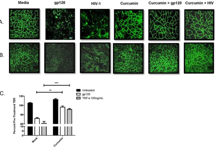
measure whether curcumin could block the barrier disrupting effects of HIV gp120. In the presence of HIV-1 or recombinant gp120, ZO-1 (Fig 1A) and occludin (Fig 1B) protein expres-sion in endometrial GEC monolayers was disrupted, which was not seen in monolayers pre-treated with curcumin. Curcumin on its own had no visible effect on TJ protein expression.
Pre-treatment with curcumin also prevented HIV gp120- and TNF-α-mediated decrease in
TER (Fig 1C), a measure of epithelial barrier integrity [4]. These results suggest that curcumin can prevent HIV-mediated mucosal barrier impairment by maintaining TJ protein expression and maintaining TER in endometrial GECs.
Curcumin prevents the induction of proinflammatory cytokines
associated with barrier disruption
Previously we have shown that epithelial cell exposure to HIV gp120 results in the upregulation
of inflammatory mediators, including TNF-α[4,5], which mediate the disruption of TJ
pro-teins. Therefore, we sought to determine whether the gp120-mediated upregulation of proin-flammatory mediators could be blocked by curcumin. At 24-hours following exposure to
recombinant gp120, TNF-α(Fig 2A) and IL-6 (Fig 2B) were significantly upregulated by
endo-metrial GECs relative to untreated GEC controls. Pre-treatment with curcumin abrogated gp120-mediated induction of proinflammatory cytokines, suggesting that curcumin may block the inflammation associated with HIV-mediated mucosal barrier disruption.
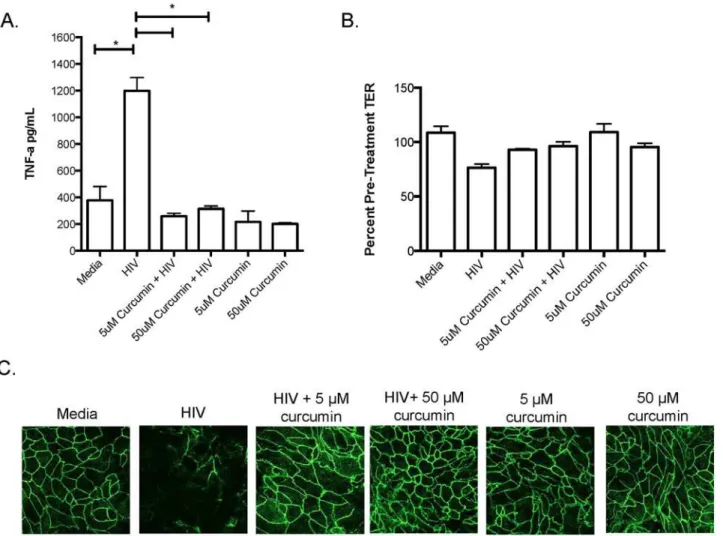
To determine whether curcumin protected the endocervix in a similar manner to endome-trium, since this site has been directly implicated in simian immunodeficiency virus (SIV)
primary human endocervical monolayers with curcumin and exposed them to HIV-1 for
24-hours and measured TNF-αproduction, TER and ZO-1 expression. Similar to endometrial
GECs, endocervical GECs exposed to HIV-1 secreted significantly elevated TNF-α, which was
inhibited by curcumin pre-treatment (Fig 3A). Furthermore, endocervical monolayer integrity, as indicated by TER and ZO-1 TJ staining, decreased following exposure to HIV-1 but
re-mained intact among cells exposed to curcumin (Fig3Band3C). These results suggest that
both endometrial and endocervical epithelial cells can be protected against the barrier-damag-ing effects of HIV-1 gp120 by curcumin.
Curcumin blocks the gp120-mediated induction of chemokines
associated with the recruitment of HIV-target cells
HIV target cells, which include CD4+T-cells, macrophage and dendritic cells (DCs), are
re-cruited to the FGT via chemokine gradients that include MCP-1, MIP-1α, IP-10, eotaxin, IL-8
or RANTES produced by resident tissue cells, such as GECs [4,5]. Exposure of endometrial
GECs to gp120 resulted in significant upregulation of IL-8, IP-10 and RANTES (Fig2C–2E)
Fig 1. Curcumin prevents tight junction disruption and breakdown of endometrial epithelial barrier integrity caused by HIV-1 gp120.Primary endometrial GECs were grown to confluency and pre-treated with 5μM curcumin or media as control for 1 hour and exposed to 105IVU of HIV-IIIB,
recombinant gp120 at 0.1μg/mL or media (mock infection). At 24 hours post exposure, GECs were fixed and stained for ZO-1 (A) or occludin (B). Imaging
was done on an inverted confocal laser-scanning microscope. Alternatively, GECs were exposed to media control (untreated), gp120 at 0.1μg/mL or
recombinant TNF-αat 100 ng/mL (positive control) for 24 hours, after which TERs were measured and the percent of pre-treatment TER was calculated (C).
Data shown represents the mean±SEM of three separate experiments. A minimum of two replicates per experimental condition was included in every
experiment. Data was analyzed by Kruskal-Wallis non-parametric analysis of variance with Dunn’s test to correct for multiple comparisons.**p<0.01,
***p<0.001. gp120: glycoprotein 120; HIV-1: human immunodeficiency virus-1; TER: trans-epithelial electrical resistance; TNF-α: tumor necrosis factor-α.
Fig 2. Curcumin prevents the gp120-mediated induction of proinflammatory cytokines or chemokines that recruit HIV-target cells.Confluent primary endometrial GECs were pre-treated with 5μM curcumin or media (untreated) for 1 hour after which the cells were exposed to recombinant gp120 at
0.1μg/mL. Apical cell culture supernatants were collected at 24 hours post-exposure and proinflammatory cytokines TNF-α(A) or IL-6 (B), as well as the
chemokines IL-8 (C), IP-10 (D), RANTES (E), MCP-1 (F), MIP-1α(G) and eotaxin (H) were measured by Magpix multi-analyte assay. Data shown represents
the mean±SEM of three separate experiments. A minimum of two replicates per experimental condition was included in every experiment. Data was
analyzed by Kruskal-Wallis non-parametric analysis of variance with Dunn’s test to correct for multiple comparisons*p<0.05,**0.01,***p<0.001. gp120: glycoprotein 120; HIV-1: human immunodeficiency virus-1; TNF-α: tumour necrosis factor-α; IL: interleukin; IP-10: interferon gamma-inducible protein-10;
RANTES: regulated on activation, normal T-cell expressed and secreted; MCP-1: monocyte chemotactic protein-1; MIP-1α; macrophage inflammatory
protein-1α; Curc: curcumin.
but not MCP-1, eotaxin or MIP-1α(Fig2F–2H). Interestingly, gp120-mediated chemokine induction was abrogated in endometrial GEC monolayers pretreated with curcumin. These re-sults show that pre-treatment with curcumin blocks gp120 mediated upregulation of chemo-kines that can recruit HIV target cells to the FGT.
Curcumin pre-treatment blocks HIV amplification in chronically infected
T-cells
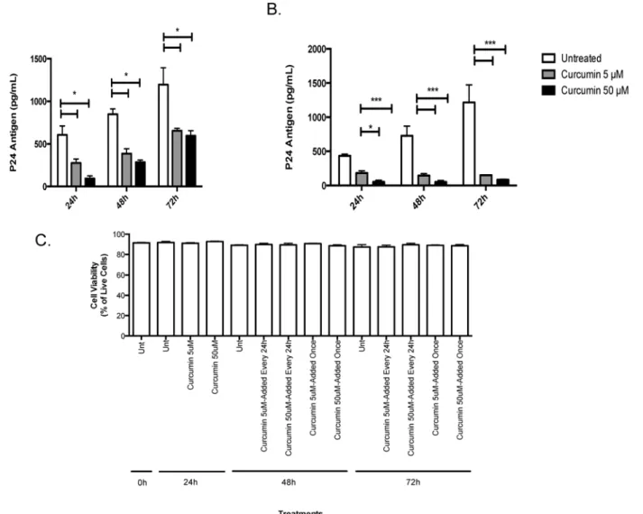
Next, we determined if curcumin could directly block HIV replication in chronically infected T-cells. Chronically infected H9 T-cells exposed to a single curcumin treatment secreted signif-icantly less virus into cell culture supernatants relative to untreated cells. After 24 hours of ex-posure, the effect of curcumin started to diminish but HIV replication remained significantly lower than untreated cells (Fig 4A). Interestingly, when H9 T-cells were exposed to curcumin once daily, HIV levels remained significantly suppressed relative to controls (Fig 4B). Both low Fig 3. Curcumin protects the endocervix against the barrier-breaking effects of HIV-1.Primary endocervical GECs were grown to confluency and pre-treated with curcumin or primary cell media as control. Monolayers of GECs were exposed to 106IVU of HIV-IIIB or media (as mock infection). At 24 hours post exposure, cell-culture supernatants were collected and TNF-αwas measured by ELISA (A). TERs were measured and the percent of pre-treatment TER
was calculated (B) and endocervical monolayers were fixed, stained for ZO-1 and analyzed by immunofluorescent microscopy (C). Imaging was done on an inverted confocal laser-scanning microscope. Data shown represents the mean±SEM of three separate experiments. Data was analyzed using a two-tailed
Mann-Whitney test for non-parametric data.*p<0.05. A minimum of two replicates per experimental condition was included in every experiment performed. HIV: human immunodeficiency virus; TNF-α: tumour necrosis factor-α.
(5μM) and high (50μM) curcumin doses appeared to be effective in controlling HIV
replica-tion, indicating that curcumin could have a potent effect on HIV replication within infected T
cells. Chronically infected T-cells remained viable (>90%) throughout the experiment and
cur-cumin-treatment, at either at 5 or 50μM concentration, or applied once or every 24-hours, did
not effect the viability of the cells (Fig 4C).
Curcumin pre-treatment blocks co-infection mediated induction of the
HIV promoter and HIV replication in T-cells
Previous studies from our group have shown that in the presence of co-infecting STIs,
specifi-cally HSV-1, HSV-2 orN.gonorrhoeae, primary GECs secrete inflammatory factors that cause
Fig 4. Curcumin prevents the amplification of HIV-1 in chronically infected T-cells.5x105chronically HIV-infected H9 T-cells were exposed once to 5 or 50μM curcumin or serum free RPMI as control. At 24, 48 or 72 hours post-treatment, supernatants were collected and HIV-1 p24-antigen was measured
using a commercial p24 ELISA kit (A). Alternatively, 5x10⁵H9 T-cells were exposed to curcumin once every 24 hours at 5 or 50
μM curcumin after which
supernatants were collected and HIV-1 p24-antigen was measured (B). Chronically infected H9 cells were treated with or without curcumin (5 and 50μM)
once every 24 hours, or just once and monitored from 24–72 hours post-treatment. Dead cells were counted following trypan blue exclusion assay using a hemocytometer (C). Data is shown as the percent of live cells in culture. Data shown represents the mean±SEM of three separate experiments. A minimum
of four replicates per experimental condition was included in every experiment performed. Data was analyzed by Kruskal-Wallis non-parametric analysis of variance with Dunn’s test to correct for multiple comparisons*p<0.05,***p<0.001. Unt: untreated.
indirectactivation of the HIV-LTR promoter in T-cells, a process synonymous with HIV replica-tion [19]. Therefore, we sought to determine whether blocking inflammatory pathways in prima-ry GECs using curcumin could abrogate indirect induction in T-cells. Cell culture supernatants collected from GECs pretreated with curcumin and exposed to co-infecting STIs were unable to indirectly activate the HIV-LTR promoter in 1G5 T-cells (Fig 5A). In contrast, supernatants col-lected from untreated GECs exposed to co-infecting microbes potently induced HIV-LTR activa-tion in 1G5 T-cells, suggesting that pre-treatment with curcumin blocks co-infecactiva-tion-mediated inflammation that may increase HIV replication. Exposing 1G5 T cells to conditioned GEC media did not affect cell viability (Fig 5B).
Fig 5. Curcumin can prevent co-infection mediated activation of the HIV-LTR promoter and induction of HIV replication in T-cells.Confluent primary endometrial GECs were pre-treated with 5μM curcumin or primary cell media (untreated) for 1 hour after which the cells were exposed to 104PFU of HSV-1,
HSV-2 or 106CFU ofN.gonorrhoeaefor two hours. The inoculum was removed and the cells were washed with PBS and subsequently replenished with fresh media. Supernatants were collected 24-hours post-exposure, inactivated to kill live virus or filter sterilized to remove bacterial cells, and subsequently incubated with 1061G5 cells for 24 hours, after which the cells were lysed and luciferase activity was measured as a readout for HIV-LTR activation (A). To assess 1G5 cell viability following exposure to conditioned GEC media, 1G5 cells were exposed for 24 hours to RPMI control (Media), primary cell media from uninfected GECs (Uninfected) and conditioned primary cell media from GECs exposed to HSV-1, HSV-2 orN.gonorrhoeaefor 24 hours. Following incubation, cells were collected and cell viability was calculated using the trypan exclusion assay. Results are reported as the percent of live cells in culture (B). 1x10⁵chronically infected H9 T-cells were pre-treated with 50
μM of curcumin for 1 hour and subsequently exposed to poly I:C, LPS or flagellin for 24
hours. Supernatants were collected and IVUs were measured using the TZM-b1 assay (C). Data shown represents the mean±SEM of three separate experiments. A minimum of two replicates per experimental condition were included in every experiment performed. Data was analyzed using a two-tailed Mann-Whitney test for non-parametric data.*p<0.05,**p<0.01,***p<0.001. HIV-LTR: human immunodeficiency virus long-terminal repeats; RLU: relative
light units; HSV: herpes simplex virus; NGO:Neisseria gonorrhoeae.; LPS: lipopolysaccharide.
In previous studies, we also demonstrated that TLR ligands, which are representative of var-ious viral and bacterial microbes, could directly activate the HIV-LTR promoter in 1G5 T-cells [19]. To determine whether curcumin treatment was capable of inhibiting the production of in-fectious virus within infected T-cells, chronically infected H9 T-cells were exposed to TLR li-gands in the presence or absence of curcumin. Supernatants were collected and HIV
replication was assessed via TZM-b1 assay, which detects infectious live virus [4]. Poly I:C (TLR3 ligand), LPS (TLR4 ligand) and flagellin (TLR5 ligand) all increased infectious HIV pro-duction from H9 T-cells, relative to unstimulated controls (Fig 5C). Interestingly, curcumin ex-posure abrogated TLR-mediated direct induction of HIV replication in T-cells.
Together our results suggest that intact co-infecting microbes, as well as conserved microbi-al ligands, are capable of inducing HIV promoter activation and production of infectious HIV virions in T-cells and that curcumin pre-treatment can abrogate such potentiation of HIV replication.
Curcumin blocks HSV-2 replication
HSV-2 infection is associated with a threefold increase in susceptibility to HIV in both men and
women [38–40] and controlling HSV-2 replication has been associated with decreased HIV
rep-lication [41]. Thus, we decided to test the anti-herpetic activity of curcumin in primary human
GECs. In cells pretreated with 5μM of curcumin, HSV-2 shed approximately 1,000-fold less virus
relative to untreated primary cell controls infected with HSV-2 (Fig 6A). At 50μM of curcumin,
no HSV-2 virus could be detected in cell culture supernatants. Since curcumin blocks various in-flammatory pathways [20], we pre-treated GECs with inhibitors of curcumin-regulated
tran-scription factors including NFκB (PDTC), p38 MAP kinase (SB203580) or c-Jun N-terminal
kinase (JNK) (SP600125), prior to infecting the cells with HSV-2. Our results show that the
NFκB inhibitor PDTC completely blocked viral shedding in primary GECs exposed to HSV-2
(Fig 6B), whereas blocking p38 MAP kinase or JNK pathways did not have any effect on HSV-2 viral replication. We also tested the toxicity of the inhibitors on primary GECs and found no sig-nificant differences with respect to cell viability between the cells treated with inhibitors com-pared to media alone treated cells or between treatments with different inhibitors (Fig 6C).
Discussion
We have described in this study the multiple pathways by which curcumin may have a benefi-cial role in protecting the FGT against HIV-1. Our results show that curcumin has pleiotropic effects and its protection of barrier function is primarily mediated by its potent anti-inflamma-tory activities. Curcumin prevented endometrial and endocervical mucosal barrier impairment by HIV-1 gp120 and abrogated gp120-mediated upregulation of proinflammatory cytokines and chemokines. Furthermore, curcumin pre-treatment of primary GECs prevented co-infec-tion mediated upregulaco-infec-tion of HIV-LTR activaco-infec-tion and replicaco-infec-tion in T-cells. Finally, we found that curcumin decreased HIV-1 and HSV-2 replication in T-cells and GECs, respectively.
In the present study, we examined primarily the anti-inflammatory activity of curcumin and how this could affect HIV interactions with the genital epithelium and T-cells in the genital tract. Previously, curcumin has been shown to exert potent anti-inflammatory activities such as
sup-pressing the production of proinflammatory cytokines, including TNF-α, IL-1βand IL-6 [20,
42]. Curcumin (at a concentration of 5 uM) has been shown to inhibit LPS-induced production
of TNF-αby the human monocytic macrophage cell line, Mono Mac 6, and reduce the biological
activity of TNF-αin a fibroblast cell assay [32].In vitro, curcumin modulates the inflammatory
response by directly binding to and downregulating the activity of cyclooxygenase-2,
observed that curcumin pre-treatment significantly decreased gp120-mediated induction of proinflammatory cytokines and chemokines and that curcumin protected the epithelial barrier
against the disrupting effects of HIV gp120 and proinflammatory cytokines, such as TNF-α.
Fur-thermore, pre-treating GECs with curcumin was sufficient to block co-infection mediated
indi-rectinduction of the HIV promoter as well as TLR-mediateddirectinduction of HIV replication
in T-cells.
In addition to the anti-inflammatory effects of curcumin, previous studies provide evidence that curcumin may block HIV by directly interfering with the HIV replication cycle. Curcu-min-loaded apotransferrin nanoparticles have been successfully used to block the synthesis of Fig 6. Inhibition of inflammatory signalling pathways decreases HSV-2 viral shedding.Primary GECs were grown to confluency and were pre-treated with media (untreated) or 5 or 50μM of curcumin (A) or PDTC the NFκB inhibitor, SB203580 a p38 MAP kinsase inhibitor or SP600125 an inhibitor of c-Jun
N-terminal kinase (JNK) (B) for 1 hour. The cells were then exposed to 104PFU of HSV-2 strain 333 for 2 hours. At 24-hours post-infection, apical cell culture supernatants were collected and the amount of shed HSV-2 was measured using a standard Vero plaque assay. To assess whether the inhibitors used in this study affected cell viability (C), primary GECs were exposed to media control, or curcumin, PDTC, SB203580 or SP600125 at 5 or 50μM for 1 hour.
Following exposure, cells were collected and cell viability was calculated using the trypan exclusion assay. Results are reported as the percent of live cells in culture. Data shown represents the mean±SEM of three separate experiments. A minimum of two replicates per experimental condition was included in
every experiment performed. Data analyzed using the Kruskal-Wallis non-parametric analysis of variance with Dunn’s test to correct for multiple comparisons.***p<0.001. HSV-2: herpes simplex virus type 2; PFU: plaque-forming units; PDTC: pyrrolidine dithiocarbamate.
HIV viral cDNA [44]. Otherin vitrostudies have demonstrated that curcumin can inhibit the enzymatic activity of recombinant, purified HIV-1 protease [45] and integrase [46], and can
di-rectly block the HIV-LTR promoter in T-cells [47–49]. Others have shown that combining
cur-cumin with existing antiretrovirals, such as the protease inhibitor indinavir (IDV), reduced viral infectivity relative to IDV alone [50]. Collectively, these results suggest that curcumin may interact and block HIV replication via multiple, perhaps, redundant pathways and that there may be utility in supplementing current antiviral therapies with curcumin to reduce HIV-1 infection and replication.
Based on our results, we posit that protecting the mucosal barrier with curcumin may play a significant role in preventing mucosal transmission of HIV. Previously, we showed that gp120 interacts with TLR2, TLR4 and heparan sulphate moieties on primary GECs resulting in
down-stream induction of TNF-α, TJ disruption and barrier impairment [5]. This mucosal barrier
disruption resulted in HIV translocation across the epithelium, presenting a mechanism by which HIV acquisition may take place [4]. In our current study, we found that curcumin pre-treatment prevented gp120-mediated disruption of the TJ proteins ZO-1 and occludin, and maintained TERs across endometrial and endocervical genital monolayers. Furthermore, pre-treating primary GECs with curcumin prevented gp120-mediated induction of
barrier-break-ing proinflammatory cytokines (TNF-α, IL-6) and HIV-target cell recruiting chemokines
(IL-8, IP-10, RANTES). Together these results suggest that curcumin and other compounds, which reduce inflammation and maintain mucosal barrier integrity, may be useful candidates to test as prophylactic formulations to prevent HIV acquisition.
Curcumin may also play a significant role in preventing or reducing chronic immune activa-tion. Microbial translocation due to mucosal barrier impairment is not only associated with HIV acquisition but is strongly believed to be one of the main contributors to chronic immune activation [51], characterized by increased inflammatory markers and immune cell activation
that persists even in HAART-treated individuals [52,53]. Immune activation is believed to be
one of the main driving forces of CD4+T-cell depletion and promoter of HIV replication [54].
Repeated exposure to HIV gp120 in mucosal tissues from shed virus or from unbound gp120 may continuously provide a source for generating inflammation in mucosal tissues that facili-tates barrier breakdown and microbial translocation. Because immune activation is typically observed during the chronic stages of infection, the window of opportunity to intervene and limit these processes likely occurs during the early stages of HIV infection. Blocking HIV-mediated barrier disruption by curcumin may likely prevent microbial translocation and the initiation of immune activation, suggesting that curcumin treatment, may also contribute to decreasing the chronic inflammatory state that contributes to HIV pathology.
By suppressing replication in chronically infected T-cells, curcumin may provide a means of controlling HIV amplification during the early phases of infection. HIV crosses the mucosal epithelial barrier to establish a small founder population that then expands locally, possibly due to an influx of newly recruited target cells caused by upregulation of chemokines, prior to systemic dissemination [55]. These early events represent a window of maximum opportunity for interventions to prevent systemic infection. Our results show that in addition to protecting the mucosal barrier, curcumin may curb HIV by limiting local propagation and expansion in T-cells and by suppressing the local chemokine environment that may play a role in target cell chemotaxis. Our results also showed that repeated administration of curcumin worked best at suppressing HIV amplification in chronically infected T-cells, suggesting that therapies that continuously release curcumin may be novel tools by which HIV viral replication can be re-duced in the FGT.
genital tract. Previous studies have characterized curcumin as a broad antimicrobial
com-pound [21,56]. The bactericidal capabilities of curcumin have been documented in the
medi-cal literature since the late 1980s [57] and have been anecdotally known for centuries.
Previous studies have shown that curcumin inhibits NFκB pathways and production of IL-6,
IL-8 and TNF-αfollowingN.gonorrhoeaestimulation of the HeLa cervical epithelial cell line,
and completely abolishes the adherence of bacteria to cells in late infection [58]. Furthermore, curcumin pretreatment significantly decreased HSV-1 infectivity and immediate early (IE) gene expression in HeLa cells [59] and provided significant protection in a mouse model of intravaginal HSV-2 challenge [60]. Using the Vero cell line, researchers also showed that cur-cumin and derivatives of the compound, containing gallium and copper metal, significantly
suppressed HSV-1 replicationin vitro[61]. Our results show that curcumin pre-treatment
sig-nificantly impairs HSV-2 replication in primary human GECs, which is a more physiological-ly relevant model than transformed or immortalized epithelial cell lines, which were almost
exclusively used in the past to examine thein vitroanti-herpetic activity of curcumin [59,61].
Parallel experiments using inhibitors of inflammatory pathways known to be regulated by
cur-cumin, showed that pre-treating cells with PDTC, an NFκB inhibitor, resulted in potent
sup-pression of HSV-2 replication, suggesting that NFκB activation may play an essential role in
HSV-2 replication in primary GECs, a notion that has been recently validated in epithelial cell lines [62]. Thus, our results suggest that curcumin could play a significant role in blocking co-infection mediated induction of HIV replication.
Our results add to the body of evidence indicating that curcumin could be a useful com-pound for decreasing HIV replication and perhaps even preventing HIV infection. However, clinical trials evaluating the efficacy of curcumin as a potential therapy for people living with HIV have had inconsistent results. In a clinical case study on three patients with HIV, two ART-naive and one who received conventional ART therapy, curcumin treatment resulted in a 10 to 10,000-fold reduction in blood viral load [63]. In this study, blood samples were tested for HIV viral load at baseline and following a regimen of 1 gram of curcumin, three times per day for 8 weeks. In contrast, a clinical trial examining the effectiveness of curcumin as an anti-viral agent in 40 AIDS patients failed to find any therapeutic benefits. No evidence of curcu-min-associated reduction in viral load was observed over the course of this short (8-week) trial, however, CD4 cells showed a slight increase in the high-dose curcumin group [64]. A possible
reason for lack of effect in some of these studies could be due to curcumin’s poor
bioavailabili-ty. Curcumin is poorly absorbed, rapidly metabolized/glucoronidated and systemically elimi-nated following oral administration [65]. Numerous efforts have been made to improve
curcumin’s bioavailability by altering these features. The use of compounds that can block the
metabolic pathway of curcumin is the most common strategy for increasing the bioavailability of curcumin, such as using piperine, a known inhibitor of hepatic and intestinal glucuronida-tion, which increased the bioavailability of curcumin in healthy human volunteers by 2,000% [66]. Other promising approaches to increase the bioavailability of curcumin in humans
in-clude the use of nanoparticles [67] or liposomes [68–70]. Other reasons why the previous
against HIV would argue that prophylactic application of curcumin may be more effective as they could prevent HIV infection on mucosal surfaces.
In conclusion, our results indicate the promising potential of anti-inflammatory compounds such as curcumin, which protect the mucosal barrier and/or limit inflammation, unlike current antiretroviral therapies that target infection and replication. Inflammation may contribute to the acquisition or spread of HIV-1 infection, as well as contribute to the sequelea of chronic HIV infection. Thus, therapeutics designed to limit inflammation in the genital tract and/or systemically, may have a significant impact on HIV infection and disease progression.
Acknowledgments
The authors would like to thank the Pre-Op Clinic Staff for their cooperation in obtaining in-formed consents, the Clinical Pathology Staff at McMaster University Medical Centre for their assistance in providing genital tract tissues and the women who donated their tissues for this study.
Author Contributions
Conceived and designed the experiments: VHF AN CK. Performed the experiments: VHF AN SED KM. Analyzed the data: VHF AN SED CK. Contributed reagents/materials/analysis tools: CK. Wrote the paper: VHF CK.
References
1. UNAIDS. UNAIDS World AIDS Day report 2011. 2011.
2. Hladik F, McElrath MJ. Setting the stage: host invasion by HIV. Nat Rev Immunol. 2008; 8(6):447–57. doi:10.1038/nri2302PMID:18469831
3. Li Q, Estes JD, Schlievert PM, Duan L, Brosnahan AJ, Southern PJ, et al. Glycerol monolaurate pre-vents mucosal SIV transmission. Nature. 2009; 458(7241):1034–8. doi:10.1038/nature07831PMID:
19262509
4. Nazli A, Chan O, Dobson-Belaire WN, Ouellet M, Tremblay MJ, Gray-Owen SD, et al. Exposure to HIV-1 directly impairs mucosal epithelial barrier integrity allowing microbial translocation. PLoS Pathog. 2010; 6(4):e1000852. doi:10.1371/journal.ppat.1000852PMID:20386714
5. Nazli A, Kafka JK, Ferreira VH, Anipindi V, Mueller K, Osborne BJ, et al. HIV-1 gp120 induces TLR2-and TLR4-mediated innate immune activation in human female genital epithelium. J Immunol. 2013; 191(8):4246–58. doi:10.4049/jimmunol.1301482PMID:24043886
6. Deeks SG, Lewin SR, Havlir DV. The end of AIDS: HIV infection as a chronic disease. Lancet. 2013; 382(9903):1525–33. doi:10.1016/S0140-6736(13)61809-7PMID:24152939
7. Poli G, Bressler P, Kinter A, Duh E, Timmer WC, Rabson A, et al. Interleukin 6 induces human immuno-deficiency virus expression in infected monocytic cells alone and in synergy with tumor necrosis factor alpha by transcriptional and post-transcriptional mechanisms. J Exp Med. 1990; 172(1):151–8. PMID:
2193094
8. Chun TW, Engel D, Mizell SB, Ehler LA, Fauci AS. Induction of HIV-1 replication in latently infected CD4+ T cells using a combination of cytokines. J Exp Med. 1998; 188(1):83–91. PMID:9653086
9. Folks TM, Clouse KA, Justement J, Rabson A, Duh E, Kehrl JH, et al. Tumor necrosis factor alpha in-duces expression of human immunodeficiency virus in a chronically infected T-cell clone. Proc Natl Acad Sci U S A. 1989; 86(7):2365–8. PMID:2784570
10. McLaren PJ, Ball TB, Wachihi C, Jaoko W, Kelvin DJ, Danesh A, et al. HIV-exposed seronegative com-mercial sex workers show a quiescent phenotype in the CD4+ T cell compartment and reduced expres-sion of HIV-dependent host factors. J Infect Dis. 2010; 202 Suppl 3:S339–44. doi:10.1086/655968
PMID:20887221
11. Chang CC, Crane M, Zhou J, Mina M, Post JJ, Cameron BA, et al. HIV and co-infections. Immunol Rev. 2013; 254(1):114–42. doi:10.1111/imr.12063PMID:23772618
13. Rotchford K, Strum AW, Wilkinson D. Effect of coinfection with STDs and of STD treatment on HIV shedding in genital-tract secretions: systematic review and data synthesis. Sex Transm Dis. 2000; 27-(5):243–8. PMID:10821594
14. Johnson LF, Lewis DA. The effect of genital tract infections on HIV-1 shedding in the genital tract: a sys-tematic review and meta-analysis. Sex Transm Dis. 2008; 35(11):946–59. doi:10.1097/OLQ.
0b013e3181812d15PMID:18685546
15. Chen A, Boulton IC, Pongoski J, Cochrane A, Gray-Owen SD. Induction of HIV-1 long terminal repeat-mediated transcription by Neisseria gonorrhoeae. AIDS. 2003; 17(4):625–8. PMID:12598784
16. Levine WC, Pope V, Bhoomkar A, Tambe P, Lewis JS, Zaidi AA, et al. Increase in endocervical CD4 lymphocytes among women with nonulcerative sexually transmitted diseases. J Infect Dis. 1998; 177-(1):167–74. PMID:9419184
17. Klotman ME, Rapista A, Teleshova N, Micsenyi A, Jarvis GA, Lu W, et al. Neisseria gonorrhoeae-in-duced human defensins 5 and 6 increase HIV infectivity: role in enhanced transmission. J Immunol. 2008; 180(9):6176–85. PMID:18424739
18. Zhang J, Li G, Bafica A, Pantelic M, Zhang P, Broxmeyer H, et al. Neisseria gonorrhoeae enhances in-fection of dendritic cells by HIV type 1. J Immunol. 2005; 174(12):7995–8002. PMID:15944306
19. Ferreira VH, Nazli A, Khan G, Mian MF, Ashkar AA, Gray-Owen S, et al. Endometrial epithelial cell re-sponses to coinfecting viral and bacterial pathogens in the genital tract can activate the HIV-1 LTR in an NF{kappa}B-and AP-1-dependent manner. J Infect Dis. 2011; 204(2):299–308. doi:10.1093/infdis/ jir260PMID:21673042
20. Aggarwal BB, Sung B. Pharmacological basis for the role of curcumin in chronic diseases: an age-old spice with modern targets. Trends Pharmacol Sci. 2009; 30(2):85–94. doi:10.1016/j.tips.2008.11.002
PMID:19110321
21. Moghadamtousi SZ, Kadir HA, Hassandarvish P, Tajik H, Abubakar S, Zandi K. A review on antibacteri-al, antivirantibacteri-al, and antifungal activity of curcumin. Biomed Res Int. 2014; 2014:186864. doi:10.1155/ 2014/186864PMID:24877064
22. Gupta SC, Patchva S, Aggarwal BB. Therapeutic roles of curcumin: lessons learned from clinical trials. AAPS J. 2013; 15(1):195–218. doi:10.1208/s12248-012-9432-8PMID:23143785
23. Vlietinck AJ, De Bruyne T, Apers S, Pieters LA. Plant-derived leading compounds for chemotherapy of human immunodeficiency virus (HIV) infection. Planta Med. 1998; 64(2):97–109. PMID:9525100
24. Di Santo R, Costi R, Artico M, Ragno R, Greco G, Novellino E, et al. Design, synthesis and biological evaluation of heteroaryl diketohexenoic and diketobutanoic acids as HIV-1 integrase inhibitors en-dowed with antiretroviral activity. Farmaco. 2005; 60(5):409–17. PMID:15910813
25. Kimpton J, Emerman M. Detection of replication-competent and pseudotyped human immunodeficien-cy virus with a sensitive cell line on the basis of activation of an integrated beta-galactosidase gene. J Virol. 1992; 66(4):2232–9. PMID:1548759
26. Smith KO. Relationship between the Envelope and the Infectivity of Herpes Simplex Virus. Proc Soc Exp Biol Med. 1964; 115:814–6. PMID:14155835
27. Westmoreland D, Rapp F. Host range temperature-sensitive mutants of herpes simplex virus type 2. J Virol. 1976; 18(1):92–102. PMID:176473
28. Aguilar-Cordova E, Chinen J, Donehower L, Lewis DE, Belmont JW. A sensitive reporter cell line for HIV-1 tat activity, HIV-1 inhibitors, and T cell activation effects. AIDS Res Hum Retroviruses. 1994; 10-(3):295–301. PMID:8018390
29. MacDonald EM, Savoy A, Gillgrass A, Fernandez S, Smieja M, Rosenthal KL, et al. Susceptibility of Human Female Primary Genital Epithelial Cells to Herpes Simplex Virus, Type-2 and the Effect of TLR3 Ligand and Sex Hormones on Infection. Biol Reprod. 2007; 77(6):1049–59. PMID:
17881767
30. Kaushic C, Nazli A, Ferreira VH, Kafka JK. Primary human epithelial cell culture system for studying in-teractions between female upper genital tract and sexually transmitted viruses, HSV-2 and HIV-1. Methods. 2011.
31. Kaushic C, Nazli A, Ferreira VH, Kafka JK. Primary human epithelial cell culture system for studying in-teractions between female upper genital tract and sexually transmitted viruses, HSV-2 and HIV-1. Methods. 2011; 55(2):114–21. doi:10.1016/j.ymeth.2011.09.022PMID:21996033
32. Chan MM. Inhibition of tumor necrosis factor by curcumin, a phytochemical. Biochem Pharmacol. 1995; 49(11):1551–6. PMID:7786295
33. Nazli A, Yao XD, Smieja M, Rosenthal KL, Ashkar AA, Kaushic C. Differential induction of innate anti-viral responses by TLR ligands against Herpes simplex virus, type 2, infection in primary genital epithelium of women. Antiviral Res. 2009; 81(2):103–12. doi:10.1016/j.antiviral.2008.10.005PMID:
34. Gillgrass AE, Tang VA, Towarnicki KM, Rosenthal KL, Kaushic C. Protection against Genital Herpes In-fection in Mice Immunized under Different Hormonal Conditions Correlates with Induction of Vagina-As-sociated Lymphoid Tissue. J Virol. 2005; 79(5):3117–26. PMID:15709031
35. Strober W. Trypan blue exclusion test of cell viability. Curr Protoc Immunol. 2001;Appendix 3:Appendix 3B.
36. Haase AT. Targeting early infection to prevent HIV-1 mucosal transmission. Nature. 2010; 464 (7286):217–23. doi:10.1038/nature08757PMID:20220840
37. Wira CR, Rodriguez-Garcia M, Patel MV. The role of sex hormones in immune protection of the female reproductive tract. Nat Rev Immunol. 2015.
38. Gupta R, Warren T, Wald A. Genital herpes. Lancet. 2007; 370(9605):2127–37. PMID:18156035
39. McClelland RS, Wang CC, Overbaugh J, Richardson BA, Corey L, Ashley RL, et al. Association be-tween cervical shedding of herpes simplex virus and HIV-1. Aids. 2002; 16(18):2425–30. PMID:
12461416
40. Barnabas RV, Celum C. Infectious co-factors in HIV-1 transmission herpes simplex virus type-2 and HIV-1: new insights and interventions. Curr HIV Res. 2012; 10(3):228–37. PMID:22384842
41. Celum C, Wald A, Lingappa JR, Magaret AS, Wang RS, Mugo N, et al. Acyclovir and transmission of HIV-1 from persons infected with HIV-1 and HSV-2. N Engl J Med. 2010; 362(5):427–39. doi:10.1056/ NEJMoa0904849PMID:20089951
42. Guo L, Xing Y, Pan R, Jiang M, Gong Z, Lin L, et al. Curcumin protects microglia and primary rat cortical neurons against HIV-1 gp120-mediated inflammation and apoptosis. PLoS One. 2013; 8(8):e70565. doi:10.1371/journal.pone.0070565PMID:23936448
43. Gupta SC, Prasad S, Kim JH, Patchva S, Webb LJ, Priyadarsini IK, et al. Multitargeting by curcumin as revealed by molecular interaction studies. Nat Prod Rep. 2011; 28(12):1937–55. doi:10.1039/ c1np00051aPMID:21979811
44. Gandapu U, Chaitanya RK, Kishore G, Reddy RC, Kondapi AK. Curcumin-loaded apotransferrin nano-particles provide efficient cellular uptake and effectively inhibit HIV-1 replication in vitro. PLoS One. 2011; 6(8):e23388. doi:10.1371/journal.pone.0023388PMID:21887247
45. Sui Z, Salto R, Li J, Craik C, Ortiz de Montellano PR. Inhibition of the HIV-1 and HIV-2 proteases by cur-cumin and curcur-cumin boron complexes. Bioorg Med Chem. 1993; 1(6):415–22. PMID:8087563
46. Mazumder A, Raghavan K, Weinstein J, Kohn KW, Pommier Y. Inhibition of human immunodeficiency virus type-1 integrase by curcumin. Biochem Pharmacol. 1995; 49(8):1165–70. PMID:7748198
47. Li CJ, Zhang LJ, Dezube BJ, Crumpacker CS, Pardee AB. Three inhibitors of type 1 human immunode-ficiency virus long terminal repeat-directed gene expression and virus replication. Proc Natl Acad Sci U S A. 1993; 90(5):1839–42. PMID:8446597
48. Barthelemy S, Vergnes L, Moynier M, Guyot D, Labidalle S, Bahraoui E. Curcumin and curcumin deriv-atives inhibit Tat-mediated transactivation of type 1 human immunodeficiency virus long terminal re-peat. Res Virol. 1998; 149(1):43–52. PMID:9561563
49. Zhang HS, Ruan Z, Sang WW. HDAC1/NFkappaB pathway is involved in curcumin inhibiting of Tat-me-diated long terminal repeat transactivation. J Cell Physiol. 2011; 226(12):3385–91. doi:10.1002/jcp. 22691PMID:21344388
50. Riva DA, Fernandez-Larrosa PN, Dolcini GL, Martinez-Peralta LA, Coulombie FC, Mersich SE. Two im-munomodulators, curcumin and sulfasalazine, enhance IDV antiretroviral activity in HIV-1 persistently infected cells. Arch Virol. 2008; 153(3):561–5. doi:10.1007/s00705-007-0023-4PMID:18175040
51. Brenchley JM, Price DA, Schacker TW, Asher TE, Silvestri G, Rao S, et al. Microbial translocation is a cause of systemic immune activation in chronic HIV infection. Nat Med. 2006; 12(12):1365–71. PMID:
17115046
52. Hazenberg MD, Otto SA, van Benthem BH, Roos MT, Coutinho RA, Lange JM, et al. Persistent im-mune activation in HIV-1 infection is associated with progression to AIDS. Aids. 2003; 17(13):1881–8. PMID:12960820
53. Redd AD, Dabitao D, Bream JH, Charvat B, Laeyendecker O, Kiwanuka N, et al. Microbial transloca-tion, the innate cytokine response, and HIV-1 disease progression in Africa. Proc Natl Acad Sci U S A. 2009; 106(16):6718–23. doi:10.1073/pnas.0901983106PMID:19357303
54. Paiardini M, Muller-Trutwin M. HIV-associated chronic immune activation. Immunol Rev. 2013; 254-(1):78–101. doi:10.1111/imr.12079PMID:23772616
55. Haase AT. Early events in sexual transmission of HIV and SIV and opportunities for interventions. Annu Rev Med. 2011; 62:127–39. doi:10.1146/annurev-med-080709-124959PMID:21054171
57. Dahl TA, McGowan WM, Shand MA, Srinivasan VS. Photokilling of bacteria by the natural dye curcu-min. Arch Microbiol. 1989; 151(2):183–5. PMID:2655550
58. Wessler S, Muenzner P, Meyer TF, Naumann M. The anti-inflammatory compound curcumin inhibits Neisseria gonorrhoeae-induced NF-kappaB signaling, release of pro-inflammatory cytokines/chemo-kines and attenuates adhesion in late infection. Biol Chem. 2005; 386(5):481–90. PMID:15927892
59. Kutluay SB, Doroghazi J, Roemer ME, Triezenberg SJ. Curcumin inhibits herpes simplex virus immedi-ate-early gene expression by a mechanism independent of p300/CBP histone acetyltransferase activi-ty. Virology. 2008; 373(2):239–47. doi:10.1016/j.virol.2007.11.028PMID:18191976
60. Bourne KZ, Bourne N, Reising SF, Stanberry LR. Plant products as topical microbicide candidates: as-sessment of in vitro and in vivo activity against herpes simplex virus type 2. Antiviral Res. 1999; 42 (3):219–26. PMID:10443534
61. Zandi K, Ramedani E, Mohammadi K, Tajbakhsh S, Deilami I, Rastian Z, et al. Evaluation of anti-viral activities of curcumin derivatives against HSV-1 in Vero cell line. Nat Prod Commun. 2010; 5-(12):1935–8. PMID:21299124
62. Qiu M, Chen Y, Cheng L, Chu Y, Song HY, Wu ZW. Pyrrolidine dithiocarbamate inhibits herpes simplex virus 1 and 2 replication, and its activity may be mediated through dysregulation of the ubiquitin-proteasome system. J Virol. 2013; 87(15):8675–86. doi:10.1128/JVI.00869-13PMID:23740985
63. Jordan WC, Drew CR. Curcumin—a natural herb with anti-HIV activity. J Natl Med Assoc. 1996; 88(6):333. PMID:8691490
64. James JS. Curcumin: clinical trial finds no antiviral effect. AIDS Treat News. 1996;(no 242: ):1–2. PMID:11363190
65. Anand P, Kunnumakkara AB, Newman RA, Aggarwal BB. Bioavailability of curcumin: problems and promises. Mol Pharm. 2007; 4(6):807–18. PMID:17999464
66. Shoba G, Joy D, Joseph T, Majeed M, Rajendran R, Srinivas PS. Influence of piperine on the pharma-cokinetics of curcumin in animals and human volunteers. Planta Med. 1998; 64(4):353–6. PMID:
9619120
67. Sasaki H, Sunagawa Y, Takahashi K, Imaizumi A, Fukuda H, Hashimoto T, et al. Innovative preparation of curcumin for improved oral bioavailability. Biol Pharm Bull. 2011; 34(5):660–5. PMID:21532153
68. Gota VS, Maru GB, Soni TG, Gandhi TR, Kochar N, Agarwal MG. Safety and pharmacokinetics of a solid lipid curcumin particle formulation in osteosarcoma patients and healthy volunteers. J Agric Food Chem. 2010; 58(4):2095–9. doi:10.1021/jf9024807PMID:20092313
69. Berginc K, Skalko-Basnet N, Basnet P, Kristl A. Development and evaluation of an in vitro vaginal model for assessment of drug's biopharmaceutical properties: curcumin. AAPS PharmSciTech. 2012; 13(4):1045–53. doi:10.1208/s12249-012-9837-9PMID:22899381