Peptidoglycan Branched Stem Peptides
Contribute to
Streptococcus pneumoniae
Virulence by Inhibiting Pneumolysin Release
Neil G. Greene1, Ana R. Narciso2, Sergio R. Filipe2, Andrew Camilli1*
1Graduate Program in Molecular Microbiology, Sackler School of Graduate Biomedical Sciences, Howard Hughes Medical Institute, and Department of Molecular Biology and Microbiology, Tufts University School of Medicine, Boston, Massachusetts, United States of America,2Laboratory of Bacterial Cell Surfaces and Pathogenesis, Instituto de Tecnologia Quimica e Biologica, Universidade Nova de Lisboa (ITQB-UNL), Oeiras, Portugal
*andrew.camilli@tufts.edu
Abstract
Streptococcus pneumoniae(the pneumococcus) colonizes the human nasopharynx and is
a significant pathogen worldwide. Pneumolysin (Ply) is a multi-functional, extracellular viru-lence factor produced by this organism that is critical for pathogenesis. Despite the absence of any apparent secretion or cell surface attachment motifs, Ply localizes to the cell enve-lope of actively growing cells. We sought to characterize the consequences of this surface localization. Through functional assays with whole cells and subcellular fractions, we deter-mined that Ply activity and its release into the extracellular environment are inhibited by pep-tidoglycan (PG) structure. The ability of PG to inhibit Ply release was dependent on the stem peptide composition of this macromolecule, which was manipulated by mutation of the
murMNoperon that encodes proteins responsible for branched stem peptide synthesis.
Additionally, removal of choline-binding proteins from the cell surface significantly reduced Ply release to levels observed in a mutant with a high proportion of branched stem peptides suggesting a link between this structural feature and surface-associated choline-binding proteins involved in PG metabolism. Of clinical relevance, we also demonstrate that a hyperactive, mosaicmurMNallele associated with penicillin resistance causes decreased
Ply release with concomitant increases in the amount of branched stem peptides. Finally, using amurMNdeletion mutant, we observed that increased Ply release is detrimental to
virulence during a murine model of pneumonia. Taken together, our results reveal a novel role for branched stem peptides in pneumococcal pathogenesis and demonstrate the importance of controlled Ply release during infection. These results highlight the importance of PG composition in pathogenesis and may have broad implications for the diverse PG structures observed in other bacterial pathogens.
OPEN ACCESS
Citation:Greene NG, Narciso AR, Filipe SR, Camilli A (2015) Peptidoglycan Branched Stem Peptides Contribute toStreptococcus pneumoniaeVirulence by Inhibiting Pneumolysin Release. PLoS Pathog 11(6): e1004996. doi:10.1371/journal.ppat.1004996
Editor:Carlos Javier Orihuela, The University of Alabama at Birmingham, UNITED STATES
Received:March 7, 2015
Accepted:June 2, 2015
Published:June 26, 2015
Copyright:© 2015 Greene et al. This is an open access article distributed under the terms of the
Creative Commons Attribution License, which permits unrestricted use, distribution, and reproduction in any medium, provided the original author and source are credited.
Data Availability Statement:All relevant data are within the paper and its Supporting Information files.
Author Summary
Pneumolysin (Ply) is a protein toxin produced byStreptococcus pneumoniaethat contrib-utes to the ability of this organism to cause invasive disease. Release of this protein from the bacterial cell is necessary for many of its functions but the underlying mechanisms driving this process are not well characterized. Previous research demonstrated that Ply localizes to the cell wall compartment. Here, we address the consequences of this localiza-tion and reveal a role for the major cell wall structural component, peptidoglycan, in inhib-iting Ply activity and release into the extracellular environment. Peptidoglycan is an essential, mesh-like sac that encases the cell, and alterations affecting its composition lead to differences in the amount of Ply released. How molecules interact with and traverse through the restrictive matrix of the cell wall and its associated structures is incompletely understood, particularly with respect to protein secretion and surface attachment. Our results argue that proper maintenance of cell wall-associated Ply is dependent on surface architecture and may be critical forS.pneumoniaepathogenesis.
Introduction
Streptococcus pneumoniae(the pneumococcus) is a Gram-positive commensal of the human nasopharynx. Though asymptomatic, nasal carriage is considered a prerequisite for the estab-lishment of invasive pneumococcal disease [1]. The pneumococcus is an extracellular pathogen that elaborates a multitude of virulence determinants that contribute to the pathogenesis of invasive pneumococcal diseases such as otitis media, pneumonia, meningitis and bacteremia, depending on the site of infection. Pneumolysin (Ply) is one such conserved, multi-functional virulence factor [2]. As a member of the cholesterol-dependent cytolysin family of pore-form-ing toxins, Ply is cytotoxic to a variety of eukaryotic host cells [3–5]. Additional activities attrib-uted to Ply include complement activation, induction of host cell signaling cascades, and stimulation of a diverse array of cytokines [6–9].
Ply must be extracellular to carry out the aforementioned functions, however, unlike all other Gram-positive cholesterol-dependent cytolysins, Ply lacks a canonical N-terminal signal peptide commonly associated with Sec-mediated secretion. Additionally, Ply does not encode any of the currently known motifs necessary for cell envelope attachment [10]. Despite these observations, previous studies have demonstrated that Ply is present in culture supernatants [11] and the cell wall compartment during growth [12]. Furthermore, these studies ruled out a role for the major pneumococcal autolysin LytA in this process, suggesting that autolysis alone could not account for extracellular Ply. Therefore, Ply export from the cytoplasm to the cell envelope has been proposed to occur via an active process that remains to be discovered [13].
A defining characteristic of the Gram-positive cell envelope is a thick layer of peptidoglycan (PG) that encompasses the cell. PG is a rigid, yet dynamic, macromolecule that confers the characteristic shape of bacterial cells and provides protection against lysis from turgor pressure. The basic structure of PG is conserved and consists of a mesh-like network of glycan strands situated circumferentially around the cell composed of alternating N-acetylglucosamine and N-acetylmuramic acid residues crosslinked through short peptides emanating from the latter sugar moiety [14]. Variation among PG types largely exists at the level of stem peptide compo-sition and the penicillin-binding protein (PBP)-catalyzed transpeptidation reactions that serve to generate crosslinks between them [15]. Pneumococcal PG is characterized by a combination of linear and branched stem peptides [16]. Formation of branched stem peptides occurs through addition of a dipeptide branch on the third position lysine residue of a nascent PG Competing Interests:The authors have declared
precursor molecule during the membrane-associated steps of PG biosynthesis. This activity is catalyzed by two gene products encoded within themurMNoperon [17–19]. Crosslink forma-tion between the dipeptide branch and an adjacent stem peptide results in formaforma-tion of a cross-bridge, thus incorporating branched stem peptides into the existing PG network. Therefore, murMNnot only affects the peptide composition of PG but also is directly involved in the structural integrity of the mature molecule.
Although the exact role of branched stem peptides in pneumococcal biology is ill-defined, it has been shown thatmurMNexpression is necessary for penicillin resistance (PenR) [20]. Fur-thermore, PG isolated from PenRstrains displays a marked shift towards a high proportion of branched stem peptides [21]. This shift is attributed to significant divergence within the coding region ofmurM[22,23] with some mutations conferring increased catalytic activity to MurM [18]. Thus,murMNis necessary for PenRand mosaicmurMalleles are commonly found in PenRisolates; however, expression ofmurMNis not sufficient for PenR[24]. The exact associa-tion between PenRandmurMNremains unclear, but it has been hypothesized that expression of low-affinity PBPs, which confer PenR, demonstrate altered substrate specificity [21], which may drive the selective pressure for mosaicmurMalleles capable of generating higher quanti-ties of the preferred branched stem peptide substrate.
In addition to its protective role, PG serves as a scaffold to which numerous secreted mole-cules are anchored including, but not limited to, a diverse array of proteins that serve a variety of functions for pneumococcal physiology and pathogenesis. Attachment of these proteins can be direct, as in the case of sortase-mediated covalent linkage to PG, or indirect, such as the non-covalent interaction between choline-binding proteins and the PG-linked wall teichoic acids [25]. Despite an extensive knowledge of protein export and the mechanisms responsible for their physical tethering to the cell surface, little is known about the traversal of secreted pro-teins not destined for attachment to the cell surface during PG maturation. Given its mesh-like structure, it has been postulated that PG acts as a barrier to the release of secreted proteins [26]. In support of this model, early observations inBacillus amyloliquefaciensdemonstrated that washed cells continue to release the secreted proteinα-amylase even after inhibition of protein synthesis and Sec-mediated secretion, suggesting the existence of a surface-associated reservoir of this protein [27].
The functional consequences of Ply localization to the cell envelope remain unexplored. In this study, we tested the hypothesis that surface-associated Ply is active and contributes to pneumococcal pathogenesis. Our results indicate that Ply activity and release into the extracel-lular milieu is inhibited by PG structure. Ply release from the cell appears to be dependent on both the incorporation of branched stem peptides in the PG layer and the action of surface-bound choline-binding proteins. Finally, we demonstrate the importance of appropriate Ply release during infection and the role of branched stem peptides in this process.
Results
and protoplast fractions correlate well with the amount of Ply detected in each fraction by Western blot analysis [12], which we confirm here (S1 Fig, wt).
Given that the washed cell sample demonstrated hemolytic activity, we sought to determine if the Ply responsible for this hemolysis remains surface-associated or is released from the cell surface. To address this question, paired whole cell samples were incubated with sheep red blood cells or buffer alone for a fixed amount of time. After this incubation, the bacterial cells were removed from the buffer alone sample by centrifugation and the cell-free supernatant was tested for hemolytic activity. The cell-free supernatant harbored the same activity as the whole cell sample indicating that all of the hemolysis observed with washed cells is due to Ply that has dissociated from the cell surface (Fig 1B). Given this, we wondered whether the low amount of Ply release relative to the total Ply present in the cell wall compartment could be explained by binding of Ply to the PG layer. Consistent with the apparent absence of any PG-binding motif, we were unable to detect binding of Ply to purified PG over a range of protein and PG concen-trations using a pull-down assay (S2 Fig). Collectively, these data suggest that cell envelope-associated Ply is either not surface-exposed or is somehow inhibited from functioning while still cell-associated.
Branched stem peptides in the PG structure inhibit Ply release
Stem peptide composition within pneumococcal PG displays a high degree of heterogeneity through the cell cycle and this diversity is further extended between different pneumococcal strains [23,28]. One feature contributing to this variation is the presence of both linear and branched stem peptides, the latter of which are formed by products of themurMNoperon (Fig 2A) [17]. MurM and MurN act sequentially to catalyze the tRNA-dependent addition of a dipeptide onto the lysine residue of a PG precursor molecule; MurM acts first to add either a Fig 1. The native cell wall inhibits Ply activity and release. (A)Washed whole cells, subcellular fractions (cell wall and protoplast), or sonicated cell lysates of wildtype pneumococcus were tested for hemolytic activity as described inMaterials and Methods. Protoplasts were lysed by sonication prior to the experiment. The hemolytic activity of each sample is represented as a percent of the total as determined for the sonicated cell lysate.(B)To determine if the activity of intact cells is due to cell-associated or secreted Ply, washed whole cells were incubated with SRBCs or buffer alone for one hour. Cells incubated with buffer alone were pelleted and the cell-free supernatant was removed and tested for hemolytic activity as described inMaterials and Methods. Of note, Ply is the only active hemolysin under these conditions; deletion ofplycompletely abolishes hemolytic activity. Columns represent the mean and error bars denote SEM of at least four(A)or two(B)biological replicates.
serine or alanine residue, which provides the substrate for the MurN-dependent addition of an alanine [17–19]. Deletion ofmurMNhas no effect on growthin vitroor the apparent amount of crosslinking within PG, yet manipulation of themurMNoperon causes drastic changes in the composition of stem peptides and the type of crosslinks that connect them [17,24]. There-fore, we reasoned that studies ofmurMNwould allow us to determine the effects of PG compo-sition and structure on Ply release.
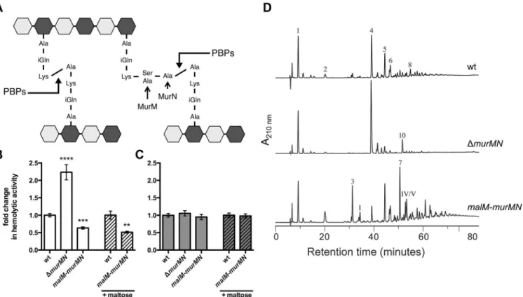
Deletion of themurMNoperon caused a two-fold increase in Ply release as measured by hemolytic activity of whole cells when compared to the wildtype (wt) (Fig 2B,ΔmurMN), sug-gesting a role for the products of this operon in controlling Ply release. Overexpression of murMhas previously been shown to favor production of branched stem peptides at the expense of linear stem peptides [29]. To test if increasing the proportion of branched stem pep-tides would yield the opposite phenotype ofΔmurMN, we created a merodiploid strain carry-ing a second copy ofmurMNdownstream of a maltose-inducible promoter [30]. In the absence of inducer,murMNoverexpression caused a decrease in hemolytic activity of washed cells to the same magnitude as themurMNdeletion (Fig 2B,malM-murMN) suggesting that basal expression from the maltose promoter is sufficient to increasemurMNtranscript levels, which was supported by quantitative reverse transcription-PCR (qRT-PCR) (S3 Fig). Fig 2. Ply release is altered bymurMN-dependent changes in PG composition. (A)Diagram of the basic pneumococcal PG structure highlighting the role of MurM and MurN. PG is a heteropolymer of glycan strands that alternate between acetylglucosamine (NAG, light grey hexagon) and
N-acetylmuramic acid (NAM, dark grey hexagon) residues crosslinked through short peptides that stem from the NAM moiety. Stem peptides can be linear or branched; the latter form is dependent on MurM and MurN. Penicillin-binding proteins (PBPs) catalyze the crosslinking reaction that links adjacent stem peptides.(B-C)Hemolytic activity of whole cells(B)or sonicated lysates(C)of wildtype (wt) and mutants either lackingmurMNor carrying a second copy of
murMNunder the control of the maltose-inducible promoter were grown with or without 0.8% maltose as indicated. Data are presented as the mean fold change in hemolytic activity compared to the wt in each condition±SEM of at least four biological replicates.**p<0.01,***p<0.0005,****p<0.0001, Student’s t-test.(D)RP-HPLC analysis of stem peptides isolated from purified PG of the wt,murMNdeletion, andmalM-murMNoverexpression strains. PG purification, stem peptide removal, and detection by RP-HPLC were performed as described inMaterials and Methods. Peptides were detected by their absorption at 210 nm. The peptide structures of indicated peaks are outlined inS4 Fig.
Supplementation of the growth medium with inducer did not further augment this decrease (Fig 2B) despite increased expression of the entire operon compared to growth without inducer as measured by qRT-PCR (S3 Fig). Thus, changes inmurMNexpression are associated with differential Ply release from the cell.
To rule out the possibility that genetic manipulation of themurMNoperon caused alter-ations in Ply production or stability, which could account for the phenotypes observed we also measured the hemolytic activity of cell lysates. Mutants lacking or overexpressing themurMN operon all harbored the same total hemolytic activity as wt (Fig 2C), supporting the notion that all strains tested contain the same amount of Ply during the course of the experiment. Further-more, we determined that Ply localization to the cell wall compartment was unaffected by dele-tion or overexpression ofmurMN(S1 Fig), indicating that the phenotype observed for washed, whole cells is not due to defects in Ply production or trafficking to the cell surface. Experiments described later will demonstrate that modest changes in the specific localization of Ply can have profound consequences.
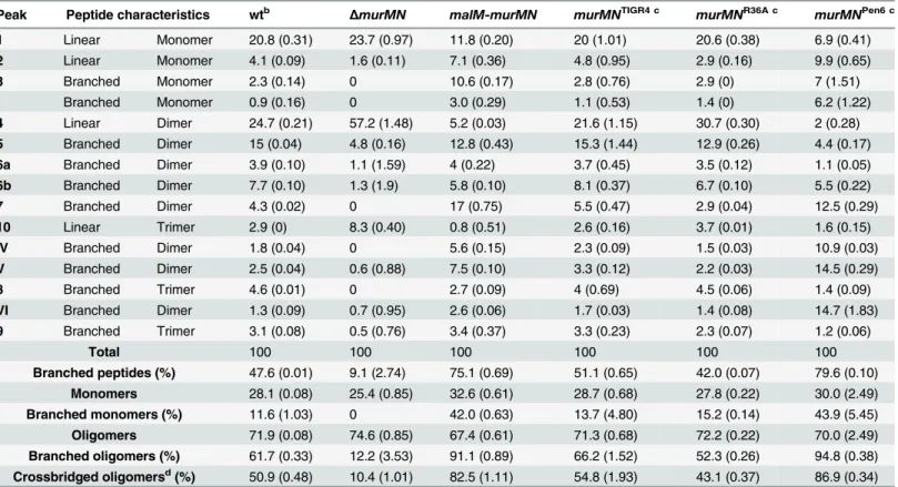
In order to verify thatΔmurMNandmalM-murMNexhibited distinct stem peptide profiles, we purified PG from each strain and analyzed its peptide composition by reversed phase-high performance liquid chromatography. The wt strain was included as a control. As depicted in Fig 2D, wt PG contained both linear and branched stem peptides. Peptide structures of assigned peaks can be found inS4 Fig. Within the monomeric species, the linear tripeptide (peak 1) represented 20.8% of the total peptide material analyzed compared to 3.2% for the branched counterparts (peaks 3 and I) (Table 1). However, dimers containing at least one branched structure (peaks 5, 6, 7, IV, V, VI) were modestly increased compared to the directly crosslinked linear dimer (peak 4) (Table 1). These data demonstrate that branched stem pep-tides can be found throughout wt PG in a manner similar to that observed in PG from other penicillin-sensitive (PenS) laboratory strains [20].
In contrast to wt, PG fromΔmurMNwas characterized by a virtual loss of branched pep-tides and a concomitant overrepresentation of linear peppep-tides (Fig 2DandTable 1). In particu-lar, linear peptides accounted for 90.9% of the total material analyzed from this strain, with the directly crosslinked dimer being the most abundant at 57.2% (Table 1). Strikingly, overexpres-sion ofmurMNcaused a drastic shift in the PG stem peptide profile compared to both wt and ΔmurMN(Fig 2D). The abundance of monomers containing a branched structure (peaks 3 and I) increased to 13.6%, approximately four-fold higher than in the wt (Table 1). Further-more, the enrichment in branched peptides was particularly noticeable in the crosslinked mate-rial of this strain. Dimers containing a branched structure (peaks 5, 6, 7, IV, V, VI) represented 55.3% of the total peptide material at the expense of the linear dimer (peak 4), which decreased five-fold compared to the wt (Table 1). Additionally, there was a near complete loss in the lin-ear trimer (peak 10) inmalM-murMN(Table 1). Given that the stem peptide profile from malM-murMNwas prepared from this strain grown without inducer, these data indicate that a modest ~1.5-fold overexpression ofmurMNis sufficient to cause profound changes to the PG layer (S3 Fig). The peptide profiles depicted inFig 2Dare wholly consistent with previously published results frommurMNdeletion and overexpression mutants in diverse strain back-grounds [20,29]. Taken together, these data strongly support a role for branched stem peptides in limiting Ply release into the extracellular environment.
Choline-binding proteins contribute to Ply release and are sensitive to
branched stem peptides
catalyze PG degradation by cleaving distinct bonds within this structure and, consequently, if not properly regulated can result in cell lysis [31]. Given the association between PG and Ply observed thus far, we hypothesized that Ply release could be due to cleavage of the cell wall by PG hydrolases. To address this possibility, we took advantage of the fact that the major pneu-mococcal PG hydrolases contain choline-binding domains and are therefore displayed on the cell surface by virtue of binding to the choline residues of teichoic acids [2]. This interaction is non-covalent and can be disrupted by the addition of exogenous choline, causing release of choline-binding proteins (CBPs) from the cell surface [32]. Thus, choline treatment would simultaneously remove multiple PG hydrolases (e.g. LytA, LytB, LytC, CbpD) from the cell sur-face as well as other CBPs that harbor distinct functions, allowing us to assess the contribution of this entire subset of proteins to Ply release.
Prior incubation with 2% choline decreased the hemolytic activity of supernatants prepared from whole cells of wt,ΔmurMN, andmalM-murMNcompared to the no choline wash control (Fig 3A). By contrast, choline treatment had no effect on the total hemolytic activity from cell lysates of either strain tested (Fig 3B). This suggests that the ability to release Ply is dependent Table 1. Stem peptide composition of selectS.pneumoniaestrains.a
Peak Peptide characteristics wtb ΔmurMN malM-murMN murMNTIGR4 c murMNR36A c murMNPen6 c
1 Linear Monomer 20.8 (0.31) 23.7 (0.97) 11.8 (0.20) 20 (1.01) 20.6 (0.38) 6.9 (0.41)
2 Linear Monomer 4.1 (0.09) 1.6 (0.11) 7.1 (0.36) 4.8 (0.95) 2.9 (0.16) 9.9 (0.65)
3 Branched Monomer 2.3 (0.14) 0 10.6 (0.17) 2.8 (0.76) 2.9 (0) 7 (1.51)
I Branched Monomer 0.9 (0.16) 0 3.0 (0.29) 1.1 (0.53) 1.4 (0) 6.2 (1.22)
4 Linear Dimer 24.7 (0.21) 57.2 (1.48) 5.2 (0.03) 21.6 (1.15) 30.7 (0.30) 2 (0.28)
5 Branched Dimer 15 (0.04) 4.8 (0.16) 12.8 (0.43) 15.3 (1.44) 12.9 (0.26) 4.4 (0.17)
6a Branched Dimer 3.9 (0.10) 1.1 (1.59) 4 (0.22) 3.7 (0.45) 3.5 (0.12) 1.1 (0.05)
6b Branched Dimer 7.7 (0.10) 1.3 (1.9) 5.8 (0.10) 8.1 (0.37) 6.7 (0.10) 5.5 (0.22)
7 Branched Dimer 4.3 (0.02) 0 17 (0.75) 5.5 (0.47) 2.9 (0.04) 12.5 (0.29)
10 Linear Trimer 2.9 (0) 8.3 (0.40) 0.8 (0.51) 2.6 (0.16) 3.7 (0.01) 1.6 (0.15)
IV Branched Dimer 1.8 (0.04) 0 5.6 (0.15) 2.3 (0.09) 1.5 (0.03) 10.9 (0.03)
V Branched Dimer 2.5 (0.04) 0.6 (0.88) 7.5 (0.10) 3.3 (0.12) 2.2 (0.03) 14.5 (0.29)
8 Branched Trimer 4.6 (0.01) 0 2.7 (0.09) 4 (0.69) 4.5 (0.06) 1.4 (0.09)
VI Branched Dimer 1.3 (0.09) 0.7 (0.95) 2.6 (0.06) 1.7 (0.03) 1.4 (0.08) 14.7 (1.83)
9 Branched Trimer 3.1 (0.08) 0.5 (0.76) 3.4 (0.37) 3.3 (0.23) 2.3 (0.07) 1.2 (0.06)
Total 100 100 100 100 100 100
Branched peptides (%) 47.6 (0.01) 9.1 (2.74) 75.1 (0.69) 51.1 (0.65) 42.0 (0.07) 79.6 (0.10) Monomers 28.1 (0.08) 25.4 (0.85) 32.6 (0.61) 28.7 (0.68) 27.8 (0.22) 30.0 (2.49) Branched monomers (%) 11.6 (1.03) 0 42.0 (0.63) 13.7 (4.80) 15.2 (0.14) 43.9 (5.45) Oligomers 71.9 (0.08) 74.6 (0.85) 67.4 (0.61) 71.3 (0.68) 72.2 (0.22) 70.0 (2.49) Branched oligomers (%) 61.7 (0.33) 12.2 (3.53) 91.1 (0.89) 66.2 (1.52) 52.3 (0.26) 94.8 (0.38) Crossbridged oligomersd(%) 50.9 (0.48) 10.4 (1.01) 82.5 (1.11) 54.8 (1.93) 43.1 (0.37) 86.9 (0.34)
aEach peak corresponds to a speci
fic peptide structure as depicted inS4 Fig. Values represent the mean abundance of the indicated species relative to the total amount of peptide material analyzed and numbers within parentheses denote the standard deviation of two independent experiments.
bTIGR4 strain
cThemurMNcoding regions and intervening sequence from each strain (TIGR4, R36A, Pen6) were introduced intoΔmurMN, replacing the chloramphenicol resistance marker at the native chromosomal locus
dA crossbridge is de
fined as a crosslink that directly incorporates the branch structure (covalent linkage between alanine from branch and fourth position alanine of adjacent peptide)
on the presence of CBPs on the cell surface. Strikingly, the magnitude by which Ply release decreased was dependent on the strain background tested (Fig 3C). The most pronounced Fig 3. Choline-binding proteins contribute to Ply and LacZ release in amurMN-dependent manner.The endogenousβ-galactosidase gene,bgaA,
was replaced withE.coli lacZexpressed from a constitutive promoter in wt,ΔmurMN, andmalM-murMNand the resulting strains were tested for hemolytic
(A-B)andβ-galactosidase(D-E)activity after incubation without or with 2% choline chloride. Strains were tested for both activities as whole cells(A, D)and sonicated lysates(B, E). The fold decrease in hemolytic(C)andβ-galactosidase(F)activity between the choline-treated and control groups of each strain was quantified. Columns represent the mean and error bars denote SEM of four biological replicates. In(A-B, D-E)the data are presented as the fold change in measured activity compared to the wt no choline wash group.*p<0.05,**p<0.01, One sample t test compared to a value of 1.0, which indicates no change.(G-H)Percent Ply and LacZ release was calculated for each strain in both conditions by dividing the whole cell activity in(A)or(D)by the sonicated lysate activity in(B)or(E), respectively. The calculated values were plotted against each other for the control(G)and choline-treated(H)groups such that each symbol represents a different strain with error bars indicating SEM. Data were fit to a straight-line model and the resulting slope was compared to a hypothetical value of 1.0, which represents equal change between the two variables measured and is indicated by the dotted line.
change was observed inΔmurMN, which decreased approximately four-fold after the choline wash relative to the control sample (Fig 3C). By comparison, wt experienced a two-fold drop in Ply release whilemalM-murMNwas modestly, yet significantly, reduced by 1.5-fold (Fig 3C). These results suggest that CBPs contribute to Ply release but this effect is sensitive to the pro-portion of branched stem peptides within PG.
Given that Ply is present in both the cell wall compartment and the cytoplasm, we wanted to address the origin of the released Ply observed in washed, whole cells. It is formally possible that the hemolytic activity of whole cells could be explained by specific Ply release from the cell wall fraction due to PG cleavage. Alternatively, the activity could be explained by lysis of a sub-population of cells, which would release Ply from both the cell wall and the cytoplasm. To dis-tinguish between these possible explanations, we reasoned that we could test for the presence of a strictly cytoplasmic marker in addition to Ply. If Ply release is the result of lysis, then we should also detect the cytoplasmic marker; if lysis is not the primary mechanism, there should be enrichment in Ply over the cytoplasmic marker. A commonly used, robust and easily detect-able cytoplasmic marker isβ-galactosidase, encoded byE.coli lacZ[33]. Therefore, we replaced the coding region of the endogenousβ-galactosidase,bgaA, with that oflacZunder the control of a constitutive promoter in the wt,ΔmurMN, andmalM-murMNstrains and tested for the presence of LacZ in whole cells and sonicated lysates with and without choline treatment.
Miller assays to detectβ-galactosidase activity were performed on the same samples used to measure hemolytic activity depicted inFig 3A and 3B. Supernatants from whole cells of ΔmurMNexpressinglacZcontained approximately twice as muchβ-galactosidase activity as wt ormalM-murMN(Fig 3D, no choline wash). Overexpression ofmurMNresulted in a mod-est decrease in LacZ release from whole cells that was not significantly different from wt (Fig 3D, no choline wash). Interestingly, choline treatment abolished the two-fold increase inβ -galactosidase activity observed in supernatants from whole cells ofΔmurMN, reducing it to lev-els comparable to that of the untreated wt sample (Fig 3D). Importantly, neither expression of murMNnor choline treatment affected the totalβ-galactosidase activity of cell lysates (Fig 3E), indicating a similar amount of LacZ was present in each strain and condition tested. Therefore, LacZ release increased upon deletion ofmurMNin a manner dependent on CBPs, whereas wt andmalM-murMNhad similar levels of LacZ release that are unaffected by CBPs (Fig 3F).
Repair of
ΔmurMN
with distinct
murMN
alleles alters Ply release and PG
composition
Given the natural diversity ofmurMalleles among clinical isolates [22,23], we hypothesized that differentmurMNalleles would have different effects on Ply release. To test this, we replaced theΔmurMNdeletion locus with themurMNcoding regions from different pneumo-coccal strains. Thus, eachmurMNallele is under transcriptional control of the native wt murMNpromoter on the chromosome. Importantly, introduction of the wtmurMNallele back intoΔmurMNrestored wt levels of Ply release (Fig 4A,murMNTIGR4), indicating that the two-fold increase observed upon deletion ofmurMN(Fig 2B) can be attributed specifically to loss of this operon and not due to a second-site mutation that may have occurred elsewhere in the genome during strain construction.
Next, we amplified themurMNcoding regions from a PenS(R36A) or PenR(Pen6) strain [20] and used these to repairΔmurMN. Purified MurM derived from a PenRstrain was previ-ously shown to harbor increased catalytic activityin vitrocompared to a PenScounterpart [18]. Expression of themurMNPen6allele caused a two-fold decrease in Ply release as compared to the wt (Fig 4A), representing a four-fold decrease compared to the parentΔmurMNstrain (compareFig 2B,ΔmurMNtoFig 4A,murMNPen6). This enhanced inhibition of Ply release was specific tomurMNPen6, as expression ofmurMNR36AphenocopiedmurMNTIGR4(Fig 4A). Thus, restoration of wt levels of Ply release was achieved with eithermurMNTIGR4or
Fig 4. Increased Ply release observed uponmurMNdeletion can be differentially restored by variantmurMNalleles.Hemolytic activity of whole cells (A)or sonicated lysates(B)of markedΔmurMNstrains expressing themurMNoperon from wt (murMNTIGR4), R36A (murMNR36A) or Pen6 (murMNPen6). The wt chromosomalmurMNpromoter controls expression of each allele. Data are presented as the mean fold change in hemolytic activity compared to the wt in
each condition±SEM of at least four biological replicates.*p<0.05, Student’s t-test.(C)RP-HPLC analysis of the stem peptides from purified PG isolated from each of the repaired strains. SeeMaterials and Methodsfor a description of the procedures for PG purification, separation of stem peptides, and analysis. Peptides were detected by their absorption at 210 nm and the chemical structures of assigned peaks are diagrammed inS4 Fig.
murMNR36A, whereas introduction of the highly activemurMNPen6inhibited Ply release to a level comparable to that observed uponmurMNoverexpression in themalM-murMNstrain.
To address whether these Ply release phenotypes were accompanied by changes in PG stem peptide composition, we purified PG from each repaired strain and analyzed the stem peptide profile as described above. Strikingly, the profiles from each strain expressing a givenmurMN allele were noticeably different than that of theΔmurMNparent strain (compareFig 4Cto ΔmurMNinFig 2D). Strains expressingmurMNTIGR4andmurMNR36Aexhibited comparable stem peptide profiles to the wt strain with respect to the presence of both linear and branched stem peptides (Fig 4C). WhileΔmurMNlacked any branched monomers (peaks 3 and I), these peptides could be detected inmurMNTIGR4andmurMNR36Aat 3.9% and 4.3%, respectively (Table 1). Additionally, expression of eachmurMNallele caused an approximate two-fold decrease in the directly crosslinked linear dimer (peak 4) compared withΔmurMN, accompa-nied by an increase in the abundance of branched dimers (peaks 5, 6, 7, IV, V, VI) to levels sim-ilar to that observed in the wt (Table 1). Thus, the stem peptide profile and Ply release
phenotype ofΔmurMNcould be restored by expression of wtmurMNor the PenS-associated allele from R36A.
Expression of themurMNPen6allele also lead to the formation of branched stem peptides, albeit to a much greater extent than observed with either of the other twomurMNalleles tested (Fig 2D). There was significant enrichment in monomers with a branched structure (peaks 3 and I), increasing from zero inΔmurMNto 13.2% (Table 1). Perhaps more striking was the complete reversal in the dimer structures; more than half of the total peptide material of the ΔmurMNparent was represented by the linear dimer, whereas there was complete replacement of this with the branched forms inmurMNPen6(Table 1). Thus, expression of the PenR -associ-atedmurMNPen6allele caused a shift towards a highly branched PG reminiscent of themurMN overexpressing strain. Of note, the stem peptide profile ofmurMNPen6is more similar to that of Pen6 itself than the wt strain described herein [20]. Thus, the pneumococcal stem peptide profile is largely dictated bymurMNexpression and activity of the resulting gene products, which is accompanied by differences in Ply release from the cell.
The incorporation of branched stem peptides is inversely correlated with
Ply release
Stem peptide analysis of the wt and variousmurMNmutants described revealed several differ-ences in discrete peptide species between each strain. We were interested in determining whether there were any global trends from this analysis that could best explain the observed differences in Ply release of each strain. As anticipated, the ratio of branched to linear stem peptides in the total material analyzed was highly dependent on the expression ofmurMN. Deletion ofmurMNcaused enrichment in linear stem peptides, whereasmurMN overexpres-sion led to a three-fold enrichment in branched stem peptides compared to wt (Fig 5A). Repair ofΔmurMNwith themurMNPen6allele caused a more pronounced enrichment in branched peptides, representing a four-fold increase over the wt (Fig 5A). The incorporation of branched stem peptides into the crosslinked material was also highly dependent onmurMNand mim-icked the trends highlighted in the total peptide material. The percentage of oligomeric species (dimers and trimers) containing a crossbridge, which is indicative of a crosslink that connects the branch structure to an adjacent stem peptide, increased to approximately 80% upon murMNoverexpression or expression ofmurMNPen6, which is up from 50% in the wt and the other strains expressing PenS-associatedmurMNalleles (Fig 5B).
of Ply released. We calculated the amount of Ply released as a percentage of the total for each of the strains described in Figs2and4, and plotted it against the total amount of branched stem peptides within each strain. Intriguingly, this analysis revealed a statistically significant, strong negative correlation (Fig 5C). We extended this analysis further by determining whether partic-ular subsets of branched peptide species are more strongly associated with Ply release. There was no significant correlation between Ply release and the percentage of monomers containing a branched structure (Fig 5D). However, the proportion of branched oligomers showed a sig-nificant negative correlation with the amount of Ply release observed (Fig 5E). These results suggest that it is not just the presence of branched stem peptides, but also their incorporation into the mature, crosslinked PG that inhibits Ply release.
Branched stem peptides are required to maintain optimal Ply release
during lung infection
To assess the contribution of branched stem peptides and Ply release to pneumococcal viru-lence, we competedΔmurMNormurMNTIGR4against wt in a murine model of pneumonia. Neither mutant demonstrated a fitness defect as determined by the competitive index (Fig 6A). However, infection withΔmurMNcaused a 125-fold decrease in the median number of recov-ered wt bacteria as compared to themurMNTIGR4competition (Fig 6B). As a control, we also performed single-strain lung infections with wt and found that the titers achieved by the wt Fig 5. Incorporation of branched stem peptides in PG inversely correlates with Ply release. (A)Ratio of branched to linear stem peptides in the PG of indicated strains.(B)Percentage of oligomers (dimers and trimers) containing a crossbridge as defined by a crosslink that directly incorporates the branch moiety.(C-E) Percent Ply release was calculated using the data from Figs2B, 2C,4A and 4Band plotted against the percentage of total branched stem peptides(C), branched monomers(D), and branched oligomers(E). Each symbol represents a given strain. Statistical dependence between percent Ply release and each variable was determined by calculating Pearson’s correlation coefficient (r). In all panels, data are presented as the
mean±SEM.
alone were 23-fold higher than those during co-infection withΔmurMN(Fig 6B). However, wt reached similar titers either alone or during competition with the repairedmurMNTIGR4strain (Fig 6B, compare wt alone tomurMNTIGR4, medians not significantly different), suggesting that the absence of branched stem peptides inΔmurMNnegatively impacts virulence.
We hypothesized that the increased Ply release in theΔmurMNstrain (Fig 2B) was detri-mental to the virulence of the co-infecting wt strain. To address whether the inhibitory effect of co-infection withΔmurMNwas Ply-dependent, we deleted theplystructural gene in the ΔmurMNbackground and competed this double mutant against wtin vivo. In support of our hypothesis, wt titers increased 53-fold compared to theΔmurMNco-infection and reached lev-els that were statistically indistinguishable from those observed during competition with murMNTIGR4(Fig 6B, compareΔmurMNΔplytomurMNTIGR4, medians not significantly dif-ferent). As a control, we also competed the singleΔplymutant against wt and were able to recover wt bacteria to approximately the same level as in co-infection withmurMNTIGR4or ΔmurMNΔply(Fig 6B). Similar to the other strains tested, neitherΔmurMNΔplynorΔply showed a fitness defect during competition with the wt (Fig 6A). Taken together, co-infection withΔmurMNlimits the ability of wt to achieve high titersin vivoand this inhibitory effect can be relieved by restoration of themurMNoperon or deletion ofply. This strongly suggests that appropriate Ply releasein vivois dependent on PG stem peptide composition and maintenance of this ability is necessary for pneumococcal virulence.
Discussion
Despite the lack of a signal peptide and a cell surface-localization motif, Ply appears to be exported through the pneumococcal membrane and associates with the cell envelope by an unresolved mechanism [12,13]. In this study, we demonstrate that exported Ply is not surface-Fig 6. Co-infection withΔmurMNdecreases wt burdens in a Ply-dependent manner.Equal amounts of the indicated strains were mixed with wt and administered into the lungs of mice via intranasal inoculation. A competitive index (CI)(A)and the recovered wt titers(B)were determined for each mouse in each sample group. As a control, single-strain infections with wt alone were performed and the titers are depicted in panel B (indicated by the gray box). Each symbol represents an individual mouse and the bars indicate the median CI(A)or median bacterial titer(B). Open symbols without or with a central dot indicate titers that are at or below the limit of detection of 100 CFU/homogenate for the mutant or wt, respectively. The median bar masks two open, central dot symbols in theΔmurMNdataset. No group in(A)has a median CI that is significantly different from 1.0 as determined by Wilcoxon Signed Rank test.*p<0.05; Mann-Whitney test.
accessible and its activity and release into the surrounding medium is dependent on enzymatic digestion of the PG layer. This may suggest that surface-localized Ply is inhibited due to an inability to fold into a pore-forming competent state while associated with the cell envelope. Alternatively, but not mutually exclusive, the accumulation of Ply at this site may be due to restricted mobility of the exported protein through the PG matrix. Our results cannot distin-guish between these two possibilities but it is clear from our experiments that Ply activity and release is dependent on PG hydrolysis.
Theoretical and experimental analysis has suggested that purified PG can accommodate globular proteins ranging from ~25–50 kilodaltons (kDa) [34]. However, this likely represents a static portrait of PG pore size, as it does not account for the dynamic remodeling of this layer that occurs during cell growth [14,35]. Ply is a 53-kDa protein, putting it on the upper bound-ary of the experimentally defined range noted above. The existence of a“periplasm-like”space delineated by the plasma membrane and cell wall matrix within the Gram-positive cell enve-lope has been proposed by several groups and experimentally investigated in different species [36–39]. It is tempting to speculate that, upon export through the membrane, Ply is transiently restricted within this“periplasm-like”space due to its limited ability to diffuse through the cell wall matrix, which may be modulated by altering the proportion of branched stem peptides in PG.
Interestingly, Ply was released from washed pneumococci in the absence of host cells (i.e. red blood cells), suggesting that this process may not strictly require a host-derived signal or direct contact. This is in contrast to the pH-dependent release of phospholipase C (PC-PLC) from the cell wall of the intracellular pathogenListeria monocytogenes[40]. PC-PLC, like Ply, lacks a sorting motif yet localizes to the cell wall compartment ofL.monocytogenesand, under native conditions, exists in a state that cannot be detected with antibodies; though digestion of the PG layer allows for complete release and exposure [40,41]. Entry into the host cell cytosol causes a rapid release of surface-associated PC-PLC only upon acidification of the intracellular environment, which is required for optimal virulence ofL.monocytogenes[40]. Attempts to detect surface-localized Ply by immunofluorescence have been unsuccessful, perhaps due to the inability of antibodies to reach the niche that Ply occupies within the cell envelope. It is important to note that we cannot rule out the possibility that a specific trigger existsin vivoto stimulate Ply release, however, it appears that this stimulus is not requiredper seand that release is at least partly dependent on PG structure and the presence of CBPs on the cell sur-face. Of note, a recent report demonstrated that phagocytosis of the pneumococcus by macro-phages results in Ply-dependent death of both host cell and bacterium. This phenomenon was potentiated in a mutant exhibiting hypersensitivity to lysozyme digestion, thus implicating this host factor in contributing to Ply releasein vivo[42].
Pneumococcal PG contains both linear and branched stem peptides, the proportions of which vary depending on the expression level ofmurMNas well as the activity of the encoded proteins (Figs2D,4CandTable 1) [17,29]. Branched stem peptide formation was inversely related to the amount of Ply released as determined through deletion and overexpression of the murMNoperon. MurM and MurN are non-essential and the pneumococcus grows similarlyin vitroboth with and without branched stem peptides [20]. Therefore, the precise role of branched stem peptides in pneumococcal biology remains an open question. Previous observa-tions demonstrated thatmurMNexpression was associated with an altered propensity to auto-lyse upon treatment with a variety of cell wall-targeting compounds suggesting that differences in stem peptide composition confer altered susceptibility to autolysis [24]. However, the under-lying mechanisms of this phenomenon are unclear.
lacking branched stem peptides (exemplified byΔmurMN) may be more sensitive to the action of a subset of CBPs capable of mediating PG hydrolysis even in the absence of any overt pertur-bation to the cell wall and/or membrane. This sensitivity can be suppressed by increasing the abundance of branched stem peptides within the PG layer. Whether variation in branched stem peptide abundance is accompanied by altered susceptibility of the PG layer to the action of PG hydrolases, including those that are CBPs, remains an interesting and unexplored possi-bility. Furthermore, these results suggest that Ply can be released specifically from the cell sur-face in the absence of lysis, which is likely the result of controlled PG remodeling by CBPs. Intriguingly, a recent report revealed a role for PG hydrolases (including pneumococcal LytA, a CBP) in limiting recognition by peptidoglycan responsive proteins of the innate immune sys-tem through controlled trimming of the PG layer [43]. This supports the hypothesis that regu-lated PG cleavage is critical during infection for additional reasons other than promoting cell growth and division.
Removal of CBPs from the cell surface caused an overall decrease in both Ply and LacZ release, especially in theΔmurMNmutant (Fig 3H). However, both proteins could be detected in equal proportions, suggesting cytoplasmic leakage due to autolysis that may be the result of incomplete removal of surface-bound CBPs or the activity of other, non-choline-binding PG hydrolases. Of note, the pneumococcus encodes numerous known and putative PG hydrolases that are likely unaffected by choline treatment [44]. Intriguingly, CBP-mediated autolysis was also recently shown to be necessary for the release of LytA amidase from the cytoplasm during logarithmic growth [45]. LytA, like Ply, lacks a N-terminal signal peptide suggesting that basal autolysis during growth can allow for the release of other leaderless extracellular proteins that may then reassociate with the cell surface. However, it is important to point out that Ply released upon autolysis was not able to bind to the surface of actively growing cells [13]. The association between PenRandmurMNled us to test whether Ply release would be affected by the expression of alteredmurMNalleles that appear to naturally arise in response to antibiotic pressure. Repair of theΔmurMNmutant with a PenR-associatedmurMNallele, but not a PenS-associated allele, caused a four-fold reduction in the amount of Ply release com-pared to theΔmurMNparent strain accompanied by significant changes to the stem peptide profile. Given that each allele was under control of the endogenousmurMNpromoter on the chromosome, these data support previous observations that variant PenR-associatedmurM alleles encode proteins with higher catalytic activity compared to a PenSMurM [18]. Also, this lends support to the model that PG containing more branched stem peptides represents a more restrictive environment for Ply release.
The biological consequences of PenRcan lead to pleiotropic effects that can cause fitness defects in different pathogens, including the pneumococcus [46–48]. The precise mechanism (s) that select formurMNmutations in the context of PenRlow-affinity PBPs are not well understood. There is some evidence suggesting that certain PBPs display different substrate specificities for transpeptidation [49,50]. Given that low-affinity PBPs demonstrate decreased affinity for penicillin, which is structurally similar to their natural substrate, it seems plausible to speculate that selection for low-affinity PBPs could create a PG structure that may pheno-copy the loss ofmurMNwith respect to both Ply release and decreased virulence. Therefore, this fitness defect may aid in selection formurMNmutations that compensate for altered PG architecture and Ply release. Whether Ply release plays a role in the selection for compensatory mutations in the context of low-affinity PBPs is an intriguing hypothesis that remains to be explored.
lung infection. Importantly, this result indicates that the altered stem peptide composition in theΔmurMNmutant does not result in a fitness defect in this strain compared to the wt. Ply is a potent inflammatory agonist that contributes to cytokine production and can cause influx of phagocytic cells [9,51]. Therefore, we propose that the increased Ply release caused by infection withΔmurMNcauses a robust, perhaps premature, immune response that results in enhanced bacterial clearance. Neither the wt nor the mutant is equipped to handle this heightened response, which results in a reduction of both strains during this co-infection. It is unclear whether the pneumococcus modulates the proportion of branched stem peptides in the PG layer during infection. Of note,murMexpression appears to be specifically upregulated in the presence of epithelial cellsin vitro[52], supporting the hypothesis that the stem peptide com-position of PG may be actively modulated under certain conditions mimicking those foundin vivo.
The finding that repair ofΔmurMNto amurMN+state restored both Ply releasein vitro and caused wt titers to reboundin vivostrongly suggests that it is the specific localization of Ply, not the amount produced, that dictates successful infection. Both of these strains areply+ and produce the same amount of total Plyin vitro. Thus, the discrepancy in bacterial loadsin vivoobserved between the groups appears to be independent of Ply production. Indeed, aΔply mutant did not exhibit a fitness defect during co-infection in this model (Fig 6A). At first glance, this appears to be in opposition to the dogma that Ply is a critically important virulence factor of the pneumococcus [53–55]. However, these classical studies were all conducted using single-strain infections with either wt or aplymutant and not a competition between the two. Indeed, our results are consistent with the observations of Bentonet al, who demonstrated that growth of aplymutant in a blood model of infection could be rescued upon co-infection with a wildtype strain [56]. The lack of attenuation observed forΔplyduring competition against the wt actually provides further support for our argument that Ply is released from the cell wall compartment during infection as it appears theΔplystrain can be complemented intransby Ply released from the wt. Based on this notion, we propose that the dynamics of Ply release in wt cells is carefully balanced so as to promote virulence without causing immune-mediated clearance.
In summary, we have demonstrated that Ply release from the pneumococcal cell wall com-partment and cytoplasm can occur in the absence of a host signal but is limited by the native PG network. Based on these observations, we propose that PG acts as a barrier that causes an accumulation of exported Ply at the cell surface that may be released upon remodeling by PG hydrolases, particularly those that are CBPs. Additionally, we identified a novel role for branched stem peptides in restricting Ply release from the cell presumably by affecting the type of crosslinks that create the PG structure. The acquisition of PenR, and other determinants nec-essary for its expression, may differentially affect Ply release and, as a result, the outcome of an infection. Finally, we demonstrate that branched stem peptides play a critical role in maintain-ing the PG barrier, which allows for precise control over Ply release. We propose that this is necessary to establish the balance between virulence and immune activation, thus suggesting that PG architecture may play an important regulatory role in the pathogenesis of diverse microbial pathogens. This is the first indication that the cell wall-associated Ply may contribute to pneumococcal virulence.
Materials and Methods
Bacterial strains and growth conditions
stocks on tryptic soy agar plates containing 5% sheep blood (Northeast Laboratory) overnight at 37°C in a 5% CO2environment. The growth from overnight plates was suspended in Todd Hewitt broth (BD Biosciences) supplemented with 0.5% (wt/vol) yeast extract (Fisher Scientific) (THY medium), diluted to an OD600of 0.02, and grown to a final OD600of 0.8 in a 37°C incuba-tor with 5% CO2for all experiments. THY medium was routinely supplemented with 0.5% Oxy-rase (OxyOxy-rase, Inc.). When necessary, the following antibiotics at the indicated concentrations were used: chloramphenicol (Cm) (4μg/mL) and spectinomycin (Spec) (200μg/mL).
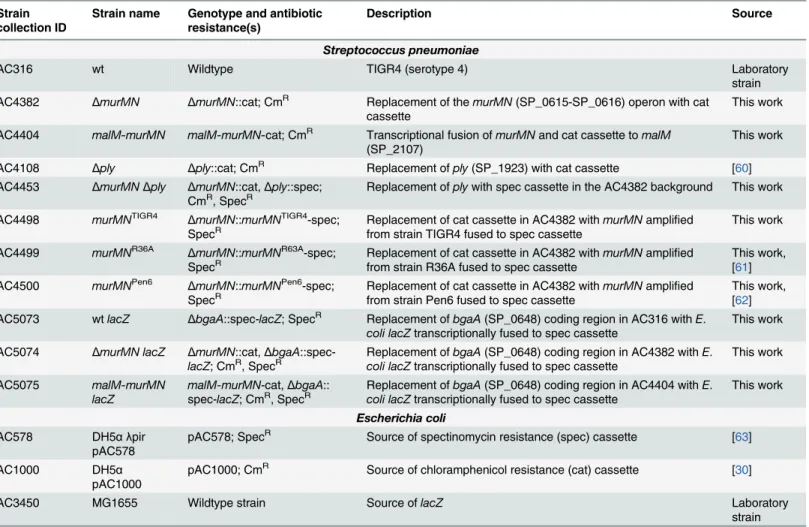
Mutant strain construction
Mutations were generated using allelic exchange with linear PCR amplicons. For deletion con-structs, linear amplicons were created via splicing by overlap extension (SOE) PCR. 1 kb of flanking sequence immediately upstream and downstream of the target gene was amplified from TIGR4 gDNA and fused to either a CmRor SpecRcassette, which were amplified from pAC1000 and pAC578 (Table 2), respectively. To repairΔmurMNwith differentmurMN alleles, the coding regions and intervening sequence ofmurMNwere amplified from gDNA prepared from the appropriate strains (Table 2) and fused to a SpecRcassette and the flanking arms of homology surrounding the nativemurMNlocus. Construction of themurMN overex-pression strain was done essentially as described [30]. Generation oflacZ-expressing strains Table 2. Strains and plasmids used in the present study.
Strain collection ID
Strain name Genotype and antibiotic resistance(s)
Description Source
Streptococcus pneumoniae
AC316 wt Wildtype TIGR4 (serotype 4) Laboratory
strain AC4382 ΔmurMN ΔmurMN::cat; CmR Replacement of themurMN(SP_0615-SP_0616) operon with cat
cassette This work
AC4404 malM-murMN malM-murMN-cat; CmR Transcriptional fusion ofmurMNand cat cassette tomalM
(SP_2107) This work
AC4108 Δply Δply::cat; CmR Replacement ofply(SP_1923) with cat cassette [60] AC4453 ΔmurMNΔply ΔmurMN::cat,Δply::spec;
CmR, SpecR Replacement ofplywith spec cassette in the AC4382 background This work AC4498 murMNTIGR4 ΔmurMN::murMNTIGR4-spec;
SpecR Replacement of cat cassette in AC4382 withfrom strain TIGR4 fused to spec cassette murMNamplified This work AC4499 murMNR36A ΔmurMN::murMNR63A-spec;
SpecR Replacement of cat cassette in AC4382 withfrom strain R36A fused to spec cassette murMNamplified This work,[61] AC4500 murMNPen6 ΔmurMN::murMNPen6-spec;
SpecR Replacement of cat cassette in AC4382 withfrom strain Pen6 fused to spec cassette murMNamplified This work,[62] AC5073 wtlacZ ΔbgaA::spec-lacZ; SpecR Replacement ofbgaA(SP_0648) coding region in AC316 withE.
coli lacZtranscriptionally fused to spec cassette This work AC5074 ΔmurMN lacZ ΔmurMN::cat,ΔbgaA
::spec-lacZ; CmR, SpecR Replacement ofcoli lacZtranscriptionally fused to spec cassettebgaA(SP_0648) coding region in AC4382 withE. This work
AC5075 malM-murMN
lacZ
malM-murMN-cat,ΔbgaA::
spec-lacZ; CmR, SpecR Replacement ofcoli lacZtranscriptionally fused to spec cassettebgaA(SP_0648) coding region in AC4404 withE. This work
Escherichia coli
AC578 DH5α λpir
pAC578
pAC578; SpecR Source of spectinomycin resistance (spec) cassette [63]
AC1000 DH5α
pAC1000
pAC1000; CmR Source of chloramphenicol resistance (cat) cassette [30]
AC3450 MG1655 Wildtype strain Source oflacZ Laboratory
strain
was performed by replacing the coding region ofbgaA(SP_0648) on the pneumococcal chro-mosome with a transcriptional fusion ofE.coli lacZto the SpecRcassette. To do so, the coding region oflacZamplified fromE.coliMG1655 gDNA (Table 2) was placed downstream of the SpecRcassette and was flanked on either side by 1 kb of sequence immediately upstream and downstream of thebgaAcoding region by SOE PCR. Transformation of the pneumococcus was carried out as described previously [57]. All mutant constructs were verified using PCR and DNA sequencing.
Hemolysis assays
Mid-exponential growth phase cells were normalized to OD600= 0.8 (approx. 108CFU/mL), collected by centrifugation, washed once in assay buffer [AB; phosphate-buffered saline (PBS), 0.1% bovine serum albumin, 10 mM dithiothreitol] and resuspended in AB. An aliquot of each sample was sonicated at 4°C at maximum amplitude for 2 minutes in a water bath sonicator (Branson, Inc.) using a 10 second on, 5 second off duty cycle. Whole cell, sonicated, or subcel-lular fractions were serially 2-fold diluted in AB in 96-well V-bottom plates (Greiner Bio-One). For each experiment, an 8% solution of triple-washed sheep red blood cells (SRBC) was freshly prepared and 50μL of this was added to each well containing 100μL of sample or control wells containing either AB alone or distilled water. To test whether the cell-free supernatant har-bored hemolytic activity, AB alone was substituted for the SRBC solution in the first hour incu-bation and then 100μL of the supernatant was transferred to a new plate to which SRBC was added as described above. Plates were incubated at 37°C for 1 hour after which the bacterial cells and any unlysed SRBC were pelleted by centrifugation at 4000 x g for 10 min. One hun-dred microliters of each supernatant was removed to a 96-well flat bottom plate and the absor-bance at 570 nm was measured. After subtracting the AB only value from each sample, the percent activity from each well was determined relative to the distilled water control, which was set to 100% hemolysis. Plotting the OD570versus the OD600of each sample yielded a sig-moidal curve, from which the linear portion of the curve was used to extrapolate the OD600at which 50% hemolysis occurred. The reciprocal of this value is defined as the number of hemo-lytic units and each whole cell sample or subcellular fractionation was divided by its paired son-icated sample to determine percent hemolytic activity.
For removal of choline-binding proteins, strains were grown and processed for hemolysis assays as described above with the following modifications. After washing away media, cells were washed once in PBS and split evenly in two separate tubes. After centrifugation, pellets were concentrated five-fold in either PBS or PBS containing 2% choline chloride (Sigma) and incubated at room temperature with agitation for 20 minutes. After centrifugation and washing once in AB, an aliquot was removed for sonication to determine total hemolytic activity and the remainder of each sample was incubated in AB at 37°C for 1 hour. Cells were collected by centrifugation and the supernatant, containing released proteins, was assayed for hemolytic activity as described above.
Miller assays
thatβ-galactosidase activity was linear down to 0.89±0.014% of the starting material (OD600= 0.8) (S5 Fig).
Peptidoglycan purification and enzymatic digestion
Cell wall material was prepared using a previously published protocol [20]. Peptidoglycan was further purified from approximately 5–20 mg of cell wall material by treatment with 48% hydrofluoric acid for 48 hours at 4°C with agitation. Hydrofluoric acid was removed through extensive washing with 100 mM Tris-HCl pH 7.0 and its absence confirmed by measuring the pH. Purified peptidoglycan was then collected by lyophilization and subjected to enzymatic digestion with purified pneumococcal LytA amidase to liberate stem peptides as described pre-viously [20].
Analysis of stem peptide composition
Stem peptides were separated and analyzed by reversed phase-high performance liquid chro-matography (RP-HPLC) as described elsewhere [20].
Subcellular fractionation
Cell wall digestion was performed as previously described [13], with some modifications. Briefly, cells from exponentially growing cultures were collected by centrifugation, washed once with 50 mM Tris-Cl, pH 7.5 and resuspended in 100μL cell wall digestion buffer [50 mM Tris-Cl, pH 7.5, 30% (w/v) sucrose, 1 mg/mL lysozyme, 300 U/μL mutanolysin, 1x protease inhibitor cocktail (Roche)]. Cell wall digestion was allowed to proceed for 2 hours at 37°C on a roller drum after which protoplasts were separated from cell wall material via centrifugation at 13,000 x g for 10 min. The supernatant was collected as the cell wall fraction. Protoplasts were resuspended in 100μL 50 mM Tris-Cl, pH 7.5.
Western blot analysis
An equivalent amount of each subcellular fraction was mixed with 2x Laemmli sample buffer and heated in a boiling water bath for 10 minutes prior to loading cell equivalents on a 10% SDS-PAGE gel. Gels were run at 125 V until the dye front reached the bottom of the gel and then proteins were transferred to a nitrocellulose membrane at 25 V for 1.5 hours. Membranes were blocked with NAP-blocker (G-Biosciences) diluted 1:2 with Tris-buffered saline contain-ing 0.1% Tween-20 (TBST) and then cut to allow for simultaneous probcontain-ing with each antibody. Primary antibodies against Ply at 1:1000 (Statens Serum Institut) and CodY at 1:1500 (a gift from A.L. Sonenshein) were diluted accordingly in NAP-blocker mixed 1:4 with TBST and applied to each membrane at room temperature for 1 hour with rocking. Membranes were washed three times for 5 minutes each with TBST. Appropriate Cy5-conjugated secondary antibody at 1:1000 (Invitrogen, Inc.) was applied to each membrane as described above for the primary and then each blot was washed as described above. Membranes were scanned with a Fuji FLA-9000 instrument and the amount of fluorescence was quantitated using MultiGauge analysis software (Fujifilm, Corp.)
Peptidoglycan binding assay
reaction volume of 30μL and allowed to incubate on a roller drum at 37°C for 2 hours. Control samples lacking either PG or lysate were performed in parallel. Insoluble PG was pelleted by centrifugation at 20,000 xgfor 30 minutes at room temperature and the supernatant (unbound fraction) was removed to a separate tube. The pellet (bound fraction) was washed once with 90μL PBS, collected by centrifugation, and then resuspended in 30μL PBS. The presence of Ply within each supernatant and pellet fraction was detected by Western blot analysis as described above with the exception that samples were resolved by SDS-PAGE using NuPAGE 4–12% Bis-Tris gels (Life Technologies) run at 200 V for 35 minutes.
qRT-PCR
RNA extraction and qRT-PCR was performed exactly as described [59] to analyzemurMand murNtranscript levels in exponentially growing cultures of wt andmalM-murMNgrown in THY medium with and without added 0.8% maltose.
Animal infections
Lung infections were carried out as described in [59] except that mice were euthanized 30–36 hours post-inoculation.
Ethics statement
All animal experiments were done in accordance with NIH guidelines, the Animal Welfare Act and US federal law. Tufts University School of Medicine’s Institutional Animal Care and Use Committee approved the experimental protocol“B2014-37”that was used for this study. All animal experiments were housed in a centralized and AAALAC-accredited research animal facility that is fully staffed with trained husbandry, technical and veterinary personnel.
Supporting Information
S1 Fig. Ply localization to the cell wall compartment is unaffected by mutations in the murMN operon. (A)The indicated strains were subjected to subcellular fractionation to sepa-rate cell wall (CW) material from protoplasts (Prt) and the amount of Ply and CodY present in each fraction was determined by Western blot analysis. A representative image of two indepen-dent experiments is shown.(B)The amount of Ply present in the cell wall fraction relative to the total amount of Ply (cell wall and protoplast) normalized to the cytoplasmic protein CodY for each of the strains analyzed was quantitated for two independent experiments and is shown relative to wt for each condition tested.
(TIFF)
S2 Fig. Ply does not bind PG in a pull-down assay. (A)A wt cell lysate was mixed with differ-ent amounts of purified wt PG (2.6 mg/mL to 0.17 mg/mL, 2-fold serial dilutions).(B)A fixed amount of purified wt PG (1.3 mg/mL) was mixed with different amounts of wt cell lysate (OD600= 3.5 to 0.2 of initial culture, 2-fold serial dilutions). PG alone (1.3 mg/mL) or lysate alone (no PG) were included as controls. Samples were incubated at 37°C on a roller drum for 2 hours. Insoluble PG was pelleted by centrifugation at 21,000 xgfor 30 minutes. The superna-tant (S) containing unbound material was removed and the pellet (P) representing the bound fraction was washed once with PBS and resuspended in an equal volume to the supernatant to normalize cell equivalents. The amount of Ply present in each sample was determined by West-ern blot analysis. In each panel, a representative image of two independent experiments is shown.
S3 Fig. Quantification of murMN expression in the malM-murMN strain grown with and without inducer.Relative expression ofmurMandmurNcompared to wt (set to 1) in malM-murMNgrown in the absence (THY) or presence (THY + 0.8% maltose) of inducer. Transcript abundance was normalized to the housekeeping gene,rplI.
(TIFF)
S4 Fig. Peptide structures of the indicated peaks in Figs2Dand4C.
(TIFF)
S5 Fig. Sensitivity of theβ-galactosidase assay used to measure LacZ release.Three indepen-dent cultures ofΔbga::spec-lacZ(AC5073) were grown to OD600= 0.8, pelleted by centrifuga-tion, resuspended in hemolysis assay buffer, and sonicated to generate whole cell lysates as described inMaterials and Methods. Each lysate was serially five-fold diluted three times to make a dilution series (1x, 0.2x, 0.04x, 0.008x).(A)β-galactosidase activity of each dilution was detected using Miller assays.(B)Miller units of each dilution plotted as a function of the per-cent starting material yields a linear relationship down to 0.89%. Each symbol represents three overlapping symbols that each depicts a biological replicate. The coefficient of determination (R2) was determined using GraphPad Prism 6 software.
(TIFF)
Acknowledgments
We thank Bharathi Patimalla for assistance with animal experiments, Alexander Tomasz for the kind gift of strains R36A and Pen6, Ankur Dalia and Mara Shainheit for helpful discussions and critical reading of the manuscript as well as David Lazinski for helpful discussions.
Author Contributions
Conceived and designed the experiments: NGG AC. Performed the experiments: NGG ARN. Analyzed the data: NGG ARN SRF. Contributed reagents/materials/analysis tools: NGG ARN SRF AC. Wrote the paper: NGG AC.
References
1. Bogaert D, de Groot R, Hermans PWM (2004)Streptococcus pneumoniaecolonisation: the key to pneumococcal disease. Lancet Infect Dis 4: 144–154. doi:10.1016/S1473-3099(04)00938-7PMID: 14998500
2. Kadioglu A, Weiser JN, Paton JC, Andrew PW (2008) The role ofStreptococcus pneumoniaevirulence factors in host respiratory colonization and disease. Nat Rev Microbiol 6: 288–301. doi:10.1038/ nrmicro1871PMID:18340341
3. Rubins JB, Duane PG, Charboneau D, Janoff EN (1992) Toxicity of pneumolysin to pulmonary endo-thelial cells in vitro. Infect Immun 60: 1740–1746. PMID:1563759
4. Zysk G, Schneider-Wald BK, Hwang JH, Bejo L, Kim KS, et al. (2001) Pneumolysin is the main inducer of cytotoxicity to brain microvascular endothelial cells caused byStreptococcus pneumoniae. Infect Immun 69: 845–852. doi:10.1128/IAI.69.2.845–852.2001PMID:11159977
5. Baba H, Kawamura I, Kohda C, Nomura T, Ito Y, et al. (2001) Essential role of domain 4 of pneumolysin fromStreptococcus pneumoniaein cytolytic activity as determined by truncated proteins. Biochem Bio-phys Res Commun 281: 37–44. doi:10.1006/bbrc.2001.4297PMID:11178957
6. Paton JC, Rowan-Kelly B, Ferrante A (1984) Activation of human complement by the pneumococcal toxin pneumolysin. Infect Immun 43: 1085–1087. PMID:6698602
7. Ratner AJ, Hippe KR, Aguilar JL, Bender MH, Nelson AL, et al. (2006) Epithelial cells are sensitive detectors of bacterial pore-forming toxins. J Biol Chem 281: 12994–12998. doi:10.1074/jbc.
M511431200PMID:16520379
pneumoniaein vitro: a novel function of pneumolysin in caspase-1 activation. Infect Immun 76: 1547–
1557. doi:10.1128/IAI.01269-07PMID:18195026
9. McNeela EA, Burke Á, Neill DR, Baxter C, Fernandes VE, et al. (2010) Pneumolysin activates the NLRP3 inflammasome and promotes proinflammatory cytokines independently of TLR4. PLoS Pathog 6: e1001191. doi:10.1371/journal.ppat.1001191PMID:21085613
10. Walker JA, Allen RL, Falmagne P, Johnson MK, Boulnois GJ (1987) Molecular cloning, characteriza-tion, and complete nucleotide sequence of the gene for pneumolysin, the sulfhydryl-activated toxin of
Streptococcus pneumoniae. Infect Immun 55: 1184–1189. PMID:3552992
11. Balachandran P, Hollingshead SK, Paton JC, Briles DE (2001) The autolytic enzyme LytA of Strepto-coccus pneumoniaeis not responsible for releasing pneumolysin. J Bacteriol 183: 3108–3116. doi:10. 1128/JB.183.10.3108–3116.2001PMID:11325939
12. Price KE, Camilli A (2009) Pneumolysin localizes to the cell wall ofStreptococcus pneumoniae. J Bac-teriol 191: 2163–2168. doi:10.1128/JB.01489-08PMID:19168620
13. Price KE, Greene NG, Camilli A (2012) Export requirements of pneumolysin inStreptococcus pneumo-niae. J Bacteriol 194: 3651–3660. doi:10.1128/JB.00114-12PMID:22563048
14. Typas A, Banzhaf M, Gross CA, Vollmer W (2011) From the regulation of peptidoglycan synthesis to bacterial growth and morphology. Nat Rev Microbiol 10: 123–136. doi:10.1038/nrmicro2677PMID:
22203377
15. Schleifer KH, Kandler O (1972) Peptidoglycan types of bacterial cell walls and their taxonomic implica-tions. Bacteriol Rev 36: 407–477. PMID:4568761
16. Garcia-Bustos JF, Chait BT, Tomasz A (1987) Structure of the peptide network of pneumococcal pepti-doglycan. J Biol Chem 262: 15400–15405. PMID:2890629
17. Filipe SR, Pinho MG, Tomasz A (2000) Characterization of themurMNoperon involved in the synthesis of branched peptidoglycan peptides inStreptococcus pneumoniae. J Biol Chem 275: 27768–27774. doi:10.1074/jbc.M004675200PMID:10869361
18. Lloyd AJ, Gilbey AM, Blewett AM, De Pascale G, Zoeiby El A, et al. (2008) Characterization of tRNA-dependent peptide bond formation by MurM in the synthesis ofStreptococcus pneumoniae peptidogly-can. J Biol Chem 283: 6402–6417. doi:10.1074/jbc.M708105200PMID:18077448
19. De Pascale G, Lloyd AJ, Schouten JA, Gilbey AM, Roper DI, et al. (2008) Kinetic characterization of lipid II-Ala:alanyl-tRNA ligase (MurN) fromStreptococcus pneumoniaeusing semisynthetic aminoacyl-lipid II substrates. J Biol Chem 283: 34571–34579. doi:10.1074/jbc.M805807200PMID:18842590
20. Filipe SR, Tomasz A (2000) Inhibition of the expression of penicillin resistance inStreptococcus pneu-moniaeby inactivation of cell wall muropeptide branching genes. Proc Natl Acad Sci USA 97: 4891–
4896. doi:10.1073/pnas.080067697PMID:10759563
21. Garcia-Bustos J, Tomasz A (1990) A biological price of antibiotic resistance: major changes in the pep-tidoglycan structure of penicillin-resistant pneumococci. Proc Natl Acad Sci USA 87: 5415–5419. PMID:2371278
22. Smith AM, Klugman KP (2001) Alterations in MurM, a cell wall muropeptide branching enzyme, increase high-level penicillin and cephalosporin resistance inStreptococcus pneumoniae. Antimicrob Agents Chemother 45: 2393–2396. doi:10.1128/AAC.45.8.2393–2396.2001PMID:11451707 23. Filipe SR, Severina E, Tomasz A (2000) Distribution of the mosaic structuredmurMgenes among
natu-ral populations ofStreptococcus pneumoniae. J Bacteriol 182: 6798–6805. PMID:11073926 24. Filipe SR, Severina E, Tomasz A (2002) ThemurMNoperon: a functional link between antibiotic
resis-tance and antibiotic tolerance inStreptococcus pneumoniae. Proc Natl Acad Sci USA 99: 1550–1555. doi:10.1073/pnas.032671699PMID:11830670
25. Bergmann S (2006) Versatility of pneumococcal surface proteins. Microbiology 152: 295–303. doi:10.
1099/mic.0.28610–0PMID:16436417
26. Dijkstra AJ, Keck W (1996) Peptidoglycan as a barrier to transenvelope transport. J Bacteriol 178: 5555–5562. PMID:8824596
27. Gould AR, May BK, Elliott WH (1975) Release of extracellular enzymes fromBacillus amyloliquefa-ciens. J Bacteriol 122: 34–40. PMID:47325
28. Laitinen H, Tomasz A (1990) Changes in composition of peptidoglycan during maturation of the cell wall in pneumococci. J Bacteriol 172: 5961–5967. PMID:2120197
29. Filipe SR, Severina E, Tomasz A (2001) Functional analysis ofStreptococcus pneumoniaeMurM