Polysomnography assessment of sleep and
wakefulness in premature newborns
Avaliação polissonográfi ca do sono e vigília de recém-nascidos prematuros
Evaluación polisomnografíca del sueño y vigilia de bebés prematuros
Nathalie Sales Llaguno
I, Mavilde da Luz Gonçalves Pedreira
II, Ariane Ferreira Machado Avelar
II,
Marta Jose Avena
II, Miriam Harumi Tsunemi
III, Eliana Moreira Pinheiro
III Universidade Federal de São Paulo, Paulista School of Nursing, Undergraduate Nursing Course. São Paulo, Brazil.
II Universidade Federal de São Paulo, Paulista School of Nursing, Department of Pediatric Nursing. São Paulo, Brazil.
III Universidade Estadual Paulista Júlio de Mesquita Filho, Departament of Biostatistics. Botucatu, São Paulo, Brazil.
How to cite this article:
Llaguno NS, Pedreira MLG, Avelar AFM, Avena MJ, Tsunemi MH, Pinheiro EM. Polysomnography assessment of sleep and wakefulness in premature newborns. Rev Bras Enferm. 2015;68(6):799-805.
DOI: http://dx.doi.org/10.1590/0034-7167.2015680616i
Submission: 06-24-2015 Approval: 08-14-2015
ABSTRACT
Objective: to describe the total sleep time and its stages, total wake time, heart rate values and oxygen saturation shown by premature infants, and the infl uence of the periods of the day on sleep and physiological parameters. Method: a descriptive study was conducted of 13 hospitalized premature infants. Data collection was performed using polysomnography and unstructured observation for 24 uninterrupted hours. Results: the newborns remained asleep for 59.6% of the day, predominantly in quiet sleep, with a higher mean heart rate during wakefulness (p<0.001). No difference was found between the variables related to sleep, physiological parameters and periods of the day, but in the morning a predominance of quiet sleep was observed (p=0.002). Conclusion: the preterm newborn presented more total sleep time than wakefulness; quiet sleep was the predominant stage, and heart rate was higher during wakefulness.
Key words: Sleep; Newborn; Neonatal Nursing.
RESUMO
Objetivos: descrever o tempo total de sono e seus respectivos estágios, o tempo total de vigília, os valores da frequência cardíaca, de saturação de oxigênio apresentados pelos recém-nascidos prematuros; verifi car a infl uência dos períodos do dia sobre o sono e os parâmetros fi siológicos. Método: estudo descritivo, conduzido com 13 recém-nascidos prematuros hospitalizados. A coleta de dados foi realizada pelo polissonígrafo e a observação não estruturada, durante 24 horas ininterruptas. Resultados: os recém-nascidos permaneceram 59,6% do dia dormindo, predominantemente em sono quieto, apresentando maior média de frequência cardíaca durante a vigília (p<0,001). Não foi evidenciada diferença entre as variáveis relativas ao sono, parâmetros fi siológicos e os períodos do dia. No período matutino houve predominância do sono quieto (p=0,002). Conclusão: os prematuros tiveram maior tempo total de sono do que vigília, o sono quieto foi o estágio predominante e a frequência cardíaca apresentou-se mais elevada durante a vigília.
Descritores: Sono; Recém-Nascido; Enfermagem Neonatal.
RESUMEN
Objectivo: describir el tiempo total de sueño y sus estagios, el tiempo total de vigilia, los valores de frecuencia cardiaca y saturación de oxígeno presentado por los bebés prematuros, y la infl uencia de los períodos del día sobre el sueño y los parámetros fi siológicos. Método: estudio descriptivo, realizado con 13 recien nacidos prematuros hospitalizados. La recolección de datos se realizó mediante polisomnografía y la observación no estructurada durante 24 horas, ininterrumpidamente. Resultados:
Nathalie Sales Llaguno E-mail: [email protected] CORRESPONDING AUTHOR
INTRODUCTION
The survival of premature infants (PI) in neonatal units has significantly increased due to scientific and technological prog-ress achieved in recent decades(1-2). However, this advance did
not prevent the occurrence of possible developmental sequelae in the hospitalized infants, since the inherent characteristics of immaturity may be associated with adverse effects of care, the unit environment, and equipment use(3). In this sense,
research-ers and health care professionals have focused on the develop-ment of preterm infants, as the early birth interrupts the matura-tional events, making them more vulnerable(1).
Sleep is an important basic human need, essential for ho-meostasis of the premature infants, and needs to be protected and promoted during hospitalization(2,4). It is defined as a
cy-clic physiological condition, characterized by different stages, based on the electroencephalogram (EEG) pattern and the presence or absence of rapid eye movement(5). During
preg-nancy, the cycles of activity and fetal rest, which can already be identified from the 28th to 32nd week, evolve to differen-tiated behavioral states as the neural mechanisms are devel-oped, becoming sleep patterns and wakefulness(2,6).
Sleep, in the neonatal period, is classified into three stages: active sleep (AS) also known as rapid eye movement (REM); quiet sleep (QS), commonly described as non-REM sleep; and indeter-minate or transitional sleep(7). The total time, as well as the
dura-tion of the stages of sleep, are important for the development of preterm infants, because, while sleeping significant neurological and physiological activities occur that are related to cardiorespi-ratory parameters, which vary with sleep and wakefulness(2,8-9).
During hospitalization in the neonatal unit, preterm infants are often exposed to excessive environmental stimuli and/or nursing care(10-11). These stimuli can lead to sleep deprivation,
which is defined as the lack of sleep in a given period or which also can be interpreted by the time spent in each pattern(4,12).
Because of the lack of studies on the sleep of premature in-fants hospitalized in neonatal units, the premise of likely sleep deprivation of preterm infants in the intermediate care nursery (ICN), and the relationship between sleep and the cardiorespi-ratory function, it is important to know about the sleep of hos-pitalized infants. This study may contribute to development of care protocols to protect and promote the sleep of newborns in neonatal units in order to provide safe care.
In this context, this study aimed to describe the total sleep time and its stages, total wakefulness time, heart rate values (HR), and percutaneous arterial oxygen saturation (SpO2) val-ues presented by hospitalized preterm infants, and to verify the influence of the periods of the day on sleep, wakefulness, and physiological parameters.
METHOD
This was a descriptive study conducted with preterm new-borns hospitalized in the ICN of a public teaching hospital in São Paulo, which provides care to the users of the Unified Health System. The care provided to the newborns is not guided by developmental care, and includes a multidisciplinary team of nurses, technicians and nursing assistants, physicians, a speech therapist, social worker, physical therapist, nutritionist, and fac-ulty, undergraduate and graduate students of nursing and medi-cine courses, among others. In the routine of the institution stud-ied there is no preventive maintenance of incubators and other equipment. All preterm infants remained inside of incubators (model 1186 A - FANEM®). The incubators remained partially
covered by fabric, throughout data collection, in order to mini-mize the incidence of light inside the equipment.
Inclusion and exclusion criteria were established for eligibil-ity of individuals, in order to prevent the influence of possible pathological and therapeutic factors on the variables investi-gated in this study. The study included clinically stable preterm infants in incubators, weighing 1200-2000 grams, and showing positive transient evoked otoacoustic emissions, performed by the speech therapist of the unit. The exclusion criteria consisted of infants using phototherapy or noninvasive mechanical venti-lation; presence of a congenital malformation or periventricular hemorrhage, grades II, III and IV; newborns treated with cortico-steroids or central nervous system (CNS) depressants 24 hours prior to the data collection; and, those whose mothers had a history of any illicit drug use during pregnancy.
The variables investigated to characterize the preterm in-fants were: sex, birth and current weight, gestational age ac-cording to the last menstrual period (LMP), corrected and chronological age. The dependent variables related to sleep were measured in relation to total sleep time and wakefulness; length of QS, AS and IS; SpO2 and HR readings(8-9).
Data analysis was performed in four different periods of the day, to verify the influence on dependent variables. Thus, the pe-riods were categorized as: morning (7:01am to 1pm); afternoon (1:01 - 7pm); night I (7:01pm -1am), and night II (1:01 -7 am).
All the national and international standards of ethics in re-search involving humans were met.
The Alice 5 (Respironics®) polysomnograph model and
unstructured observation were used for data collection. The polysomnograph was installed by technicians specialized in polysomnography from the Sleep Institute / Association for Research Incentive Fund, and the devices of that instrument were placed with the newborns in the afternoon period and removed the next day. The polysomnographic recordings of 24 uninterrupted hours were obtained, and included the tranquilo (p=0,002). Conclusión: el prematuros tuvieran más tiempo de sueño total que vigilia, el sueño tranquilo fue el estagio predominante y la frecuencia cardíaca fue mayor durante la vigília.
monitoring of the EEG and electrocardiogram (EKG), thoracic and abdominal movements involving respiratory rate (RR) and SpO2. So, this instrument, which is considered to be the gold standard for assessment of sleep in children(13), provided the
data for sleep analysis, wakefulness, and the HR and SpO2 readings of the investigated premature infants.
The evaluation of the records generated by the polysomno-graph was performed by a pediatric neurologist, a specialist in Sleep Medicine of the Sleep Institute / Association for Research Incentive Fund, and conformed to the guidelines recommended by the American Academy of Sleep Medicine (AASM)(14). An
un-structured observation during the 24 uninterrupted hours was performed by two nurses previously trained by the researcher, concomitantly with the polysomnograph recordings. Each ob-server followed a script that only indicated the variables that should be recorded. Recordings were made only as changes occurred. To help the sleep specialist in the interpretation of recordings generated by the polysomnograph, the observers recorded the time and described the changes in the infant be-havior. From the moment and tracing obtained by polysomno-graph, the neurologist could interpret the data, relating the char-acteristics presented to the behavioral records obtained at that particular moment as: open/closed eyes, presence or absence of crying, eye movements, upper and lower limb movements, as recommended by the AASM. The observers also registered the hour and the environmental changes in the unit, such as the presence of noise, professionals, students around the incubator, lighting. In addition, the manipulation of premature infant by professionals, families and students was registered, to provide support to the researchers for the interpretation of the results of some dependent variables proposed for the study.
After evaluation of the polysomnograph, data were compiled in a spreadsheet and analyzed using the Statistical Package for the Social Sciences (SPSS) software, version 17.0. The de-scriptive analysis of quantitative variables was performed using mean, standard deviation, median, quartiles, and interquartiles, minimum and maximum. The paired t-test was used to perform the statistical analysis of some variables such as: total sleep time and its respective patterns, heart rate and SpO2 readings at dif-ferent times, namely in the morning, afternoon, night I and II periods. The comparison between the median times for sleep, and its respective stages, as well as wakefulness, HR and SpO2 readings was performed using the non-parametric Friedman test, as each preterm infant was analyzed in four periods.
The same test was used for comparing the median values of HR and SpO2 readings between the different sleep patterns for each period, as these variables were evaluated in the same infant. In both cases, the choice of the Friedman’s test was due to this dependency and the sample size.
RESULTS
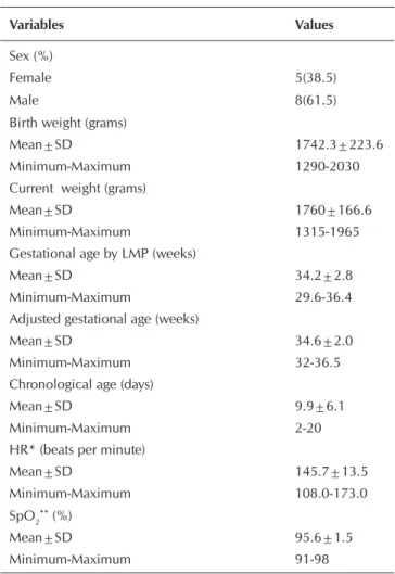
Due to time and resources available for the research, the non-probability sample consisted of 13 preterm infants. The prema-ture infants were predominantly male, classified as late preterm according to gestational age by LMP, and adjusted, with a mean chronological age of 9.9 days, low birth and current weight, and
mean of HR and SpO2 during the 24 hours of 145.7 beats per minute (bpm) and 95.6%, respectively (Table 1).
Regarding the analysis of the 1440 minutes of recordings, Table 2 show the mean values of total sleep time, wakeful-ness, and the duration of the different sleep patterns, heart rate and SpO2 of the investigated newborns.
The preterm infants remained longest in QS, followed by the AS and IS. Regarding the proportion of total sleep time of the 24 hours analyzed, this value represents 14.3 (59.6%) hours, of the obtained recordings.
In order to evaluate whether the care routines at different pe-riods of the day had influence on sleep and physiological param-eters of premature infants, Table 3 shows the median and inter-quartile ranges of total sleep time, sleep patterns and wakefulness, HR and SpO2 in the morning, afternoon, night I and II periods.
Table 3 shows no statistically significant difference between the four periods of the day and the total sleep time, the different patterns, and the wakefulness, as well as HR and SpO2. Howev-er, the highest median values of total sleep time of the premature infants occurred in the morning and night II periods. Regarding the different sleep patterns exhibited by the premature infants, the major median total time of AS was presented during the
Table 1 - Characteristics of preterm infants, São Paulo, São Paulo, Brazil, 2013
Variables Values
Sex (%)
Female 5(38.5)
Male 8(61.5)
Birth weight (grams)
Mean±SD 1742.3±223.6
Minimum-Maximum 1290-2030 Current weight (grams)
Mean±SD 1760±166.6
Minimum-Maximum 1315-1965 Gestational age by LMP (weeks)
Mean±SD 34.2±2.8
Minimum-Maximum 29.6-36.4 Adjusted gestational age (weeks)
Mean±SD 34.6±2.0
Minimum-Maximum 32-36.5 Chronological age (days)
Mean±SD 9.9±6.1
Minimum-Maximum 2-20
HR* (beats per minute)
Mean±SD 145.7±13.5
Minimum-Maximum 108.0-173.0 SpO2**(%)
Mean±SD 95.6±1.5
Minimum-Maximum 91-98
Notes:*HR – Heart rate; **SpO
afternoon and night I periods. The IS pattern was higher during night I, and QS prevailed during the morning shift.
Table 4 shows the results of the supplementary analysis of the sleep patterns of the preterm infants in different periods of the day.
During the morning shift, a predominance of QS in preterm infants was observed, and a statistically significant difference was identified when comparing to other patterns. In relation to the other periods of the day, there was no difference in the three sleep patterns of the newborns studied.
Relating the physiological parameters studied to the sleep and wakefulness of the premature infants, a significant differ-ence in the HR mean (p <0.001) was identified. The highest mean occurred during wakefulness (155.44 ± 13.1), compared to sleep (142.7 ± 14.3). A differences was also identified when comparing the HR with the sleep of preterm infants in the four periods of the day (p = 0.012), being the highest median value found during the night II (148.63). No statistically significant dif-ference with respect to SpO2 (p = 0.247) was found.
Table 2 - Mean values of premature newborns for total sleep time, patterns of sleep and wakefulness, in minutes, heart rate and oxygen saturation, São Paulo, São Paulo, Brazil, 2013
Variables Mean±SD Minimum-Maximum
Total sleep time 860.7±131.1 730.5-1149.5
Total AS+time 275.6±83.9 129.5-436
Total QS++ time 359.0±167.0 222.0-835.5
Total IS+++ time 222.0±79.4 51.0-293.5
Total wakefulness time 577.1±129.8 290.0-709.5
Notes: +SA – Active sleep; ++QS – Quiet sleep; +++IS – Indeterminate sleep.
Table 3 - Comparison of the medians of total sleep time, total time of active sleep, quiet and indeterminate sleep and wakeful-ness, in minutes, heart rate and oxygen saturation presented by premature infants, according to the different periods of the day, São Paulo, São Paulo, Brazil, 2013
Variables Morning
Median (II)
Afternoon Median (II)
Night I Median (II)
Night II Median (II)
Friedman P value
Total sleep time 227.0 (59.3) 211.0 (68.5) 202.0 (79.0) 216.0 (86.8) 0.748
Total AS# time 55.5 (47.5) 70.5 (28.0) 71.0 (50.3) 65.5 (57.5) 0.801
Total QS## time 83.5 (50.3) 74.5 (58.3) 63.5 (35.8) 70.5 (69.0) 0.070
Total IS### time 59.0 (35.8) 57.0 (22.3) 66.5 (19.3) 51.5 (43.8) 0.595
Total wakefulness time 133.0 (59.3) 149.0 (68.5) 143.5 (87.5) 144.0 (86.8) 0.845
HR#### (bpm) 148.9 (14.56) 147.03 (19.0) 144.1 (15.69) 147.85 (16.95) 0.338
SpO2##### (%) 96.5 (3.0) 96.0 (1.88) 96.0 (2.50) 96.0 (1.75) 0.992
Notes: II – Interquartil range; #AS – Active Sleep; ##QS – Quiet Sleep; ###IS – Indeterminate sleep; ####HR – Heart Rate; #####SpO
2 – Peripheral capillary oxygen
saturation.
Table 4 - Comparison of median total time of sleeping patterns presented by premature infants in minutes, according to periods of day, São Paulo, São Paulo, Brazil, 2013
Variables Total AS§time
Median (II)
Total QS§§ time
Median (II)
Total IS§§§ time
Median (II)
Friedman P value
Morning 55.5 (47.5) 83.5 (50.3) 59.0 (35.8) 0.002
Afternoon 70.5 (28.0) 74.5 (58.2) 57.0 (22.2) 0.412
Night I 71.0 (50.2) 63.5 (35.7) 66.5 (19.2) 0.165
Night II 65.5 (57.50) 70.5 (69.0) 51.5 (43.7) 1.000
DISCUSSION
The results show that premature newborns had a sleep time less than that described in the literature. One study indicated that preterm infants need 22 hours of sleep/day, while anoth-er research study suggested a mean of 17 hours panoth-er day(2,15).
These findings becomes alarming, as the recordings were made for 24 uninterrupted hours, and sleep is characterized as the predominant behavioral state of premature newborns and is required to meet their developmental demands.
Sleep deprivation in preterm infants can change the HR, in-crease pain perception and apneic events(4). Moreover, it can
trigger infectious processes, increasing metabolic rate and lead-ing to consequent weight loss; it can also cause stress, irritability, crying, with consequent increased intracranial pressure, bleed-ing and delay of discharge(10,16). The relationship of long-term
sleep deprivation with psychomotor, cognitive and personality development is not clearly understood in human beings(17).
The absence of routines in the NICU that promote individu-alized care of newborns may have contributed, in part, to the sleep deprivation of premature newborns; a high level of noise and excessive numbers of professionals and students around the incubators was identified, including several manipulations of the newborns by professionals, families and students for cares, performing procedures and also to interact with the child.
Research that aimed to evaluate the sleep of hospitalized newborns demonstrated that, by implementing some care ac-tions with a focus on developmental care, such as reducing the noise level, protecting the newborn from direct lighting, and promotion of twilight periods, showed that newborns slept longer(18-19). Thus, it is imperative to rethink the
philoso-phy of care and actual routines in order to promote sleep, and therefore protect the newborn, through actions based on care protocols involving continuing education for the multidisci-plinary team.
Quiet sleep was the predominant pattern in the investi-gated period. During QS, the basal metabolism that favors the replenishment of energy reserves decreases, preparing the body for AS, and also for awakening, and promotion of cell repair(6,20). The prevalence of QS in this study may be
re-lated to the maturation of premature infants evaluated, as the mean gestational age was 34.2 weeks, which is considered late prematurity.
In general, studies indicate that the premature newborn should sleep about 80% of the total time in AS, decreasing as his gestational age increases; during this sleep pattern, brain ac-tivation occurs, which is essential for the structural maturation of the CNS(2,6,19,21). There are numerous cellular activities in the
brain during AS, which favor the development of the sensorineu-ral system, the process of learning, memory and preservation of brain plasticity(21). In this sense, the literature states the
im-portance of nursing care in terms of negative or positive effects upon the states of sleep and wakefulness of the newborn, which may favor the more rapid decline of the AS in neonates(22).
Although the researchers recognize the different health care demands of morning, afternoon, night I and II periods of the unit where the study was conducted, the results indicate
little difference between the median values of total sleep time of the premature infant when the four periods of the day were compared. Thus, this result does not discard the necessity of the multidisciplinary team to focus its attention on sleeping protection of newborns hospitalized in the neonatal unit, based on their behavioral states, favoring sleep organization, as the premature newborns are in critical periods of develop-ment, particularly cerebral(22).
The analysis of sleep and wakefulness of preterm infants demonstrated an influence on HR. Our findings confirm those of other studies that demonstrated the influence of sleep on cardiorespiratory parameters(8-9,23). The HR showed a
signifi-cant decrease in sleep as compared to the wakeful state. Thus, this result is expected, since during periods of wakefulness most spontaneous activity of the newborns was verified, as well as episodes of crying related to manipulation.
Significant differences were also observed in HR in pre-term newborns during sleep in the four periods analyzed. This result indicates the need for further studies, as a lack of infor-mation is observed in the literature and may be related to the types of care for newborns on different shifts.
In this study, no significant difference was identified be-tween the SpO2 readings of the premature infants in rela-tion to sleep and wakefulness; during sleep, however, it was observed that this reading was higher during sleep. Studies indicate an elevation of SpO2 in QS as compared to AS, dem-onstrating that with increased brain activity there is a greater oxygen consumption(8,24). These results can be explained by
the anatomical and physiological structure of premature in-fants.The acinar region of the lungs of these newborns is not fully developed, and depending upon their gestational age, the amount of surfactant and true alveoli responsible for gas exchange are insufficient. These aspects trigger areas of resid-ual atelectasis, resulting in a ventilation-perfusion imbalance during periods of increased activity in the premature infants. Thus, changes in tissue oxygenation of preterm infants can be identified through pulse oximetry(8).
The fact that the variables of sleep and wakefulness were not jointly evaluated along with those related to the NICU environ-ment and the handling of the infants can be considered limita-tions of the study. The sample size can be considered another limitation; however, the records were analyzed during 24 unin-terrupted hours, which reveals an unprecedented characteristic of research regarding the evaluation of sleep and wakefulness of hospitalized preterm infants in the neonatal unit.
CONCLUSION
REFERENCES
1. Zomignani AP, Zambelli HJL, Antonio MARGM. [Cere-bral development in preterm newborn infants]. Rev Paul Pediatr [Internet]. 2009[cited 2015 Jun 19];27(2):198-203. Available from: http://www.scielo.br/pdf/rpp/v27n2/13. pdf Portuguese.
2. Calciolari G, Montirosso R. The sleep protection in the preterm infants. The Journal of Maternal-Fetal and Neona-tal Medicine [Internet]. 2011[cited 2015 Jun 19];24(1):12-4. Available from: http://www.tandfonline.com/doi/abs/1 0.3109/14767058.2011.607563?journalCode=ijmf20#. VeS1lZeurfc
3. Hu W, Kotagal S. Sleep in the pediatric intensive care unit. Sleep Medicine Reviews [Internet]. 2014[cited 2015 Jun 19];18:101-2. Available from: http://www.sciencedirect. com/science/article/pii/S1087079213001111
4. Bertelle VMD, Sevestre AMD, Laou-Hap KRN, Nagahap-itiye MCHN, Sizun JMD. Sleep in the neonatal intensive care unit. J Perinat Neonat Nurs [Internet]. 2007[cited 2015 Jun 19];21(2):140–8. Available from: http://www. ncbi.nlm.nih.gov/pubmed/17505234
5. Fernandes RMF. The Normal Sleep. Medicina (Ribeirão Pre-to) [Internet]. 2006[cited 2015 Jun 19];39 (2):157-168 Avail-able from: http://revista.fmrp.usp.br/2006/vol39n2/1_o_so no_normal1.pdf
6. Graven, S. Sleep and Brain Development. Clin Perinatol [Internet]. 2006[cited 2015 Jun 19];33:693-706. Available from: http://www.sciencedirect.com/science/article/pii/ S0095510806000546
7. Sukumaran TU. Pediatric Sleep Project. Indian Pediatrics [Inter-net]. 2011[cited 2015 Jun 19];48(11): 843-4. Available from: http://www.indianpediatrics.net/nov2011/nov-843-844.htm
8. Elder DE, Campbell AJ, Larsen PD, Galletly D. Respira-tory variability in preterm and term infants: Effect of sleep state, position and age. Respiratory Physiology & Neuro-biology [Internet]. 2011[cited 2015 Jun 19];175(2);234–8. Available from: http://www.sciencedirect.com/science/ article/pii/S1569904810004258
9. Witcombe NB, Yiallourou SR, Walker AM, Horne RSC. Blood Pressure and Heart Rate Patterns During Sleep Are Al-tered in Preterm-Born Infants: Implications for Sudden Infant Death Syndrome. Pediatrics [Internet].2008[cited 2015 Jun 19];122(6);1242-8. Available from: http://www.researchgate. net/publication/23560157_Blood_Pressure_and_Heart_Ra te_Patterns_During_Sleep_Are_Altered_in_Preterm-Born_Inf ants_Implications_for_Sudden_Infant_Death_Syndrome
10. Sousa MM, Silveira BL, Machado LCS, Santana MCCP, Flores NGC. Heart rate variability in high-risk newborns In the presence of noise. Rev CEFAC [Internet]. 2014[cit-ed 2015 Jun 19];16(1):99-104. Available from: http:// www.scielo.br/pdf/rcefac/v16n1/en_1982-0216-rcef ac-16-1-0099.pdf
11. Sousa MWCR, Silva WCR, Araújo SAN. Quantification of manipulations in neonatal intensive care unit: proposal of protocol elaboration. ConScientiae Saúde [Internet].
2008[cited 2015 Jun 19];7(2):269-74. Available from: http://www.uninove.br/pdfs/publicacoes/conscientiae_ saude/csaude_v7n2/cnsv7n2_3o.pdf
12. Antunes HKM, Andersen ML, Tufik S, Mello MT. [Sleep deprivation and exercise]. Rev Bras Med Esporte [Inter-net]. 2008[cited 2015 Jun 19];14(1):51-56. Available from: http://www.scielo.br/pdf/rbme/v14n1/a10v14n1. pdf Portuguese.
13. Das S, Mindell J, Millet GC, Ofner D, Beck SE, Mason TBA, et al. Pediatric Polysomnography: the patient and family perspective. J Clin Sleep [Internet]. Med 2011[cited 2015 Jun 19];7(1):81-7. Available from: http://www.ncbi. nlm.nih.gov/pmc/articles/PMC3041616/pdf/jcsm.7.1.81. pdf
14. International Classification of Sleep Disorders: Diagnostic and Coding Manual. 2. Westchester: American Academy of Sleep Medicine; 2005.
15. Nunes ML. [Sleep disorders].J Pediatr [Internet]. 2002[cit-ed 2015 Jun 19];78(1):S63-72. Available from: http:// www.scielo.br/pdf/jped/v78s1/v78n7a10.pdf Portuguese.
16. Gaíva MAM, Marquesi MC, Rosa MKO. Sleeping pattern of a newborn hospitalized in the intensive care unit: nurs-ing care. Ciênc Cuid Saúde [Internet]. 2010[cited 2015 Jun 19];9(3):602-9. Available from: http://pesquisa.bvs.br/ brasil/resource/pt/lil-655761
17. Ednick M, Cohen AP, McPhail GL, Beebe D, Simakajorn-boon N, Amin RS. A Review of the Effects of Sleep Dur-ing the First Year of Life on Cognitive, Psychomotor, and Temperament Development. Sleep [Internet]. 2009[cited 2015 Jun 19];32(11):1449-58. Available from: http:// www.ncbi.nlm.nih.gov/pmc/articles/PMC2768951/pdf/ aasm.32.11.1449.pdf
18. Shahheidari M, Homer C. Impact of the Design of Neo-natal Intensive Care Units on Neonates, Staff, and Fami-lies. J Perinat Neonat Nurs [Internet]. 2012[cited 2015 Jun 19];26(3):260-6. Available from: http://www.ncbi.nlm. nih.gov/pubmed/22843008
19. Vandenberg KA. Individualized developmental care for high risk newborns in the NICU: a practice guideline. Early Human Development [Internet]. 2007[cited 2015 Jun 19];83(7):433-42. Available from: http://www.sciencedire ct.com/science/article/pii/S0378378207000515
20. Kuhn P, Zores C, Langlet C, Escande B, Astruc D, Dufour A. Moderate acoustic changes can disrupt the sleep of very preterm infants in their incubators. Acta Pædiatrica [Inter-net]. 2013[cited 2015 Jun 19];102:949-54. Available from: http://onlinelibrary.wiley.com/doi/10.1111/apa.12330/pdf
on_to_Promote_Sleep_in_Premature_Infants_A_Pilot_Study
22. Allen KA. Promoting and protecting infant sleep. Adv Neo-natal Care [Internet]. 2012[cited 2015 Jun 19];12(5):288– 91. Available from: http://www.ncbi.nlm.nih.gov/pmc/ articles/PMC3439810/pdf/nihms-395479.pdf
23. Yiallourou SR, Sands SA, Walker AM, Horne RSC. Matu-ration of heart rate and blood pressure variability during sleep in term-born infants. Sleep [Internet]. 2012[cited
2015 Jun 19];35(2):177-86. Available from: http:// www.ncbi.nlm.nih.gov/pmc/articles/PMC3250356/pdf/ aasm.35.2.177.pdf