* Corresponding author.
E-mail: [email protected] (J.M. Budel).
0102-695X/$ - see front matter © 2014 Sociedade Brasileira de Farmacognosia. Published by Elsevier Editora Ltda. All rights reserved. http://dx.doi.org/10.1016/j.bjp.2014.10.002
Original article
Pharmacobotanical study of the leaf and stem of Mikania
lanuginosa for its quality control
Mariana Amorin
a, Josiane P. de Paula
b, Rosi Z. da Silva
b, Paulo V. Farago
b, Jane M. Budel
b,*
aPrograma de Pós-graduação em Ciências Farmacêuticas, Universidade Federal do Paraná, Curitiba, PR, Brazil bDepartamento de Ciências Farmacêuticas, Universidade Estadual de Ponta Grossa, Ponta Grossa, PR, Brazil
A R T I C L E I N F O
Article history: Received 2 June 2014 Accepted 7 October 2014
Keywords: Anatomy Cipó-cabeludo Herbal drugs Tampering
A B S T R A C T
Mikania lanuginosa DC, Asteraceae, is popularly known as "cipó-cabeludo" in Brazil due to a remarkable number of trichomes on its leaves and stems. It shows antimicrobial activity against Staphylococcus aureus, S. epidermidis and Bacillus cereus. This species can be confused with M. microlepis Baker and M. hirsutissima DC for substitution and tampering purposes. The aim of this study was to investigate the morpho-anatomy of leaf and stem of M. lanuginosa to obtain pharmacobotanical data that may contribute to its identification and taxonom-ic definition from other species of Mikania. The leaves and stems were investigated using scanning electron microscopy and light microscopy techniques. Mikania lanuginosa shows a uniseriate epidermis covered by a thin and smooth cuticle. The epidermal cells present sinuous anticlinal walls on both sides and anomocytic stomata were observed. A few glan-dular trichomes and numerous non-glanglan-dular trichomes were identified on both surfaces. The mesophyll is dorsiventral, the midrib has a biconvex contour and the petiole shows a circular shape in a cross-section. The stem has a circular shape. These pharmacobotanical features described for M. lanuginosa support data for its identification and taxonomic delim-itation from other Mikania species, and are a contribution for the quality control of herbal drugs.
© 2014 Sociedade Brasileira de Farmacognosia. Published by Elsevier Editora Ltda. All rights reserved.
Introduction
The Asteraceae family presents numerous species used in popular medicine, many of which have been chemically and pharmacologically studied (Di Stasi and Lima, 2002). Mikania
Willd is one of the major genera of Asteraceae and belongs to the Eupatorieae tribe. It comprises 430 species spread across the warm tropical and subtropical regions of the American and Asian continents. In Brazil, approximately 198 species can be found from North to South, mainly in São Paulo, Rio de Janeiro,
Minas Gerais, and Rio Grande do Sul (King and Robinson 1987; Ritter and Miotto, 2005; The Plant List, 2014).
Many species of Mikania are known by the popular names “guaco”, as M. glomerata Spreng and M. laevigata Sch. Bip ex Baker, and “cipó-cabeludo” including M. hirsutissima DC and
532
Mariana Amorin et al. / Rev Bras Farmacogn 24(2014): 531-537(Muellas-Serrano et al., 2000; Paul et al., 2000; Gasparetto et al., 2010; Pérez-Amador et al., 2010; Czelusniak et al., 2012; Rios et al., 2014; Mourão et al., 2014).
Several compounds have been identified in Mikania such as coumarins, terpenoids, flavonoids, stigmasterol and a large number of glycosides. Among the sesquiterpenoids, sesquiterpene lactones have been isolated. In addition, various resins can be observed at high concentrations of diterpenes, mainly derived from primarane, labdane, and caurane skeletons (Vilegas et al., 1997; Aguinaldo et al., 2003; Bolina et al., 2009; Pérez-Amador et al., 2010; Rios et al., 2014). Mikania lanuginosa is a climbing plant that occurs in Southeast and South regions of Brazil, particularly in Minas Gerais, Rio de Janeiro, São Paulo, Paraná, and Santa Catarina. It is popularly known as "cipó-cabeludo" due to the large number of non-glandular trichomes that are observed in its stems and leaves. Thus, it is sometimes confused with M. microlepis Baker, known as "guaco-piloso" or "guaco-de-praia" and also M. hirsutissima, which is the official species listed as "cipó-cabeludo" in the Brazilian Pharmacopoeia (Farm. Bras., 1959). Considering their morphological similarities, these species are often used as substitutes or for tampering purposes (Oliveira et al., 1994; Rodrigues et al., 1996; Silva et al., 2002), which can compromise the therapeutic efficacy in comparison to exclusive use of the official species, or even pose as a health risk for the patient. In that sense, further morpho-anatomical studies involving M. lanuginosa
are required to provide additional information to avoid this reported problem.
A previous paper on this species showed that kaurenoic acid and other fractions obtained from M. lanuginosa have antimicrobial activity against Staphylococcus aureus, S. epidermidis, and Bacillus cereus (Silva et al., 2002). Considering this medicinal potential, a morpho-anatomical characterization can be a simple and fast tool to provide quality control.
Taking into consideration these previous reports, the aim of this work was to investigate the anatomical characteristics of the stem and leaf of M. lanuginosa, which can be used therapeutically. This paper was devoted to discuss the pharmacobotanical features of M. lanuginosa in order to identify this medicinal plant, as well as to compare it to other species of Mikania.
Materials and methods
Plant material
Aerial parts of M. lanuginosa DC, Asteraceae, were collected at the Campos Gerais region of Paraná (24° 18’ S and 49° 37’ W) in September, 2006. The plant material containing inflorescences was used to prepare a voucher specimen that was identified and stored at the Herbarium of the State University of Ponta Grossa under the number HUPG 10438.
Anatomical analyses
The leaves and stems of M. lanuginosa were obtained at 5 cm from the apex of the plant of at least five specimens, placed
in a solution of FAA 70 (Johansen, 1940), and stored in ethanol 70% (Berlyn and Miksche, 1976). Free-hand longitudinal and cross-sections were prepared. These materials were stained using Astra blue and basic fuchsin (Roeser, 1972), and toluidine blue (O'Brien et al., 1964) to produce semi-permanent slides. In order to confirm the presence of particular compounds, microchemical analyses were carried out using the following reagents: hydrochloric phloroglucin for lignin (Foster, 1949), Sudan III for lipophilic compounds (Sass, 1951), ferric chloride for staining phenolic compound (Johansen, 1940) and lugol to observe starch (Berlyn and Miksche, 1976). Photomicrographs were taken with the Olympus BX-40 light microscope equipped with a digital camera.
The scanning electron microscopy of the leaf and stem surface was performed in high vacuum and high accelerating voltage (15 kV) using a Jeol JSM-6360LV microscope. For this procedure, the samples were previously dehydrated using increasing amounts of ethanol and the critical point of CO2. Then, they were submitted to metallization with gold (Souza, 1998). This procedure was carried out at the Electron Microscopy Center of the Federal University of Paraná (UFPR).
Results
Leaf
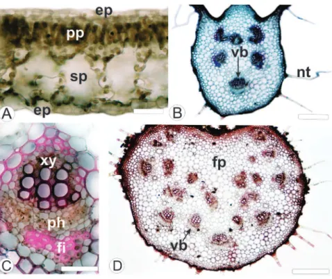
The leaf of Mikania lanuginosa DC, Asteraceae (Fig. 1A), shows epidermal cells with sinuous anticlinal walls (Fig. 1B) on both sides. The leaf has anomocytic stomata predominantly on the abaxial side (Fig. 1C). In the cross-sections it was identified a uniseriate epidermis with smooth and thin cuticle. It also presents numerous non-glandular trichomes (Fig. 1D, E, F) and a few numbers of glandular ones (Fig. 1E, F).
The non-glandular trichomes are either short or long and they are uniseriate and formed of five cells, with an acute apical cell (Fig. 1D-F, 2A-D). The glandular trichomes are capitate, multicellular, uniseriate, and composed of 8 cells (Fig. 2E, F). They can have curved appearance (Fig. 2F) and sometimes stuck cells can occur (Fig. 2E).
The mesophyll is dorsiventral and is formed by 1-2 layers of palisade parenchyma and four layers of spongy parenchyma (Fig. 3A). Small collateral vascular bundles are observed immersed into the mesophyll, associated with secretory ducts. The midrib is biconvex; however the convexity is more conspicuous on the abaxial surface. It presents three layers of angular collenchyma underlying the coating system on the adaxial side and 2 strata on the abaxial surface. Six vascular bundles are disposed in an open arc (Fig. 3B) and these structures show a collateral arrangement (Fig. 3C). The secretory ducts are located near vascular bundles.
Figure 1 – Leaves of Mikania lanuginosa DC, Asteraceae. A. M. lanuginosa in habit. B. Adaxial side of epidermis in surface view. C. Abaxial side of epidermis in surface view, showing stomata (SEM - Scanning Electron Microscopy). D. Abaxial side of epidermis, presenting trichomes (SEM). E. Adaxial surface, midrib region, showing glandular and non-glandular trichomes (SEM). F. Abaxial surface exhibiting glandular and non-glandular trichomes near the border. st: stomata; gt: glandular trichome; nt: non-glandular trichome. Bar = 5 cm (A); 20 µm (B, C); 50 µm (E, F); 10 µm (D).
534
Mariana Amorin et al. / Rev Bras Farmacogn 24(2014): 531-537Figure 3 – Leaf of Mikania lanuginosa DC, Asteraceae, in cross-section. A. Dorsiventral mesophyll. B. Midrib showing collateral vascular bundles. C. Detail of the collateral vascular bundle. D. Petiole showing collateral vascular bundles. ep: epidermis; fi: fiber; fp: ground parenchyma; nt: non-glandular trichome; ph: phloem; pp: palisade parenchyma; sp: spongy parenchyma; vb: vascular bundle; xy: xylem. Bar = 20 µm (A); 50 µm (C); 100 µm (B, D).
Discussion
The leaf of Mikania lanuginosa DC, Asteraceae, demonstrates epidermal cells covered by a thin and smooth cuticle. According to Oliveira et al. (1994), a smooth cuticle is usual in
Mikania species. However, M. congesta DC showed an epidermis with a striated cuticle. In addition, the investigated taxon has epidermal cells with sinuous anticlinal walls in surface view. Epidermal cells with sinuous anticlinal walls were also found in several species of Mikania (Oliveira et al., 1999; 2000; Oliveira and Akisue, 2005; Milan et al., 2006; Budel et al., 2009; Gasparetto et al., 2010). Nevertheless, epidermal cells with polygonal shape have been reported for M. hookeriana DC and
M. confertissima Schultz Bip ex Baker (Oliveira et al., 1994). Therefore, M. lanuginosa presents a similar epidermal pattern to its genus.
Trichomes are epidermal structures used for the characterization of drugs composed by leaves, their fragments and related powders (Glória and Guerreiro, 2003). Curved multicellular uniseriate glandular trichomes are typically verified in Mikania (Oliveira et al., 2000), as previously reported in M. conferta (Oliveira et al., 1999), M. glomerata (Neves and Sa, 1991, Oliveira et al., 1994; Gasparetto et al., 2010), M. hirsutissima
(Oliveira and Akisue, 2005), M. malacolepsis B. L. Rob. (Rodrigues et al., 1996), M. microlepis, M. hookeriana, M. hatschbachii G. Barroso (Oliveira et al., 1994), M. cordifolia (Oliveira et al., 2000),
Stem
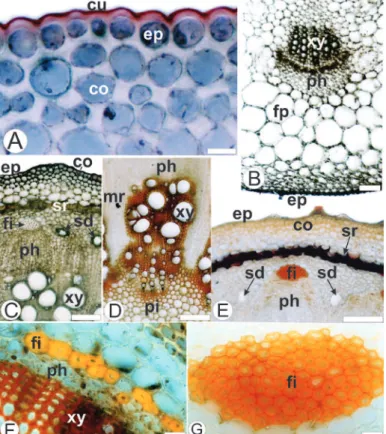
In the transection, the stem presents a circular shape. The epidermis appears in a single series with thickened cuticle (Fig. 4A, B). Non-glandular trichomes are observed. The angular collenchyma is adjacent to the epidermis. The sclerenchyma formed by thickened cells containing lignin leads to a sclerenchymatous ring (Fig. 4A-C).
The endodermis is formed by one layer of cells (Fig. 4C). The vascular cylinder shows collateral bundles (Fig. 4A, C) and layers of cambium (Fig. 4C, F). Secretory ducts appear close to the phloematic system and between the vascular bundles (Fig. 4D). Perivascular fibers caps adjoin to the phloem are observed (Fig. 4A, C). The pith is composed by relatively small parenchymatic cells with thin walls. Medullary rays are also observed (Fig. 4A, E).
Microchemical analyses
Figure 4 – Stem of Mikania lanuginosa DC, Asteraceae, in cross-section. A. Partial aspect of the stem. B. Detail of the cells of the sclerenchymatous ring. C. Detail of Figure 3A. D. Detail of a secretory duct. E. Detail of xylem and medullary ray in cross-section. F. Detail of the vessel elements. ca: cambium; en: endodermis; ep: epidermis; co: collenchyma; fi: fiber; mr: medullary ray; ph: phloem; pi: pith, sd: secretory duct; sr: sclerenchymatous ring; ve: vessel element; xy: xylem. Bar = 20 µm (D); 50 µm (B, F); 100 µm (C, E); 200 µm (A).
536
Mariana Amorin et al. / Rev Bras Farmacogn 24(2014): 531-537and M. laevigata (Budel et al., 2009; Gasparetto et al., 2010). In addition, glandular trichomes with stuck cells are described for M. lanuginosa.
In contrast to the observations in the present work, multicellular biseriate glandular trichomes have been described for many species of Asteraceae (Budel and Duarte, 2008a; Souza et al., 2013; Youssef et al., 2013) including
Mikania: M. congesta DC, M. microlepis (Oliveira et al., 1994), M. glomerata (Neves and Sá, 1991; Milan et al., 2006; Gasparetto et al., 2010), and M. laevigata (Budel et al. 2009). Apple-shaped glandular trichomes with 1-3 cells on both epidermal sides were described by Oliveira et al. (1994) for M. smilacina DC. These particular structures are not observed in M. lanuginosa. Additionally, sessile glandular trichomes are common in the genus (Holmes, 1993). However, they are not encountered in this study.
Both long and short conical non-glandular trichomes, which are described for M. lanuginosa, were previously reported to other members of Asteraceae (Budel et al., 2006; Farago et al., 2006; Budel and Duarte, 2008b), including various representatives of Mikania (Oliveira et al., 1994; Rodrigues et al., 1996; Oliveira et al., 1999; 2000; Oliveira and Akisue, 2005). Uniseriate non-glandular trichomes composed of up to twenty cells were observed mainly on the abaxial surface of M. hookeriana DC. Additionally, M. confertissima differed from the general pattern by showing no trichomes on either of its leaf surfaces (Oliveira et al., 1994).
Anisocytic and anomocytic stomata predominate in Asteraceae (Metcalfe and Chalk, 1950) and were observed for numerous species of Mikania (Oliveira et al. 2000; Oliveira and Akisue, 2005; Milan et al., 2006; Budel et al., 2009; Gasparetto et al. 2010). For M. lanuginosa, exclusively anomocytic stomata are verified.
Mikania lanuginosa shows dorsiventral mesophyll and this arrangement is commonly reported in Mikania (Rodrigues et al., 1996; Oliveira et al., 1999; Oliveira et al., 2000; Oliveira and Akisue, 2005; Milan et al., 2006; Budel et al., 2009; Gasparetto et al., 2010).
The midrib of M. lanuginosa is biconvex with collateral vascular bundles, angular collenchyma, perivascular fiber caps, and secretory ducts. These features correspond with those reported for M. laevigata (Budel et al., 2009; Gasparetto et al., 2010), M. glomerata (Neves and Sá, 1991; Oliveira and Akisue, 2005; Milan et al., 2006), M. malacolepsis (Rodrigues et al., 1996), M. conferta (Oliveira et al., 1999), and M. cordifolia
(Oliveira et al., 2000).
The stem of M. lanuginosa shows a circular contour similar to M. glomerata (Neves and Sá, 1991) and M. laevigata (Budel et al., 2009). However, M. malacolepsis (Rodrigues et al., 1996) and M. cordifolia (Oliveira et al., 2000) show a hexagonal shape.
According to Oliveira et al. (2000), thickened stems of
Mikania revealed the presence of a sclerenchymatous ring in their cortical region, which is a usual feature of M. scandens (L.) Willd and M. hirsutissima (Oliveira and Akisue, 2005). In this study, M. lanuginosa also showed a sclerenchymatous ring. In addition, secretory ducts located near to the vascular bundles are also observed in M. lanuginosa. These particular structures were previously reported for Mikania spp. (Neves and Sá, 1991; Oliveira et al., 1999; Oliveira et al., 2000; Budel et al., 2009).
In summary, uniseriate epidermis covered by a thin and smooth cuticle, curved and capitate glandular trichomes, long and short uniseriate non-glandular trichomes with acute apical cell, biconvex midrib, rounded petiole with vascular bundles dispersed in the ground parenchyma, circular stem with angular collenchyma and sclerenchymatous ring can be used as relevant anatomical structures to distinguish
M. lanuginosa from other Mikania species. Several of these features can be also verified during powder analysis as anatomical markers, except for the contour of midrib, petiole, and stem.
Authors’ contributions
MA contributed in running the laboratory work. RZS contributed in collecting the plant material and its identification. JPP and PVF contributed in performing the scanning electron microscopy (SEM) analysis and providing a critical reading of the manuscript. JMB created the project, supervised the laboratory work, and wrote the paper. All the authors have read the final manuscript and approved the submission.
Conflicts of interest
The authors declare no conflicts of interest.
Acknowledgment
The authors thank the Electron Microscopy Center of the Universidade Federal do Paraná for providing SEM images and Araucaria Foundation for financial support.
R E F E R E N C E S
Aguinaldo, A.M., Padolina, W.G., Abe, F., Yamauchi, T., 2003. Flavonoids from Mikania cordata. Biochem. Syst. Ecol. 31, 665-668.
Berlyn, G.P., Miksche, J.P., 1976. Botanical microtechnique and cytochemistry. Ames: Iowa State University, p. 121. Bolina, R.C., Garcia, E.F., Duarte, M.G.R., 2009. Estudo
comparativo da composição química das espécies vegetais Mikania glomerata Sprengel e Mikania laevigata Schultz Bip. ex Baker. Rev. Bras. Farmacogn. 19, 294-298.
Budel, J.M., Duarte, M.R., 2008a. Estudo farmacobotânico de folha e caule de Baccharis uncinella DC., Asteraceae. Lat. Am. J. Pharm. 27, 740-746.
Budel, J.M., Duarte, M.R., 2008b. Estudo farmacobotânico de partes vegetativas aéreas de Baccharis anomala DC., Asteraceae. Rev. Bras. Farmacogn. 18, 761-768. Budel, J.M., Duarte, M.R., Farago, P.V., Takeda, I.J.M., 2006.
Caracteres anatômicos de folha e caule de Calea uniflora Less., Asteraceae. Rev. Bras. Farmacogn. 16, 53-60.
Czelusniak, K.E., Brocco, A., Pereira, D.F., Freitas, G.B.L., 2012. Farmacobotânica, fitoquímica e farmacologia do guaco: revisão considerando Mikania glomerata Sprengel e Mikania laevigata Sch. Bip. ex Baker. Rev. Bras. Plantas Med. 14, 400-409.
Di Stasi, L.C., Lima, C.A.H., 2002. Plantas medicinais na Amazônia e na Mata Atlântica. São Paulo: UNESP, p. 463-465.
Farago, P.V., Budel, J.M., Duarte, M.R, Jurgensen, I., Takeda, I.J.M., 2006. Anatomia da folha e do caule de Calea longifolia (Asteraceae). Acta Farm. Bonaerense 25, 512-517.
Farm. Bras., 1959. Farmacopéia dos Estados Unidos do Brasil 2nd ed. São Paulo: Indústria Gráfica Siqueira.
Foster, A.S. 1949. Practical plant anatomy. 2 ed. Princeton: D. Van Nostrand.
Gasparetto, J.C., Campos, F.R., Budel, J.M., Pontarolo, R., 2010. Mikania glomerata Spreng e M. laevigata Sch. Bip. ex Baker, Asteraceae: estudos agronômicos, genéticos, morfoanatômicos, químicos, farmacológicos, toxicológicos e uso nos programas de fitoterapia do Brasil. Rev. Bras. Farmacogn. 20, 627-640.
Glória, B.A., Guerreiro, S.M.C., 2003. Anatomia vegetal. Viçosa: UFV. Holmes, W.C., 1993. The genus Mikania (Compositae Eupatoriae)
in the Greater Antilles. Texas: Brit Press.
Johansen, D.A., 1940. Plant microtechnique. New York: McGraw Hill Book, p. 41, 193.
King, R.M., Robinson, H., 1987. The genera of the Eupatorieae (Asteraceae). St. Louis: Missouri Botanical Garden, v. 22. Metcalfe, C.R., Chalk, L., 1950. Anatomy of dicotyledons: leaves,
stem, and woods in relation to taxonomy with notes on economic uses. Oxford: Clarendon Press v. 2.
Milan, P., Hayashi, A.H., Appezzato-da-Glória, B., 2006. Comparative leaf morphology and anatomy of three Asteraceae species. Braz. Arc. Biol. Technol. 49, 135-144. Mourão, V.B., Giraldi, G.M., Neves, L.M., de Gaspi, F.O., Rodrigues,
R.A., Alves, A.A., Esquisatto, M.A., Mazzi, M.V., Mendonça, F.A., dos Santos, G.M., 2014. Anti-hemorrhagic effect of hydro-alcoholic extract of the leaves of Mikania glomerata in lesions induced by Bothrops jararaca venom in rats. Acta Cir. Bras. 29, 30-37.
Muellas-Serrano, S., Nogal, J.J., Martinez-Diaz, R.A., Escario, J.Á., Martinez-Fernandez, A.R., Gómez-Barrior, A., 2000. In vitro screening of American plant extracts on Trypanosoma cruzi and Trichomonas vaginalis. J. Ethnopharmacol. 71, 101-107. Neves, L.J., Sá, M.F.A., 1991. Contribuição ao estudo de plantas
medicinais Mikania glomerata Spreng. Rev. Bras. Farm. 72, 42-47.
O'Brien, T.P., Feder, N., McCully, M.E., 1964. Polychromatic staining of plant cell walls by toluidine blue O. Protoplasma 59, 368-373.
Oliveira, F., Akisue, G., 2005. Fundamentos de farmacobotânica. 2 ed. São Paulo: Atheneu.
Oliveira, F., Rodrigues, R.F.O., Bastos, D.H.M., Pereira, F.H. 2000. Caracterização morfohistológica e verificação da atividade microbiológica da espécie vegetal Mikania cordifolia (Lf) Willd. Lecta 18, 33-63.
Oliveira. F., Rosa, F.O.R., Edna, T.M.K., 1999. Estudo
farmacognóstico da almécega-da-praia – Mikaniaconferta Gardn. Lecta 17, 43-68.
Oliveira, F., Saito, M.L., Garcia, L.O., 1994. Morfologia externa das partes aéreas e anatomia foliar das espécies brasileiras de Mikania Willdenow secção Globosae Robinson – visão farmacognóstica. Lecta 12, 23-65.
Paul, R.K., Jabbar, A., Rashid, M.A., 2000. Antiulceractivity of Mikania cordata. Fitoterapia 71, 701-703.
Pérez-Amador, M.C., Ocotero, V.M., Balcazar, R.I., Jiménez, F.G., 2010. Phytochemical and pharmacological studies on Mikania micrantha H.B.K. (Asteraceae). Int. J. Exp. Bot. 79, 77-80. Rios, E.V., León, A., Chavez, M.I., Torres, Y., Ramírez-Apan,
M.T., Toscano, R.A., Bravo-Monzón, Á.E., Espinosa-Garcia, F.J., Delgado, G., 2014. Sesquiterpene lactones from Mikania micrantha and Mikania cordifolia and their cytotoxic and anti-inflammatory evaluation. Fitoterapia 94, 155-163.
Ritter, M.R., Miotto, S.T.S., 2005. Taxonomia de Mikania Willd. (Asteraceae) no Rio Grande do Sul, Brasil. Hoehnea 32, 309-359.
Rodrigues, R.F.I.O., Oliveira, F., Kato, E.T.M., 1996. Morfodiagnose da droga conhecida como cipó-almécega – Mikania
malacolepsis Robinson. Rev. Farm. Bioquim. 32, 37-44. Roeser, K.R., 1972. Die Nadel der Schwarzkiefer-Massenprodukt
und Kunstwerk der Natur. Mikrokosmos 61, 33-36. Sass, J.E., 1951. Botanical microtechnique. 2. ed. Ames: Iowa State
College, p. 97.
Silva, R.Z., Rios, E.M., Silva, M.Z., Leal, L.F., Yunes, R.A., Miguel, O.G, Cechinel-Filho, V., 2002. Investigação fitoquímica e avaliação da atividade antibacteriana da Mikania lanuginosa DC. (Asteraceae). Visão Acad. 3, 59-64.
Souza, J.P., Santos, V.L.P., Franco, C.R.C., Bortolozo, E.A.F.Q., Farago, P.V., Matzenbacher, N.I., Budel, J.M., 2013. Baccharis rufescens Spreng. var. tenuifolia (DC.) Baker: contribuição ao estudo farmacognóstico. Rev. Bras. Plantas Med. 15, 566-574. Souza, W., 1998. Técnicas básicas de microscopia eletrônica aplicadas
às Ciências Biológicas. Rio de Janeiro: Sociedade Brasileira de Microscopia Eletrônica, p. 1-44.
The Plant List, 2014. Version 1.1. Published on the Internet; http://www.theplantlist.org/, accessed June 2014). Vilegas, J.H.Y., Marchi, E., Lanças, F.M. 1997. Determination of
coumarin and kaurenoic acid in Mikania glomerata (“Guaco”) leaves by capillary gas chromatography. Phytochem. Analysis 8, 74-77.