312 Rev Bras Hematol Hemoter. 2011;33(4):312-314
Rosai-Dorfman disease with spontaneous resolution: case report of a child
1
Universidade Federal do Ceará – UFC, Fortaleza, CE, Brazil 2
Pediatric Oncology Service, Hospital do Câncer do Ceará, Instituto do Câncer do Ceará – ICC, Fortaleza, CE, Brazil
3
Department of Pathology, Universidade Federal do Ceará – UFC, Fortaleza, CE, Brazil 4
Department of Surgery, Universidade Federal do Ceará – UFC, Fortaleza, CE, Brazil Felipe Barbosa Lima1
Pedro Samuel de Valões Barcelos1
Ana Paula Nunes Constâncio2
Cleto Dantas Nogueira3
Antônio Aldo Melo-Filho4
Rosai-Dorfman disease is a self-limiting condition caused by histiocyte proliferation within the sinusoids of lymph nodes and in extranodal tissue. It is a rare disease, particularly in children, that progresses with extensive lymphadenopathy. This paper reports on the case of a 2-year-old child with progressive cervical lymphadenopathy associated with persistent fever and radiological findings suggestive of lymphoma. Histopathological and immunohistochemistry studies of a lymph node biopsy established the diagnosis of Rosai-Dorfman disease. Both lymphadenopathy and fever resolved spontaneously.
Keywords: Histiocytosis, sinus; Histiocytosis, non-Langerhans-cell; Lymphatic diseases; Lymph nodes; Humans; Child
Introduction
Rosai-Dorfman disease (RDD) or sinus histiocytosis with massive lymphadenopathy is characterized by non-neoplastic proliferation of histiocytes/phagocytes in the sinusoids of lymph nodes and in extranodal tissues. Although rare in children, RDD can mimic malignant lymphoproliferative disorders. The objective of this paper was to report on a case of RDD in a 2-year-old child and review the literature.
Case report
A two-year-old girl presented with a 2-month history of progressive left cervical lymphadenomegaly without signs of inflammation and with fever over the preceding two weeks. The lab tests revealed a high blood sedimentation rate (BSR: 103 mm/h), neutrophilic leukocytosis (28 x109/L) and anemia (Hb = 8.6 g/dL). Serology (IgG and IgM)
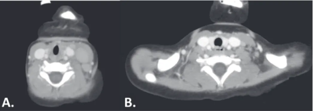
was negative for Epstein-Barr, cytomegalovirus, herpes type I and II, toxoplasmosis and Hepatitis B and C. Diffuse medullary hyperplasia was observed on the myelography, whereas cervical computed tomography (CT) demonstrated submandibular lymphadenomegaly on the right measuring 12 x 9 mm and on the left (Grade II, III, IV and V) measuring up to 25 x 16 mm, which was suggestive of lymphoma (Figure 1).
Conflict-of-interest disclosure: The authors declare no competing financial interest
Submitted: 11/20/2010 Accepted: 1/20/2011
Corresponding author:
Antônio Aldo Melo Filho
Department of Surgery, Universidade Federal do Ceará, UFC
Rua Professor Costa Mendes 1608, 3º andar Rodolfo Teófilo
60430-140 – Fortaleza, CE, Brazil Phone: 55 85 3366-8062 [email protected]
www.rbhh.org or www.scielo.br/rbhh
DOI: 10.5581/1516-8484.20110083
Case Report
Rev Bras Hematol Hemoter. 2011;33(4):312-314 313 revealed a lymph node with subverted architecture and
greatly dilated medullary sinuses due to an excess of mononuclear cells, blood cells and foam cells. In addition, histiocytes containing phagocytosed cells (emperipolesis) and few eosinophils were observed. The less affected parts of the lymph node displayed prominent reactive lymphoid follicles with medullary substance containing plasmocytes, mature lymphocytes and foamy cell aggregates (Figure 2A). The immunohistochemical examination was positive for PS100, CD68, CD45 and CD31 and negative for CD1a, thus compatible with RDD (Figure 2B). Staging CT of the chest evidenced pretracheal mediastinal lymphadenomegaly (prevascular, right hilar, subcarinal and aortopulmonary window) with lymph nodes of up to 29 x 19 mm. Abdominal and pelvic CT revealed enlarged retroperitoneal and mesenteric lymph nodes of up to 17 x 16 mm. A conservative approach (clinical observation) was adopted. Seven months later, CT scans confirmed clinical findings of significant reductions in lesion size (Figure 1B). A cervical spine CT showed carotid-jugular lymph nodes measuring up to 17 mm. Chest CT showed nodes measuring 19 mm (paratracheal) and 20 mm (left hilar). Abdominal CT evidenced enlarged paraaortic lymph nodes of up to 13mm (largest diameter). Twelve months after diagnosis, the patient is clinically well and is followed up in the outpatient clinic.
Discussion
RDD is a rare proliferative histiocytic disease with a benign course. The condition was first described in 1965 but only recognized as a clinical and pathological entity in 1969 through a publication by Rosai and Dorfman.(1) Found
worldwide and affecting individuals with an average age of 20.6 years, RDD is slightly more common among men (1.4:1) and is significantly more common among Caucasians and blacks than among Asians.(2) In children it is a rare cause of
rapidly progressive lymphadenopathy, which sometimes mimics malignancy.(3)
The disease typically presents with extensive cervical lymphadenomegaly, most often bilateral and painless (87%).
At first lymph nodes are mobile and discrete, but over time they become adherent and tend to develop into a large multinodular mass. The axillary (23.7%), inguinal (25.7%) and mediastinal (14.5%) regions may be affected, though not as severely as the cervical region.(4) Our patient presented
painless unilateral cervical lymphadenomegaly with compromised lymph nodes in the mediastinum (pretracheal, prevascular, aortopulmonary window, right hilar and subcarinal), retroperitoneum and mesentery. Extranodal tissue involvement is documented in 43% of RDD patients, especially of the skin, soft tissues, upper airway, bones, urogenital system, lower airway and oral cavity.(4-7) In addition,
RDD may be associated with other conditions, including other forms of histiocytosis.(8)
Up to 30% of RDD patients report fever,(9) frequently
associated with a high BSR and polyclonal hypergamma-globulinemia (up to 90% of cases), anemia and neutrophilic leukocytosis.(5) Our patient presented with fever (preceding
two weeks), a high BSR, neutrophilic leukocytosis (28 x109/L)
and anemia (Hb = 8.6 mg/dL).
The etiology, pathogenesis and natural history of RDD remain obscure. Some authors have suggested a role for human herpesvirus six (HHV-6) supported by reports of peculiar patterns of expression of HHV-6 antigens in abnormal histiocytes from RDD patients.(10) Other authors believe RDD
may be the consequence of an exacerbated response of the immune system to infection by the Epstein-Barr virus, cytomegalovirus, Brucella or Klebsiella.(11,12)
The diagnosis of RDD is based on the clinical history and confirmed by histopathological examination. Specimens may be obtained by open surgical biopsy or fine needle aspiration. The latter is by many considered a sensitive and reliable diagnostic method and has the advantage of being possible in the outpatient setting.(13) Excised lymph nodes
are often grayish with capsular fibrosis or pericapsular fibroadipose tissue. In general, the architecture of the lymph node is subverted and changes are observed in patients with long-standing lymphadenopathy. Usually the sinusoids of the lymph nodes are markedly expanded due to lymphatic stasis and display a mixed cell population, including Figure 2 - Histological sections of excised lymph node showing increased volume. A. Histiocyte containing numerous lymphocytes (emperipolesis) (hematoxylin and eosin at 400x magnification); B. Immunohistochemical staining for protein S100 expression (400x magnification)
314 Rev Bras Hematol Hemoter. 2011;33(4):312-314 lymphocytes, plasmocytes and histiocytes. The most
characteristic cells in the sinuses are histiocytes of accentuated phagocytic appearance. These cells are large and irregular with abundant eosinophilic and sometimes vacuolated cytoplasm, usually displaying a round or oval nucleus with well-defined and delicate membranes and a single prominent nucleolus. Mitosis is rarely observed. However, histiocytes with foamy cytoplasm may be predominant in the cellular environment. The most important histological finding in RDD is histiocytes with a variable number of phagocytosed cells, usually lymphocytes, plasmocytes or erythrocytes. Some cells, especially lymphocytes, remain viable inside the vacuoles, giving rise to a phenomenon known as lymphophagocytosis or emperipolesis, defined as the presence of intact lymphocytes inside other cells, in this case, histiocytes. The most useful marker of histiocytes in RDD is the expression of protein S100. Histiocytes may also express pan-macrophage antigens (CD68, HAM 56, CD14, CD64 and CD15), phagocytosis-related antigens (CD64 or the Fc receptor for IgG) and lysosomal activity (lysozyme and alpha-1-antitrypsin). In addition, histiocytes are negative for CD1a and contain no Birbeck granules.(5) The anatomopathological evaluation of
our patient revealed, among other things, emperipolesis, positivity for PS100 and CD68 and negativity for CD1a. The differential diagnosis of RDD includes histiocytosis of Langerhans cells, histiocytic sarcoma, lysosomal storage diseases (such as Gaucher's disease), classic Hodgkin's lymphoma, melanoma and metastatic carcinomas and infections caused by Histoplasma and mycobacteria involving the lymph node.(5)
Due to its low incidence, no standard treatment has yet been defined for RDD. However, since the condition is self-limiting, it is often unnecessary to intervene, except when the airways are obstructed or vital organs are compressed. Several forms of therapy have been described involving corticosteroids, chemotherapy combined with periwinkle alkaloids, anthracyclines, antimetabolics and alkylating agents, interferon, antibiotics, radiotherapy and partial or total surgical resection.(12) A review of the literature revealed that
50% of patients with RDD require no treatment and that 82% of untreated patients experience spontaneous and complete disease regression.(12) Some reports describe disease control
in children without therapy.(14,15) In this case, after careful
analysis of the biopsied specimen, a conservative approach was adopted with progressive reduction of the lymphadenomegaly.
The course of RDD is unpredictable. Episodes of remission and exacerbation may occur for several years. In approximately 70% of cases the disease is permanent but stable, 20% experience spontaneous and permanent remission and 10% suffer from progressive and generalized disease.(4)
References
1. Rosai J, Dorfman RF. Sinus histiocytosis with massive lymph-adenopathy: a newy recognized benign clinicopathological entity. Arch Pathol. 1969;87(1):63-70.
2. Brenn T, Calonje E, Granter SR, Leonard N, Grayson W, Fletcher CD, et al. Cutaneous Rosai-Dorfman disease is a distinct clinical entity. Am J Dermatopathol. 2002;24(5):385-91.
3. Duval M, Nguyen VH, Daniel SJ. Rosai-Dorfman disease: An uncommon cause of massive cervical adenopathy in a two-year-old female. Otolaryngol Head Neck Surg. 2009;140(2):274-5. 4. Foucar E, Rosai J, Dorfman RF. Sinus histiocytosis with massive
lymphadenopathy (Rosai-Dorfman disease): review of entity. Semin Diagn Pathol. 1990;7(1):19-73.
5. McClain KL, Natkunam Y, Swerdlow SH. Atypical cellular disorders. Hematology Am Soc Hematol Educ Program. 2004:283-66.
6. Landim FM, Rios HO, Costa CO, Feitosa RG, Rocha Filho FD, Costa AA. [Cutaneous Rosai-Dorfman disease]. An Bras Dermatol. 2009;84(3):275-8. Portuguese.
7. Zhang JT, Tian HJ, Lang SY, Wang XQ. Primary intracerebral Rosai-Dorfman disease. J Clin Neurosci. 2010;17:1286-88. 8. Sachdev R, Shyama J. Co-existent Langerhans cell histiocytosis
and Rosai-Dorfman disease: a diagnostic rarity. Cytopathology 2008;19(1):55-8.
9. Bist SS, Varshney S, Bisht M, Pathak VP, Kusum A, Gupta N. Rosai Dorfman syndrome - a rare clinical entity. Indian J. Otolaryngol. Head Neck Surg. 2007;59:184-6.
10. Luppi M, Barozzi P, Garber R, Maiorana A, Bonacorsi G, Artusi T, et al. Expression of human herpesvirus-6 antigens in benign and malignant lymphoproliferative diseases. Am J Pathol. 1998;153 (3):815-23.
11. Lu D, Estalilla OC, Manning JT Jr, Medeiros LJ. Sinus histiocytosis with massive lymphadenopathy and malignant lymphoma involving the same lymph node: a report of four cases and review of the literature. Mod Pathol. 2000;13(4):414-9.
12. Pulsoni A, Anghel G, Falcucci P, Matera R, Pescarmona E, Ribersani M, et al. Treatment of sinus histiocytosis with massive lymphadenopathy (Rosai-Dorfman Disease): report of a case and literature review. Am J Hematol. 2000;69(1):61-71.
13. Kumar B, Karki S, Paudyal P. Diagnosis of sinus histiocytosis with massive lymphadenopathy (Rosai-Dorfman Disease) by fine needle aspiration cytology. Diagn. Cytopathol. 2008;36(10):691-5. 14. Stones DK, Havenga C. Sinus histiocytosis with massive
lymphadenopathy. Arch Dis Child. 1992;67(4):521-3.
15. Dearth J, Hunter D, Kelly D, Crist W. Sinus histiocytosis with massive lymphadenopathy. CA Cancer J Clin. 1980;30(1):55-8.
xxx