C
C
ABSTRACT
RESUMO
ASSOCIATED DENTAL ANOMALIES: CASE REPORT
MÚLTIPLAS ANOMALIAS DENTÁRIAS ASSOCIADAS: CASO CLÍNICO
Daniela Gamba GARIB1, Nildiceli Leite Melo ZANELLA2, Sheldon PECK3
1- DDS, MSc, PhD, Associate Professor of Orthodontics, UNICID, São Paulo, Brazil; Research Fellow, Department of Developmental Biology, Harvard School of Dental Medicine, Boston, USA.
2- DDS. MSc, PhD, Department of Pedodontics, FOB/USP, São Paulo; Dentist of Health Municipal Secretary of Bauru, São Paulo, Brazil. 3- DDS, MScD, Associate Clinical Professor, Department of Developmental Biology, Harvard School of Dental Medicine, Boston, USA.
Corresponding address: R: Rio Branco no 19-18 - Cep.: 17014480 - Bauru - São Paulo - Brazil - E-mail: [email protected]
Received: June 9, 2005 - Modification: September 26, 2005 - Accepted: September 30, 2005
ertain human dental anomalies frequently occur together, supporting the accumulated evidence of the shared genetic control of dental developmental disturbances. The present study reports a rare and interesting case of a 12-year-old girl with an association of multiple dental abnormalities, including agenesis, tooth malposition and delayed development. The etiology and treatment planning are discussed with reference to the literature. The clinical implications of genetically controlled patterns of dental anomalies are important in the establishment of early diagnosis and appropriate orthodontic intervention.
Uniterms: Agenesis; Dental anomalies; Orthodontics.
ertas anomalias dentárias humanas freqüentemente ocorrem concomitantemente, contribuindo para validar as evidências do controle genético nos distúrbios de desenvolvimento. O presente estudo relata um caso raro e interessante de uma jovem de 12 anos de idade, com associação de anormalidades dentárias múltiplas, incluindo agenesia, ectopia e atraso no desenvolvimento dentário. A etiologia e tratamento planejados são discutidos com referência à literatura. As implicações clínicas do padrão de anomalias dentárias geneticamente controladas são importantes no estabelecimento do diagnóstico precoce e de adequada intervenção ortodôntica.
Unitermos: Agenesia; Anomalias dentárias, Ortodontia.
INTRODUCTION
Tooth agenesis is the most common developmental anomaly of the human dentition, occurring in 25% of the population1. The third molar (M3) represents the tooth most affected with agenesis2, 3, having a prevalence rate of 20.7%2. In contrast, permanent second molar (M2) agenesis is a rare occurrence, found in only 2 of 6,000 consecutive orthodontic patients (0.03%)4.
Excluding the third molars, the prevalence rate of tooth agenesis is reported as 4.3 to 7.8%4, 5. The mandibular second premolar (MnP2) is the tooth most often absent, with a relative frequency of 2.2 to 4.1%4, 5. In fact, the MnP2 is highly variable developmentally. Besides the high prevalence of agenesis, the MnP2 often shows significantly retarded development, especially when there is agenesis of other permanent teeth6. Despite the fact that the mean initial calcification age for MnP2 is 3 years (varying from 2y3m to
3y7m)7, its development can be suppressed until 6 years8, and some published reports show radiographic appearance of the MnP2 after the age of 9 and even at 13 years old9, 10. In addition, the MnP2 accounts for approximately 24% of all impacted teeth, excluding the third molars11. The most frequent malposition reported for the unerupted MnP2 is distoangular development, with a prevalence rate of 0.2% in dental clinic patients12. This malposition was found to be associated with agenesis of the contralateral MnP213.
Case report
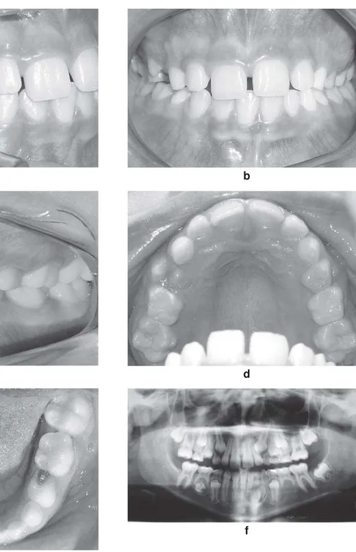
A girl aged 12.1 years was referred to orthodontic treatment by her pediatric dentist, due to retention of the mandibular left permanent lateral incisor (32). At diagnosis, the patient presented with a Class I facial pattern and Class I malocclusion in the late mixed dentition, with retention of the mandibular left deciduous lateral incisor and well-aligned arches. The mandibular dental midline was deviated to the left (Figure 1).
Initial panoramic dental radiograph (Figure 1f) showed a
cystic formation at the dental follicle of the unerupted 32, suggesting a dentigerous cyst, which besides leading to its retention seemed to negatively influence formation of the mandibular left permanent canine (33), whose eruption was delayed compared to the fully erupted contralateral canine. However, retention of the 32 was not the only irregularity observed. Other notable radiographic features were tooth agenesis of the mandibular left second premolar, distoangular position and delayed development of its unerupted antimere, agenesis of the mandibular right permanent second molar, and absence of all third molars.
FIGURE 1- (a-f) Initial intraoral photographs and panoramic radiograph
a b
c d
e
The M3 agenesis were confirmed using the later panoramic radiographs taken after age 14, because that appears to be a critical age for M3 formation23, 28. The dental age was delayed as observed in the majority of cases with dental agenesis6, 14.
Treatment was initiated by placement of a lingual arch, extraction of the mandibular left deciduous lateral incisor and canine, and surgical exposure followed by orthodontic traction of 32 with closed-eruption technique and cantilever springs. Extraction of the deciduous mandibular right second molar was also requested in an attempt to stimulate spontaneous correction of the eruption pathway of the mandibular right second premolar10, 29. The post-surgical histopathological report confirmed the initial assumption of dentigerous cyst on the mandibular left lateral incisor.
After 6 months of treatment, the mandibular left lateral incisor reached the oral cavity, and a pre-adjusted mandibular fixed partial appliance was then bonded for midline correction and simultaneous space recovery for 33, which was still able to erupt due to its incomplete root formation. Eighteen months after deciduous mandibular right second molar extraction, the succeeding permanent tooth still displayed the same magnitude of distoangular position and delayed development, and thus its orthodontic traction was performed. After 3 months of traction, the mandibular right second premolar was upright, and after 5 months it erupted in the oral cavity. In fact, a minor periodontal surgery was required at completion of orthodontic traction to expose the crown of that tooth, since it did not perforate the oral mucosa, but rather only stretched it. Meanwhile, the 33 erupted spontaneously. Thereafter, maxillary and mandibular fixed appliance was bonded, and corrective orthodontics was accomplished for further 1 year and 3 months. The deciduous mandibular left second molar was maintained up to spontaneous exfoliation, for later replacement by a dental implant.
The appliance was removed after 3 years and 2 months of therapy, (Figure 2). At this stage, there was still incomplete root formation of the mandibular right second premolar, and the permanent maxillary right second molar (17) was unerupted (Figure 2f). A retainer was designed to avoid extrusion of the 17 at completion of eruption, and this retainer will be kept until completion of growth, when a dental implant will be placed at mandibular right second molar area.
DISCUSSION
Besides dentigerous cyst causing retention of 32 and delayed development of the neighboring tooth, 33, this interesting case displays the concomitant occurrence of five remarkable dental anomalies: agenesis of the third molars, agenesis of the mandibular right permanent second molar, agenesis of the mandibular left second premolars, and distoangular position and delayed development of the mandibular right second premolar. A considerable amount of evidence exists suggesting that genes play a fundamental role in the etiology of many dental anomalies of clinical
significance. It has also been inferred that a common genetic defect may give rise to different phenotypic manifestations, including tooth agenesis, delayed development and ectopia10, 13, 14, 22, 23, 25, 30-32, which were observed in the present case report, along with microdontia. Published evidence also suggested that, when a third molar or second premolar is absent, agenesis of the remaining teeth is more likely to occur24, 28.
Tooth agenesis clearly has a genetic basis. Grahnen16 conducted a study in children with tooth agenesis and found that up to half of their siblings and parents also had tooth agenesis, a high prevalence when compared to the expected population rate. A twin study interestingly found a high concordance rate for tooth agenesis in monozygotic twins, while all dizygotic twin pairs were discordant18. These studies suggested that the mode of transmission could be explained by a single autosomal dominant gene with incomplete penetrance. Recently, Vastardis, et al.26, analyzing a large family with agenesis of all second premolars and third molars, identified a mutation in the MSX1 gene on chromosome 4. It is further suggested that delayed eruption, as well as microdontia, constitute a partial expressivity of the same gene leading to tooth agenesis15, 33.
In a sample with unilateral MnP2 agenesis, Shalish, et al.13 observed that the contralateral MnP2 tooth bud presented a mean increase of 10º in the distoangular position compared to a control group, and thus this alteration in tooth positioning would constitute a different phenotype of the same genetic defect that led to tooth agenesis occurrence. When measured on the panoramic radiograph, the mean angle formed between the inferior mandibular border and the long axis of the unerupted MnP2 corresponded to 85.5º in the control group and 75.6º in the experimental group. A similar situation was observed in the present case report, yet more severe. The initial mandibular right second premolar angulation was 25º. On the basis of literature reports stating that MnP2 malposition usually self-corrects and the tooth erupts spontaneously in the dental arch10, 12, 13, 29, a more passive approach was initially adopted, with extraction of the preceding deciduous tooth. However, after a 6-month follow-up, there was an improvement of only 5º in the tooth bud angulation, and after one year and a half, the period expected for spontaneous correction of the ectopic eruption pathway29, the tooth bud displayed a mild distal movement, yet still exhibited an angulation of 31º. The remarkable distoangular malposition of the right MnP2 may have contributed to conservative approach failure, even though Collett29 has reported a good response to the same intervention in a case of a MnP2 with its long axis lying transversely.
deciduous second molar followed by further removal of the mesial portion would led to continuing space closure with no or minor collateral effects36, 37. However, the mesial drift of the mandibular first and second molar, considering the third molar absence, could leave de maxillary second molar with no antagonist. On the other hand, a longitudinal follow-up has demonstrated that, in cases with agenesis of premolars, the deciduous molars may be kept in the oral cavity for a long period of time38-40. Bjerklin and Bennet38 investigated subjects with agenesis of mandibular second premolars and retained mandibular second molars from 11 years of age until the third decade of life. During the
observation period, only 2 of the 59 primary teeth were exfoliated, and beyond the age of 20 years no teeth were lost. Besides, 45% of the deciduous molars showed no infra-occlusion and in the teeth affected, the infra-infra-occlusion increased by less than 1 to 1.4 mm until the age of 18 years. Complementing this study, Sletten, et al.40 evaluated longitudinally the retained mandibular deciduous molars in adults (from 36.1 + 12.9 until 48.5 + 13. 9 years of age). Of the 28 retained deciduous molars, 24 (86%) continued to function. Only 4 (14%) were lost at a mean age of 51 years because of caries or periodontal breakdown. Considering the results and the fact that the average shortening of all
FIGURE 2- (a-f) Final intraoral photographs and panoramic radiograph.
a b
c d
e
deciduous root lengths was negligible (0.16mm), the authors concluded that retention of healthy deciduous mandibular second molars is a viable treatment alternative.
The only negative aspect of mandibular deciduous second molar maintenance is the impossibility to finalize the treatment with a Class I molar relationship at the same side (Figure 2c). However, extraction of the deciduous tooth to allow the molar relationship adjustment from Class II to Class I might lead to significant alveolar bone loss until growth completion for implant placement41. After being informed on all these issues, the patient and her family decided to keep the deciduous second molar, and were aware of the future prosthetic replacement need in case the deciduous tooth was exfoliated. The one-year follow-up after the treatment demonstrated that the deciduous molar remained unchanged.
The clinical implications of genetically controlled patterns of dental anomalies are very important. Peck, et al.23found certain dental anomalies occurring frequently in anatomically specific groupings, patterned according to anterior and posterior orofacial genetic fields. Further, the early diagnosis of one dental abnormality may alert the professional to the possible development of others the same patient or other family members13, 33, sometimes facilitating appropriate orthodontic intervention.
REFERENCES
1- Bredy E, Erbring C, Hübenthal B. The incidence of hypodontia with the presence and absence of wisdom teeth. Dtsch Zahn Mund Kieferheilkd Zentralbl 1991;79:357-63.
2- Lynham A. Panoramic radiographic survey of hypodontia in Australian Defense Force recruits. Aust Dent J 1990;35:19-22.
3- Garn SM, Lewis AB. The relationship between third molar agenesis and reduction in tooth number. Angle Orthod. 1962;32(14-8).
4- Rose JS. A survey of congenitally missing teeth, excluding third molars, in 6000 orthodontic patients. Dent Pract Dent Rec. 1966;17(3):107-13.
5- Rolling S. Hypodontia of permanent teeth in Danish schoolchildren. Scand J Dent Res. 1980;88(5):365-9.
6- Garn S, Lewis A, Vicinus J. Third molar polymorphism and its significance to dental genetics. J Dent Res. 1963;42:1344-63.
7- Moorrees CF, Fanning EA, Hunt EE, Jr. Age Variation of Formation Stages for Ten Permanent Teeth. J Dent Res. 1963;42:1490-502.
8- Ravn JJ, Nielsen HG. A longitudinal radiographic study of the mineralization of 2nd premolars. Scand J Dent Res. 1977;85:232-6.
9- Coupland MA. Apparent hypodontia. Br Dent J. 1982;152(11):388.
10- Alexander-Abt J. Apparent hypodontia: a case of misdiagnosis. Am J Orthod Dentofacial Orthop 1999;116(3):321-3.
11- Thilander H, Thilander B, G P. Treatment of impacted teeth by surgical exposure: a survey study. Swed Dent J 1973;66:519-25.
12- Matteson SR, Kantor ML, Proffit WR. Extreme distal migration of the mandibular second bicuspid. A variant of eruption. Angle Orthod 1982;52(1):11-8.
13- Shalish M, Peck S, Wasserstein A, Peck L. Malposition of unerupted mandibular second premolar associated with agenesis of its antimere. Am J Orthod Dentofacial Orthop 2002;121(1):53-6.
14- Baba-Kawano S, Toyoshima Y, Regalado L, Sado B, Nakasima A. Relationship between congenitally missing lower third molars and late formation of tooth germs. Angle Orthod. 2002;72(2):112-7.
15- Garn SM, Lewis AB. The gradient and the pattern of crown-size reduction in simple hypodontia. Angle Orthod. 1970;40:51-8.
16- Grahnen H. Hypodontia in the permanent dentition. Odontol Revy 1956;7:1100.
17- Leonardi R, Peck S, Caltabiano M, Barbato E. Palatally displaced canine anomaly in monozygotic twins. Angle Orthod. 2003;73(4):466-70.
18- Markovic M. Hypodontia in twins. Swed Dent J. 1982 (Suppl);15:153-62.
19- Peck L, Peck S, Attia Y. Maxillary canine-first premolar transposition, associated dental anomalies and genetic basis. Angle Orthod. 1993;63(2):99-109; discussion 110.
20- Peck S, Peck L, Hirsh G. Mandibular lateral incisor-canine transposition in monozygotic twins. ASDC J Dent Child. 1997;64(6):409-13.
21- Peck S, Peck L, Kataja M. Prevalence of tooth agenesis and peg-shaped maxillary lateral incisor associated with palatally displaced canine (PDC) anomaly. Am J Orthod Dentofacial Orthop. 1996;110(4):441-3.
22- Peck S, Peck L, Kataja M. Mandibular lateral incisor-canine transposition, concomitant dental anomalies, and genetic control. Angle Orthod. 1998;68(5):455-66.
23- Peck S, Peck L, Kataja M. Concomitant occurrence of canine malposition and tooth agenesis: evidence of orofacial genetic fields. Am J Orthod Dentofacial Orthop. 2002;122(6):657-60.
24- Symons AL, Stritzel F, Stamation J. Anomalies associated with hypodontia of the permanent lateral incisor and second premolar. J Clin Pediatr Dent. 1993;17(2):109-11.
25- Symons AL, Taverne AA. A family case report: disturbances in tooth form and eruption of the second premolar. Aust Orthod J. 1996;14(3):168-71.
26- Vastardis H. The genetics of human tooth agenesis: new discoveries for understanding dental anomalies. Am J Orthod Dentofacial Orthop. 2000;117(6):650-6.
27- Weide van der Y, Prahl-Andersen B, Bosman F. Tooth formation in patients with oligodontia. Angle Orthod. 1993;63(1):31-7.
28- Garn SM, Lewis AB. The relationship between third molar agenesis and reduction in tooth number. Angle Orthod. 1962;32(1):14-18.
29- Collett AR. Conservative management of lower second premolar impaction. Aust Dent J. 2000;45(4):279-81.
30- Uner O, Yucel-Eroglu E, Karaca I. Delayed calcification and congenitally missing teeth. Case report. Aust Dent J. 1994;39(3):168-71.
31- Weide van der YS, Prahl-Andersen B, Bosman F. Tooth formation in patients with oligodontia. Angle Orthod. 1993;63(1):31-7.
33- Mossey PA. The heritability of malocclusion: part 2. The influence of genetics in malocclusion. Br J Orthod. 1999;26(3):195-203.
34- Janson G, Dainesi EA, Henriques JF, de Freitas MR, de Lima KJ. Class II subdivision treatment success rate with symmetric and asymmetric extraction protocols. Am J Orthod Dentofacial Orthop. 2003;124(3):257-64; quiz 339.
35- Fines CD, Rebellato J, Saiar M. Congenitally missing mandibular second premolar: treatment outcome with orthodontic space closure. Am J Orthod Dentofacial Orthop. 2003;123(6):676-82.
36- Northway W. Hemisection: one large step toward management of congenitally missing lower second premolars. Angle Orthod. 2004;74(6):790-7.
37- Valencia R, Saadia M, Grinberg G. Controlled slicing in the management of congenitally missing second premolars. Am J Orthod Dentofacial Orthop. 2004;125(5):537-43.
38- Bjerklin K, Bennett J. A long-term survival of lower second primary molars in subjects with agenesis of the premolars. Eur J Orthod. 2000;22:245-55.
39- Ith-Hansen K, Kjaer I. Persistence of deciduous molars in subjects with agenesis of the second premolars. Eur J Orthod. 2000;22(3):239-43.
40- Sletten DW, Smith BM, Southard KA, Casko JS, Southard TE. Retained deciduous mandibular molars in adults: a radiographic study of long-term changes. Am J Orthod Dentofacial Orthop. 2003;124(6):625-30.