T
A
ABSTRACT
RESUMO
MICROSCOPIC ANALISYS OF POROUS
MICROGRANULAR BOVINE ANORGANIC BONE
IMPLANTED IN RAT SUBCUTANEOUS TISSUE
ANÁLISE MICROSCÓPICA DO OSSO BOVINO INORGÂNICO MICROGRANULAR
IMPLANTADO EM SUBCUTÂNEO DE RATOS
Willian Fernando ZAMBUZZI1, Rodrigo Cardoso de OLIVEIRA1, Danilo ALANIS2, Renato MENEZES3, Ariadne LETRA4, Tânia Mary CESTARI4, Rumio TAGA5, José Mauro GRANJEIRO6
1- PhD Student, Department of Biochemistry, Institute of Biological Sciences de Biologia (IB - UNICAMP / Campinas – SP, Brazil). 2- DDS, Private Practice.
3- DDS, Graduate Student (Doctor degree), Bauru Dental School, University São Paulo.
4- PhD Student, Department of Biological Sciences, Bauru Dental School, University of São Paulo. 5- Chair Professor, Department of Biological Sciences, Bauru Dental School, University of São Paulo.
6- Associated Professor, Department of Cell and Molecular Biology, Institute of Biology, Fluminense Federal University (UFF, Niterói, RJ).
Corresponding address: Prof. Dr. José Mauro Granjeiro, Depto de Biologia Celular e Molecular; Instituto de Biologia, Universidade Federal Fluminense – UFF, Outeiro de São João Baptista, s/n, Campus do Valonguinho, Centro/Niteroi – RJ, CEP:24.020-150. E-mail: [email protected]
Received: March 21, 2005 - Modification: May 30, 2005 - Accepted: July 7, 2005
he tissue response to porous bovine anorganic bone implanted in rat connective tissue was evaluated by subjective light microscopy analysis. Forty rats were divided into two groups: control (empty collagen capsules) and test (collagen capsule filled with 0.1g biomaterial) and killed 10, 20, 30 and 60 days after implantation. At 10 days, intense chronic inflammatory infiltrate consisting mainly of macrophages and inflammatory multinucleated giant cells (IMGC) was observed. Neutrophils, plasma cells and lymphocytes were present in discrete amounts and slowly disappeared along the repair process. Porosity of the material was filled by reaction connective tissue exhibiting IMGC. The fibrosis was more intense after 60 days and clearly higher than the control group. Thus, the material did not cause any severe adverse reactions and did not stimulate the immune system. Based on the results it could be concluded that deproteinized bovine cancelous bone was well tolerated by rat connective tissue. Uniterms: Bone marrow; Histological analysis; Biocompatibility; Bone graft; Xenograft.
resposta tecidual ao osso inorgânico bovino medular implantado em subcutâneo de rato foi avaliada por análise subjetiva através de microscopia de luz. Quarenta ratos foram divididos em 2 grupos: controle (cápsulas vazias de colágeno) e teste (cápsulas de colágeno contendo 0,1g do biomaterial) e mortos 10, 20, 30 e 60 dias após a implantação. Histologicamente, aos 10 dias, observou-se infiltrado inflamatório crônico composto por macrófagos e Células Gigantes Multinucleadas Inflamatórias (IMGC). Neutrófilos, plasmócitos e linfócitos estavam presentes de maneira discreta, desaparecendo durante o processo de reparo tecidual. A porosidade do material foi preenchida pelo tecido conjuntivo reacional mostrando as IMGC. A fibrose foi mais intensa aos 60 dias e evidentemente superior ao grupo controle. Entretanto, o material não causou reações adversas severas, não estimulando a resposta imunológica. Baseado nos resultados encontrados, concluímos que o osso inorgânico bovino medular foi bem tolerado pelo tecido conjuntivo de rato.
INTRODUCTION
The increasing demands of bioactive materials for orthopedic, maxillofacial and oral surgeries have instigated several researchers to develop new graft materials. Autogenous bone was the primary choice due to its great biologic advantages. However, inconveniences such as hospitalization, susceptibility to infections and the occurrence of continuous progressive resorption1,2 stimulated the search for an ideal substitute for autogenic bone grafts.
Among the many polymers and plastics developed until now, polymethylmetacrilate3, polylatic acid and polyglycolic acid4 have been widely used in oral, orthopedic, neurological, plastic reconstructive and maxillofacial surgeries. In the last four decades, inorganic-derived materials such as hydroxyapatite (HA) and tricalcium phosphate (TCP) were developed, receiving great attention as alternative reconstructive materials for bone grafts, by their biocompatibility, bioactivity and osteocondutivity in host tissue environments5-11.
At present, biomaterials research should be focused on regeneration of tissues instead of replacement. Bioactive scaffolds are needed to engineer in vitro living cellular constructs to tissue regeneration. In this way, deproteinized bovine cortical or cancellous bone could provide a supporting and osteoconductive structure and amount of calcium and phosphate, essential for new bone tissue formation12,13.
Deproteinization of xenogenic graft could be achieved by chemical and heat treatment, which affects surface properties14.
Therefore, the purpose of this study was to evaluate the biological response to deproteinized bovine microgranular cancellous bone implanted in subcutaneous tissue of rats.
MATERIAL AND METHODS
Material
Porous microgranular bovine anorganic bone (BAB), thermally deproteinized at 100ºC (GenOx®, Baumer, Health Ministry Register nº 103.455.00001) was gently provided by Baumer S.A., Mogi Mirim, São Paulo, Brazil.
The sterilized material (0.1g) was stored in clear pharmaceutically-graded collagen capsules for posterior implantation in rat subcutaneous tissue.
Experimental and Surgical Procedures
Forty male adult rats (Rattus norvegicus), weighing approximately 160g each, were randomly divided into two groups: control (empty collagen capsules) and BAB (collagen capsule filled with 0.1g of BAB).
Trichotomy in the dorsal region of each rat and asepsis with iodized alcohol were followed by a linear incision in the skin. Surgical pouches were created in subcutaneous tissue
where the capsules were then implanted. All the animals received a single intramuscular dose of 0.5 mL of Garamycin (0.52 mg/mL) and Brexine (0.14 mg/mL).
The animals received a normal diet consisting of granular food and water “ad libitum” throughout the experiment and were sacrificed after 10, 20, 30 and 60 days. Samples were immediately removed, fixed by immersion in 10% buffered formalin for 24 hours, dehydrated in ethanol, clarified in xylol and embedded in Histosec (paraffin + synthetic resin). Histological 6-mm thick cuts were obtained and stained by hematoxylin-eosin (HE).
The intensity of inflammatory infiltrate and fibrosis around implant particles were analyzed by light microscopy. The semi-quantitative tissue analysis using scores was conducted according to the following criteria: absent (0); mild (1); moderate (2); severe (3). Statistical non-parametric analysis was performed using Kruskal-Wallis Test plus Dunn´s multiple Comparisons Test with a probability level of 0.05.
RESULTS
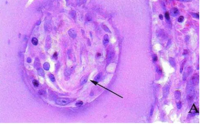
In the 10-day group, large macrophage-like cells with clear material in the cytoplasm suggesting phagocytosis of the collagen capsules (Figure 1) and blood vessels were observed. In the BAB group there was intense fibroblastic and angioblastic proliferation, and mild fibrosis. Some particles adjacent to the implant were surrounded by inflammatory multinucleated giant cells (IMGC). Penetration of blood vessels and cells (Figure 2A) into the pores of the implant particles was also observed.
After 20 days, the control group presented inflammatory infiltrate with moderate proliferation of blood vessels and fibroblasts. In the BAB group there was chronic inflammatory infiltrate, angiogenesis and IMCGs, and a decrease in the number of macrophages. Comparing to the 10-day period, there was a significant decrease of the inflammatory infiltrate and increase of cellular density around implant particles (Figure 2B).
The control group, at the 30-day period, presented a significant increase in the amount of collagen fibers and in the number of blood capillaries when compared to previous periods. In the test group, there was a decrease in inflammatory infiltrate and in number of macrophages. However, the number of IMCGs, fibroblastic and angioblastic proliferation was maintained as at the 20-day period.
DISCUSSION
Hundreds of thousands of bone graft surgeries are performed annually for the treatment of bone loss, with autogenous bone considered the preferred graft. In dentistry, a great number of cases demands such surgical procedures, where many times the graft is obtained from a donor area inside the oral cavity15,16.
The inconvenience and risks of using autogenous bone grafts1,2 have stimulated the search for development alternative materials with properties of biocompatibility and osteoconduction. Xenogenic derived materials have been investigated as biomaterials since 1960´s 17, including clinical trials for the treatment of bone defects18.
In this study the tissue response to a biomaterial derived of cancellous bovine bone (porous inorganic bone) produced in a Brazilian laboratory was evaluated after implantation in rat subcutaneous tissue. Throughout the experimental periods, the presence of multinucleated giant cells and chronic inflammatory infiltrate, and the absence of PMNs, plasma cells and eosinophils, were observed after 10 days post implantation, the biologically designed pores of the material were already filled with reparative tissue and fibrosis increased around implant particles through the
experimental periods.
Adequate mechanical, chemical and thermal processing of the xenografts allows the production of materials with appropriate biocompatibility and osteoconduction.
Hürzeler, et al. (1997)19 implanted a porous inorganic bone, commercially named Bio-Oss, in experimental bone defects in monkeys, and observed significant bone formation, attributing osteoconductive properties to the material. In 1999, Stephan, et al. 20, studied the same biomaterial in cultured rat osteoblasts and noted that these cells erode the surfaces of the implant particles facilitating their adhesion and proliferation.
The presence of IMCGs does not necessarily imply in the lack of biocompatibility of the material. There are many scientific hypotheses regarding the nature of inflammatory multinucleated giant cells (IMGCs) attracted to subcutaneous grafts of mineralized particles21. Glowacki22 (1986) identified those cells as osteoclasts, with ruffled borders. Other authors, however, do not support these results 21. Thus, according to Bagi and Miller23 (1989), two types of multinucleated cells may be observed surrounding mineralized bone grafts. One type has a typical morphology of an IMGC and evidence of bone degradation, and another, in smaller number, has cytological characteristics of an
FIGURE 1- Photomicrography showing subcutaneous
tissue response to empty collagen capsules (control group) at 10 days post implantation. Magnifications: x40 Hematoxylin and Eosin
FIGURE 3- Photomicrography showing subcutaneous
tissue response to xenogenic bone graft at 60 days post-implantation, specially the fibrosis (arrows) around the particles. Magnifications: x40, Hematoxylin and Eosin
FIGURE 2- Photomicrographies showing subcutaneous
osteoclast but without the genuine ruffled borders. For these authors resorption may occur by means of either an IMGC or an osteoclast. In the present study we verified the presence of such IMGCs in contact with the implanted material, however, the presence of brush-like borders was not observed. These findings are supported by Kelly and Shineider21 (1994) that showed IMGCs recruitment in response to mineralized homograft but without osteoclast characteristics.
Porosity of the material certainly contributed to an increase in the surface area, allowing a larger number of inflammatory cells and, latter, of fibroblasts in contact with the material. At the initial periods, especially in the first 10 days, we found few cells in the central area of the granulation tissue, probably due to the large amount of compacted material used. This may have retarded the penetration of the resident connective tissue cells and blood vessels and favored the attraction of PMNs in this period.
Interconnected biological pores may favor rapid development of the bone24, once they form a kind of bone gallery where inductive factors and cells migrate to. Alloplastic materials that mimic these characteristics have been developed as synthetic carriers for growth factors25 and for bioengineering26.
Bovine bone-derived anorganic matrix is a natural, porous xenogenic hydroxyapatite, approved by the Food and Drug Administration (FDA) for the use as a filling material27. Thermal deproteinization of the bovine bone allows the elimination of protein and yet maintains the natural biological surface structure of the hydroxyapatite which provides for cell adhesion and proliferation20.
These xenografts may be associated to bioactive molecules enhancing their potential as osteoconductive biomaterials for the treatment of bone loss27.
CONCLUSION
In summary, tissue response was characterized by the presence of IMGCs during every experimental period, reaching its peak at 30 days. These giant cells were observed in close contact with the material at 30 and 60 days. Despite the moderate level of chronic inflammatory infiltrate, we did not detect a significant presence of PMNs, lymphocytes or plasma cells, discarding a significant immune reaction in response to the graft material. Based in these observations, one could conclude that anorganic porous bovine bone is biocompatible and favors the cells movement/migration into material surface and pores.
ACKNOWLEDGEMENTS
The authors would like to thank the financial support: FAPESP (02/03928-0), the staff of FOB-USP: Ovídio dos Santos Sobrinho, Gilmar Vicente da Silva, Luiz Carlos da Silva, Erasmo Gonçalves da Silva and Daniele Santi Ceolin, for the technical aid provided during the accomplishment of this study.
REFERENCES
1- Joshi A, Kostakis GC. An investigation of post-operative morbidity following iliac crest graft harvesting. Br Dent J. 2004;196:167-71.
2- Dyer PV. What do patients think of iliac crest graft harvesting? Br Dent J. 2004;96:155.
3- Marzola C, Toledo Filho JL, Zorzetto DLG, Pastori CM. Implantes de Biohapatita+Osseobond+Membrana Reabsorvível Dentoflex + Aglutinante Dentoflex. Apresentação de casos clínicos. Rev Bras Ciênc Estomatol. 1996;1:51-63.
4- Athanasiou KA, Niederauer GG, Agrawal CM. Sterilization, toxicity, biocompatibility and clinical applications of polylactic acid/ polyglycolic acid copolymers. Biomaterials. 1996;17:93-102.
5- De Groot K. Bioceramics consisting of calcium phosphate salts. Biomaterials. 1980;1:47-50.
6- Jarcho M. Calcium phosphates ceramics as hard tissues prosthetics. Clin Orthop Rel Res. 1981;157:259-78.
7- Damien CJ, Parsons JR. Bone graft and bone graft substitutes: a review of current technology and applications. J Appl Biomater. 1990;2:187-208.
8- Granjeiro JM, Taga EM, Oliveira DT, Taga MS, Trebacchetti C, Maeda L, et al. Características físico-químicas de Hidroxiapatita para uso clínico. RGO. 1992;40:130-4.
9- Burstein FD, Cohen SR, Hudgins R, Boydston W. The use of porous granular hydroxyapatite in secondary orbitocranial reconstruction. Plast Reconstr Surg. 1997;100:869-74.
10- Proussaefs P, Lozada J, Kleinman A, Rohrer MD. The use of ramus autogenous block grafts for vertical alveolar ridge augmentation and implant placement: a pilot study. Int J Oral Maxillofac Implants. 2002;17:238-48.
11- Tadic D, Epple MA. Thorough physicochemical characterisation of 14 calcium phosphate-based bone substitution materials in comparison to natural bone. Biomaterials. 2004;25:987-94.
12- Damien CJ, Parsons JR, Prewett AB, Huismans F, Shors E, Holmes RE. Effect of demineralized bone matrix on bone growth within a porous HA material: a histologic and histometric study. J Biomater Appl. 1995;9:275-88.
13- Sciadini MF, Dawson JM, Johnson KD. Evaluation of bovine-derived bone protein with a natural coral carrier as a bone-graft substitute in a canine segmental defect model. J Orthop Res. 1997;15:844-57.
14- Wenz B, Oesch B, Horst M. Analysis of the risk of transmitting bovine spongiform encephalopathy through bone grafts derived from bovine bone. Biomaterials. 2001;22:1599-606.
15- Santoro F, Maiorana C, Rabagliati M. Long-term results with autogenous onlay grafts in maxillary and mandibular atrophy. J Long Term Eff Med Implants. 1999;9:215-22.
16- Dortbudak O, Haas R, Bernhart T, Mailath-Pokorny GJ. Inlay autograft of intra-membranous bone for lateral alveolar ridge augmentation: a new surgical technique Oral Rehabil. 2002;29:835-41.
17- Scopp IW, Kassouny DY, Morgan FH. Bovine bone (Boplant). J Periodontol. 1966;37:400-7.
19- Hürzeler M, Quiñones C, Kirsh A, et al. Maxillary sinus audmentation using different grafting materials and dental implants in monkeys. Clin Oral Implants Res. 1997;8:476-86.
20- Stephan E, Jiang D, Lynch S, Bush P, Dziak R. Anorganic bovine bone supports osteoblastic cell attachment and proliferation. J Periodontol. 1999;70:364-9.
21- Kelly JD, Schneider GB. Morphological and Histochemical comparison of cells elcitedly ectopic bone implants and tibial osteoclasts. Amer J Anat. 1991;192:45-9.
22- Glowacki J. Osteocastic feature of cells that resorb bone implants in rats. Calcif. Tissue Int. 1986;39:327-31.
23- Bagi CM, Miller SC. Osteoclast features of cells that resorb demineralized and mineral-containing bone implants in rats. Scanning Microsc. 1989;3:963-8; discussion 969-70.
24- Mushipe MT, Revell P, Shelton JC. Cancelous bone repair using bovine trabecular bone matrix particules. Biomaterials. 2002;23:365– 70.
25- Gallardo J. Preparation and in vitro evaluation of porous silica gels. Biomaterials. 2002;23:4277–84.
26- Taboas JM. Indirect solid free form fabrication of local and global porous, biomimetic and composite 3D polymer-ceramic scaffolds. Biomaterials. 2003,24:181-94.