Contents lists available atScienceDirect
Immunobiology
journal homepage:www.elsevier.com/locate/imbio
Systemic Immunological changes in patients with distinct clinical outcomes
during
Mycobacterium tuberculosis
infection
Tatiane Figueiredo Morais-Papini
a,b, Jordana Grazziela Alves Coelho-dos-Reis
a,
Ana Paula Barbosa Wendling
a, Lis Ribeiro do Vale Antonelli
c, Pryscilla Fanini Wowk
d,f,
Vânia Luiza Deperon Bonato
d, Valéria Maria Augusto
e, Silvana Elói-Santos
a,e,
Olindo Assis Martins-Filho
a, Cláudia Martins Carneiro
b,g, Andréa Teixeira-Carvalho
a,⁎aGrupo Integrado de Pesquisas em Biomarcadores, Centro de Pesquisas René Rachou
–FIOCRUZ, Belo Horizonte, Minas Gerais, Brazil bPós-graduação em Ciências Farmacêuticas (CIPHARMA), Universidade Federal de Ouro Preto, Ouro Preto, Minas Gerais, Brazil cLaboratório de Biologia e Imunologia Parasitária, Centro de Pesquisas René Rachou
–FIOCRUZ, Belo Horizonte, Minas Gerais, Brazil dDepartamento de Bioquímica e Imunologia, Escola de Medicina, Universidade de São Paulo, Ribeirão Preto, Brazil
eDepartamento de Propedêutica Complementar, Faculdade de Medicina da Universidade Federal de Minas Gerais, Belo Horizonte, Minas Gerais, Brazil fInstituto Carlos Chagas
–FIOCRUZ, Curitiba, Paraná, Brazil
gLaboratório de Imunopatologia, Núcleo de Pesquisas em Ciências Biológicas, Universidade Federal de Ouro Preto, Ouro Preto, Minas Gerais, Brazil
A R T I C L E I N F O
Keywords:
Tuberculosis Immune response Clinical outcomes Immunophenotyping
A B S T R A C T
Background: The lung lesions in an individual infected with tuberculosis (TB) are surprisingly variable and independent of each other. However, there is no circulating biomarker yet able to segregate patients according to the extent of lung lesions.
Materials and methods:In this study, the phenotypic and functional profile of leukocytes of patients with active pulmonary tuberculosis (TB) and controls (CO) were fully scrutinized by immunophenotyping assays andin vitro
short-term whole blood culture. The TB group was subdivided according to the extent of lung lesions as uni-lateral (UNI) and biuni-lateral (BI).
Results:The results show that TB group display an altered leukocyte profile in the peripheral blood with sig-nificant lower counts of NK-cells, CD3+CD56+CD16+/−NKT-cells, CD4+
T-cells, CD19+B-cells when compared to CO. Increased CD4+T-cells and CD8+T-cell activation was observed by the upregulation of activation markers
(HLA-DR) as well as of chemokine receptors (CCR2, CCR3, and CXCR4). In addition, TB group presented a significant decrease proportion of CD14LowCD16+ monocytes despite the increase in HLA-DR expression. Regarding the severity of the disease, in the BI group a reduction in frequency of CD19+CD5+B-cells and
expression of HLA-DR in CD14LowCD16+monocytes was observed. Furthermore, the extent of lung lesions
influences the production of molecules as observed by significantly larger production of IL-4 by neutrophils, total T-cells, CD4+T-cells, CD8+T-cells and CD19+B-cells in UNI as compared to BI. By contrast, in BI group the
frequency of high producers of both IL-17+CD4+T-cells and IL-17+CD8+T-cells were signi
ficantly increased than UNI, suggesting the deleterious role of these subsets during active pulmonaryMtbinfection.
Conclusion:The immunophenotypic characterization of unilateral and bilateral active TB performed in the present study indicates that the extent of lung lesion could be associated with afine-tuning between im-munological responses during untreatedMtbinfection.
1. Introduction
Tuberculosis (TB) is caused byMycobacterium tuberculosis(Mtb) and remains as a highly prevalent infectious disease worldwide (Lee et al., 2015). In 2015, 10.4 million new active cases of TB were notified and
1.5 million people died from TB, including 400,000 HIV-positive in-dividuals (WHO, 2016).
Heightened morbidity and mortality associated with this disease are consequences of complex processes triggered by Mtb. The vaccine composed of the bacillus Calmette Guérin (BCG) has contributed for
http://dx.doi.org/10.1016/j.imbio.2017.05.016
Received 22 December 2016; Received in revised form 5 April 2017; Accepted 23 May 2017
⁎Corresponding author at: Grupo Integrado de Pesquisas em Biomarcadores, Centro de Pesquisas René Rachou
–FIOCRUZ, Belo Horizonte, Minas Gerais. Av. Augusto de Lima, 1715, Barro Preto, Belo Horizonte, Minas Gerais, Brazil.
E-mail address:andreat@cpqrr.fiocruz.br(A. Teixeira-Carvalho).
0171-2985/ © 2017 Elsevier GmbH. All rights reserved.
decreasing lethality. However, BCG has been proven to be of in-adequate efficacy against the most frequent outcome of pulmonary tuberculosis in adults (Fine, 2005). Regarding the current therapy for the disease, the number of multi-drug resistant (MDR) isolates is on the rise in many areas of the world (Korbel et al., 2008).
Variations in the host immune response against the pathogen, as the type and intensity, may influence the status ofMtbinfection in active or latent tuberculosis, which differ in the type of treatment needed and the risk of its transmission (Lyadova and Panteleev, 2015).
Mtbis an intracellular pathogen that infects preferentially myeloid cells such as macrophages (Russell et al., 2009). Therefore, it is sug-gested that development of a cellular immune response, with special participation of Th1-lymphocytes, is critical for disease control (Dorhoi et al., 2011) by decreasing the growth and spread ofMtbin the host cells (O’Garra et al., 2013). Other immune cells, such as B-lymphocytes, appear to exert protective role in tuberculosis (Maglione et al., 2007) through the production of cytokines such as IL-12 and IL-4, which di-rects the response to proinflammatory or regulatory profiles, respec-tively (Maglione et al., 2007). In addition, NK-cells seem to be able to limit pathogen growth by lysis of Mtb-infected cells mediated by the perforin and granzyme pathway (Sia et al., 2015).
Studies that proposed to phenotypically and functionally char-acterize the innate and the adaptive immune cells in tuberculosis are complex and not completely conclusive (da Silva et al., 2015). There-fore, the present study aimed at analyzing innate and adaptive immune cell subsets byex vivoimmunophenotyping and short-term culture with Mtb(H37Rv) antigen. Ourfindings suggest that unilateral and bilateral TB are composed of a different immunophenotypic profile, which is associated with a fine-tuning of immune responses during untreated Mtbinfection.
2. Population, material and methods
2.1. Study population
This study was conducted in Belo Horizonte and surrounding cities, at Minas Gerais state, Brazil. Twenty-one patients with active tu-berculosis (TB) were included in the study and had confirmed diagnosis by examination of sputum smear and culture. The severity of illness of patients with TB, directly related to the extension of lung involvement, was assessed through the analysis of chest radiographs and patients were segregated as bearing lesions classified as unilateral (UNI) with involvement of only one pulmonary lobe, and bilateral (BI) with in-volvement of both pulmonary lobes (Kobashi et al., 2007). A control group (CO) of twenty-eight co-inhabitants of the same endemic area with no evidence of disease by clinical examination and negative cul-ture forMtbwere enrolled in this study. For the immunophenotyping assays, the study groups were constituted by: CO (n = 21), TB (n = 15), UNI (n = 05) and BI (n = 08). Two patients from the TB group had no chest radiograph and thus were excluded from the ana-lysis. For thein vitroshort-term whole blood culture, the groups were: CO (n = 09), TB (n = 08), UNI (n = 03) and BI (n = 05). Hematolo-gical parameters, smoking, alcohol, body mass index (BMI), blood glucose and C-reactive protein were described inTable 1. All subjects included in this evaluation were HIV-negative. This study was carried out in full accordance with all International and Brazilian accepted guidelines and was approved by the Ethics Committee for Research at Federal University of Minas Gerais (UFMG protocol # 228/03).
2.2. Blood sample
Venous peripheral blood sample of 5 mL was collected from each subject using heparin (whole blood culture) or EDTA (ex vivo) as an-ticoagulant. Blood samples from TB patients were collected im-mediately prior to the chemotherapy treatment.
2.3. Mycobacterium tuberculosis antigen
Mtbantigen (H37Rv Mtb-Ag) was provided by the Microbiology Laboratory (Departamento de Bioquímica/USP/Brazil). The Mtbwas cultured in tubes with Loweinstein Jensen medium and incubated at 37°C until evidence of bacterial growth. Colonies were inactivated at 80°C for 1 h and sonicated in 2 cycles of 20 s at 40 Hz in an ice bath. The suspension was then sterilized using gamma radiation (dose of 5000 Gray for 2:15 h). The protein concentration was measured by the Lowry method.
2.4. Immunophenotyping assays
For the realization ofex vivoimmunophenotyping assays, peripheral blood leukocytes from TB patients (TB) and controls (CO) were col-lected. EDTA blood aliquot of 50μL was transferred to 5 mL polystyrene tubes (Falcon–BD, USA) containing aliquots of monoclonal antibodies labeled withfluorochromes: FITC (anti-CD4, anti-CD14, anti-CD5), PE (anti-HLA-DR, anti-CD54, anti-CD25, anti-CD18, anti-CD62, anti-CD69, anti-CD23) and TC (anti-CD8, anti-CD19, anti-CD16). The samples were homogenized and incubated for 30 min at room temperature, protected from light. Red blood cell lysis was performed by adding 2 mL of lysis solution to the cell suspension (FACS Lysing Solution- Becton Dickinson) and incubated for 10 min at room temperature. Subsequently, cell suspension was washed twice with 1 mL of phos-phate buffered saline-PBS (0.015 M, pH 7.4) and centrifuged for 10 min, 18°C at 400 ×g (Beckman, Model J-6B, USA). The samples were fixed with 200μL of FACS fixative solution (10 g/L Table 1
Demographic, clinical and laboratorial features of the study population
Groups
Parameters Control (CO) Pumonary
tuberculosis (TB)
Genre
Male 16 (57.1%) 14 (66.7%)
Female 12 (42.9%) 7 (33.3%)
Age
42 (19–62) 44 (21–70)
Pulmonay disease Severity
Unilateral – 08
Bilateral – 11
Hematological Profile
RBC (mm3)1 5,200 (4,900
−5,600) 5,000 (4,000−5,000) Platelets (mm3)2 233,000
(190,000−321,000)
377,000
(288,000−488,500) Hemoglobin (g/dL)3 15.0 (13.9
–16.6) 12.3 (11.5–13.2) Leukocytes (mm3)4 6,620 (4,965
−7,900) 8,540 (6,540−10,190) Neutrophils (mm3)5 3,970 (2,560
−5,227) 6,170 (4,260−8,085) Eosinophils (mm3) 170 (95
–245) 140 (80–405) Monocytes (mm3)6 520 (415
–700) 680 (520–940) Lymphocytes
(mm3)7
1,910 (1,515−2,155) 1,360 (830−1,665)
Alcoholism
Yes 16 (57.1%) 09 (42.9%)
No 12 (42.9%) 12 (57.1%)
Smoking
Yes 13 (46.4%) 14 (66.7%)
No 15 (53.6%) 07 (33.3%)
BMI (weight/ height2)8
26.2 (24.2–30.6) 20.0 (17.0−21.0)
C-reactive protein (mg/L)9
5.0 (3.0−10.1) 54.0 (21.5–76.0)
glycemia (mg/L) 82.0 (76.0−90.0) 80.0 (74.0−92.5)
paraformaldehyde, 1% sodium cacodylate, 6.67 g/L sodium chloride, pH 7.2). After 15 min at 4°C, samples were acquired using FACScan flow cytometer (BD, San Jose, CA, USA).
2.5. In vitro short-term whole blood culture
The short-term whole blood cultures were performed as described by Luiza-Silva et al., 2011modified as follows: 1 mL aliquots of he-parinized peripheral blood were dispensed into duplicates of 14 mL polypropylene tubes (Falcon®, B.D. Pharmingen, San Diego, CA) and
incubated for 24 h at 37°C in a 5% CO2humidified atmosphere in the presence of 1 mL of RPMI 1640 (GIBCO–Grand Island, NY) (non-sti-mulated culture) or in the presence ofMtb-Agdiluted in RPMI at afinal concentration of 15μg/mL (stimulated culture). After priming, 10μg/
mL of Brefeldin A (BFA) (Sigma–Chemical Company–St Louis, MO) were added and the samples were re-incubated for an additional 4 h at 37°C in a 5% CO2humidified atmosphere. Following incubation, the cultures were treated with 2 mM EDTA, (Sigma–Chemical Company– St Louis, MO) and maintained at room temperature for 15 min. The duplicates were pooled into a 15 mL polypropylene tubes and then washed with 6 mL of FACS buffer (0.015 M PBS, supplemented with 0.5% bovine serum albumin—BSA and 0.1% sodium azide) by cen-trifugation at 600gfor 7 min at room temperature. After centrifugation, the supernatant was discarded and the cell pellet was resuspended in 1 mL of FACS buffer. The cell suspension was immediately submitted to immunophenotyping and intracytoplasmic cytokine analysis by flow cytometry. Positive control cultures were performed using Phorbol 12-Myristate 13-Acetate-PMA, ionomycin and BFA atfinal concentration of 25 ng/mL, 1μg/mL and 10μg/mL, respectively. These cultures were
characterized by high levels of activation and cytokine production were used in all immunophenotyping and intracytoplasmic cytokine analysis to confirm cell viability of all blood samples (data not shown).
2.6. Intracytoplasmic cytokine staining
Aliquots of whole blood cultures were stained with anti-human cell surface TC-labeled monoclonal antibodies (CD4, CD8, anti-CD14, anti-CD16 and anti-CD19) for 30 min at room temperature. After membrane staining, erythrocyte lysis and leukocytesfixation, cell sus-pension was permeabilized with FACS perm-buffer (FACS buffer sup-plemented with 0.5% saponin) and aliquots incubated for 30 min at room temperature, in the dark, withfluorescent anti-cytokine mono-clonal antibodies, including PE-labeled anti-IL-6, anti-IFN-γ, anti-TNFα,
anti-IL-4, anti-IL-17, anti-IL-8 and anti-IL-10. After intracytoplasmic cytokine staining, the leucocytes were washed with FACS perm-buffer and FACS buffer and fixed in FACSfixative solution. Immunostained samples were acquired using a FACScanflow cytometer and analyzed using Flowjo software (Tree Star, San Jose, CA, USA). A total of 20,000 events/tube were acquired and stored for further analysis. Distinct gating strategies were used to analyze the cytokine-expressing leuko-cytes subsets, including neutrophils, monoleuko-cytes and lympholeuko-cytes (B-cells, CD4+T-cell and CD8+T-cell subsets). The results werefirst ex-pressed as percentages of cytokine-positive cells for different gated leukocyte subpopulations analyzed in this study. These values were used to obtain the cytokine index, calculated as the ratio between the percentage of cytokine-positive cells observed in the stimulated culture (Mtb-Ag) by the percentage of cytokine positive cells observed in the unstimulated culture (control).
2.7. Flow cytometric acquisition and analysis
Flow cytometry acquisition was performed in a BD FACScanflow cytometer (Becton Dickinson, San Jose, CA, USA). After acquiring 30,000 events/tube, distinct gating strategies were used to analyze the frequency of different leucocyte subsets according to surface molecules defined by specific clusters of differentiation (CD). Samples werefirst
gated on lymphocytes using the morphometric parameters side-scatter (SSC low) and forward-scatter (FSC low). Subsequently, T and B lym-phocytes were identified. CD4+-T and CD8+-T lymphocytes subsets were defined within the lymphocyte gate and the expression of che-mokine (CC and CXC) receptors (CCR2, CCR3, CCR5, CXCR3 and CXCR4), cell adhesion markers (CD62L, CD18, CD54) and cell activa-tion markers (CD25 and HLADR) were evaluated. Thus, the phenotype of CD4+T and CD8+T-lymphocytes were defined as: CCR2+CD4+/ CD8+, CCR3+CD4+/CD8+, CCR5+CD4+/CD8+, CXCR3+CD4+/ CD8+, CXCR4+CD4+/CD8+; CD62negCD4+/CD8+, CD18+CD4+/ CD8+, CD54+CD4+/CD8+; CD4+CD25+and HLADR+CD4+/CD8+. The reason for testing chemokine receptors on CD4+T-cells and CD8+ T-cells is that these surface molecules can shape the migration potential of T-lymphocytes and induce polarization towards protective or pa-thogenic responses (Kim et al., 2001).
B-lymphocyte subsets were defined by expression of CD19+. Within this subset, cell activation markers CD23, CD5 and CD69 were assessed. Gates were created from CD19 vs. CD23 or CD5 and B- lymphocyte subsets were defined as: CD23+CD19+, CD23LowCD19+, CD23HighCD19+and CD5+CD19+. In addition, the density of expres-sion of CD69 on CD5+CD19+B-lymphocytes was determined by the geometric meanfluorescence intensity (MFI). NK and NKT subsets were defined by the absence or presence of CD3, CD56 and CD16 expression as follows: gates were created from CD3 versus SSC for selection of populations CD3−and CD3+. Subsequently, gates were created from CD56 vs. CD16 and NK-cells were defined as: NK (CD56+CD16+/
−CD3−) and (CD56+
CD16+/−CD3+
), respectively. NK-cells were evaluated according to the expression intensity of CD56, which were classified as: CD56negCD16+, CD56dimCD16+ and CD56brightCD16− (Fig. S1). The analysis of the subpopulations of circulating monocytes were defined by expression of HLA-DR, CD14 (receptor for binding of lipopolysaccharide complex and protein) and CD16 (low affinity re-ceptor for IgG). For this purpose, a gate was created in HLA-DR+and intermediate SSC and, subsequently, gates were created from CD14 versus CD16. The circulating monocytes were defined as: classical monocytes (CD14+CD16−), proinflammatory monocytes (CD14+CD16+) and patrol monocytes (CD14LowCD16+). Activation each subset was assessed by the expression intensity of HLA-DR, de-termined by the geometric mean intensity offluorescence (MFI). The software FlowJo version 7.0 (TreeStar, San Diego, CA, USA) was uti-lized forflow cytometric data analysis.
2.8. Phenotypic and functional signature analysis
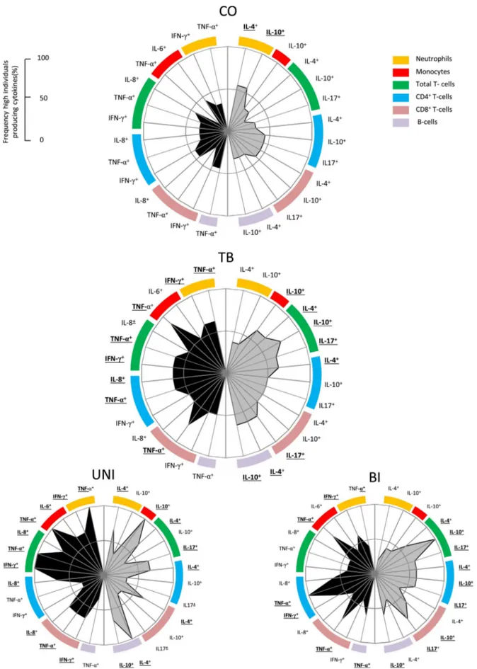
The Phenotypic and functional signatures of patients with TB (UNI and BI) as well as controls (CO) werefirst calculated by assessing the percentages of low and high cytokine producers, as previously sug-gested byLuiza-Silva et al. (2011), using the global median as the cut offvalues to segregate low and high producers. The percentage of TB patients and CO volunteers showing high frequency was calculated for each leukocyte subset. For thein vitrostudy, Mtb-Ag/control indices were calculated. The ascendant curve of high frequency subjects on control group was then used as the reference curve to identify changes in the overall immune patterns from the other groups. Radar charts (Campi-Azevedo et al., 2016; Coelho-dos-Reis et al., 2013) were further used to summarize the cytokine signatures in a range of leukocyte subsets of innate [neutrophils (orange), monocytes (red)] and adaptive immunity [lymphocytes T (green), lymphocytes CD4 (blue), lympho-cytes CD8 (salmon) e lympholympho-cytes B (lilac)]. Each axis represents the frequency (%) of volunteers showing high cytokine-producing cell in-dices or high frequency of a specific cell subset.
2.9. Statistical analysis
cases, significance was considered at p≤0.05. All tests were provided by GraphPad Prism version 5.0 (San Diego, CA). The analysis of cyto-kine signatures was performed using the cytocyto-kine signature from con-trol group as the reference curve, and significant differences were considered when the indices were superior to the 50th percentile as compared to the one in the reference signature (CO).
3. Results
3.1. Patients with pulmonary tuberculosis display altered leukocyte profile in the peripheral blood
The profile of circulating leukocytes in peripheral blood of patients with pulmonary tuberculosis was evaluated.Fig. 1shows theex vivo phenotypic results as percentages (Fig. 1A), as well as absolute numbers (Fig. 1B) of NK-cells, NKT-cells, CD4+T-cells, CD8+T-cells and B-cells from peripheral blood of CO and TB groups. Regarding NK-cells and CD8+T-cells, no differences were observed between the groups (Fig. 1A). Data analysis demonstrated that there is significant decrease in both proportions and absolute numbers of the CD3+CD56+CD16+/− NKT- cell and CD19+B-cell subsets in TB as compared to CO. A statis-tically significant increase was observed in the CD4+T-cells in the TB group as compared to CO group. On the other hand, the opposite was observed when the absolute of CD4+T-cell numbers were assessed. Despite no differences were observed in the frequency of NK-cells (Fig. 1A), TB patients displayed decreased absolute number compared to CO (Fig. 1B). These data suggest that the TB group has selective alterations in CD3+CD56+CD16+/−cells, CD4+-T as well as CD19+-B lymphocytes.
3.2. Monocyte subsets display distinct activation profile during tuberculosis
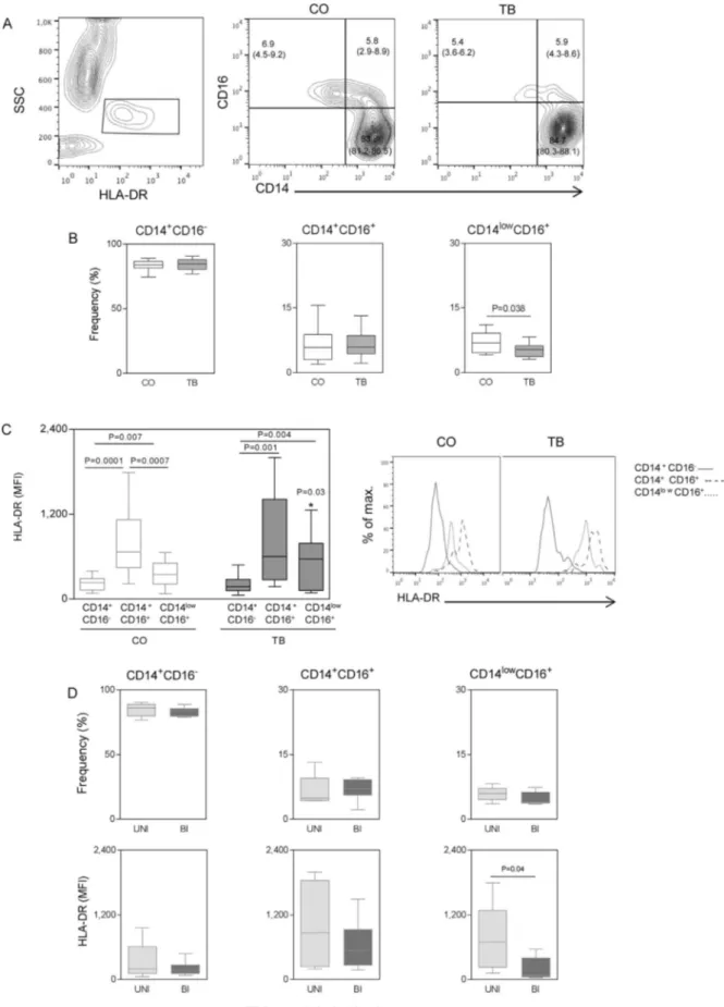
In order to verify if subsets of monocytes and their HLA-DR
expression could be related toMtbinfection, the phenotypic profile of three monocyte subsets was evaluated. For that, classic (CD14+CD16−), proinflammatory (CD14+CD16+) and patrolling (CD14LowCD16+) monocyte subsets were analyzed (Fig. 2A). TB pre-sented a significant decrease in the frequency of patrolling monocytes when compared to CO (Fig. 2B). No differences were observed in classic and proinflammatory monocytes. Analysis of the HLA-DR on each subset indicated that the proinflammatory monocytes displayed the highest levels of expression in both CO (classic and patrolling) and TB (Classic) (Fig. 2C). Higher HLA-DR expression was observed in patrol-ling monocytes from TB when compared to CO. No differences in HLA-DR expression were observed between classic and proinflammatory monocytes.
Mtb-infected patients were segregated according to disease severity as unilateral (UNI) and bilateral (BI) based on the extent of pulmonary lesions observed by the analysis of chest radiographs. The results show that despite similar frequencies of distinct monocyte subsets, patrolling monocytes from BI display a significant decrease in HLA-DR expression compared to UNI. Thesefindings indicate that the activation profile of patrolling monocytes is not only altered in TB patients but also de-creased significantly in patients with bilateral TB.
3.3. Phenotypic analysis of circulating CD4+T-cell and CD8+T-cell subsets of patients with pulmonary tuberculosis
We next investigated the percentage of circulating CD4+T-cells and CD8+T-cells expressing the chemokine receptors CCR2, CCR3, CCR5, CXCR3 and CXCR4 as well as the cellular activation markers CD62L, CD54, CD18, CD25 and HLA-DR in CO and TB groups. The results displayed inFig. 3A showed that there is a statistically significant in-crease in the percentage of CCR2+CD4+T-cells, CCR3+CD4+T-cells, CD18+CD4+T-cells and HLA-DR+CD4+T-cells in TB when compared to CO. In regards to the CD8+T-cells (Fig. 3B), there is statistically Fig. 1.Patients with pulmonary tuberculosis display altered leukocyte profile in the peripheral blood. (A) Percentage and total number (B) of circulating NK-cells (CD3+CD56+CD16+/
−), CD4+T-cells, CD8+T-cells and CD19+B-cells in Control-CO (n = 20), Tuberculosis-TB (n = 15) groups, obtained by
Fig. 2.Circulating monocytes profile in patients with pulmonary tuberculosis. (A) FACS plots showing the distribution of circulating subtypes of monocytes defined by markers HLA-DR, CD14 and CD16 in Control-CO (n = 20) and Tuberculosis-TB (n = 15) groups, with the respective values of the median and Q2 and Q3 quartiles. (B) Monocyte subsets CD14+CD16−, CD14+CD16+and CD14lowCD16+are shown in box plot graphics, with median and interquartile range. Data were analyzed using the Mann-Whitney test. (C) On the left, expression of
HLA-DR (Median Fluorescence Intensity−MFI) in subsets of monocytes in the group CO (white bars) and TB (black bars). Data were analyzed using thet-test. On the right, FACS histogram graphs showing HLA-DR expression in CD14+CD16−(continuous line), CD14+CD16+(dashed line) and CD14lowCD16+(dotted line) subsets of monocytes. (D) Left,
per-centage of subsets of monocytes CD14+CD16−, CD14+CD16+and CD14lowCD16+in TB group patients with unilateral radiological abnormalities-UNI (n = 05) and bilateral-BI
significant increase in the percentage of CCR2+CD8+T-cells, C-CR3+CD8+T-cells, CXCR4+CD8+T-cells and HLA-DR+CD8+T-cells in TB versus CO. On the whole, the analysis of circulating CD4+T-cells and CD8+T-cells suggest that TB patients displayed higher activation and migration pattern in these lymphocyte subsets.
3.4. TB patients with bilateral pulmonary lesion display lower percentages of CD19+CD5+B-lymphocytes
Fig. 4shows the percentage of CD19+B-cells and subsets according CD23 expression (CD19+CD23+, CD19+CD23Low and CD19+CD23High) in patients with pulmonary tuberculosis (Fig. 4A). The data allowed the identification of distinct frequency of B subsets in TB. There is a significantly reduced percentage of CD19+CD23+and percentage of CD19+CD23Highcells in the TB group compared to CO. The percentage of CD19+CD23Low cell subset did not differ sig-nificantly between groups. No significant differences were observed in the CD19+B-cell subsets when UNI and BI were compared.
In addition, we evaluated the percentage of CD19+CD5+B-cells as well as the activation of these cells by CD69 expression. The data de-monstrated that TB patients displayed significantly lower percentages of CD19+CD5+cells when compared to CO. The activation of these cells did not differ significantly between groups. The percentage of CD19+CD5+cells were higher in UNI when compared to BI. The results reveal that the innate B-cell subsets such as CD19+CD5+-cells may be important for the protective mechanisms triggered during the lung le-sion formation.
3.5. Ex vivo immunophenotypic signature analysis reveal increase and decrease in innate and adaptive immune components in TB patients with distinct radiological involvement
The phenotypic leukocyte signatures in the peripheral blood of CO and TB were determined (Fig. 5). For that, continuous values were transformed in categorical data and trend curves for the CO group (reference curve) were plotted and overlaid on the histograms of TB, UNI and BI groups. It is possible to observe that both the innate and adaptive immunity curves of TB show a peak above the reference curve, indicating that there is an important role of acquired immunity with emphasis on CD4+T-cell and CD8+T-cell subsets during TB. On the other hand, there are clear gaps at the end of both the innate and adaptive immunity curves, composed mainly by CD4+T-cells and
B-cells. However, when comparing UNI and BI groups, there was a mixed profile in relation to the innate and adaptive immunity; thefirst of which resembles the profile of the individuals in the group CO, and the second, the TB group. Thesefindings reveal that the overall innate and adaptive immune profiles of TB patients with different lung lesion commitment are diversified, which may be relevant for the foundation of such clinical patterns.
3.6. Production of proinflammatory and regulatory cytokines of the innate and adaptive immunity are distinct in tuberculosis patients with different extent of lung lesions
The production of intracytoplasmic chemokine/cytokines by circu-lating leukocytes of patients with pulmonary tuberculosis was analyzed after short-term whole blood culture in the presence or absence of Mtb-Ag (Fig. 6). Categorical data for chemokine/cytokine-producing neu-trophils, monocytes, total T-cells, CD4+T-cells, CD8+T-cells and CD19+B-cells are displayed as radar graphs built with the frequency of high producers (%) from each group (CO, TB, UNI and BI). Left wing indicates molecules (black) associated with inflammation and right wing indicates molecules (gray) associated with regulation of the im-mune response. It is possible to observe that the chemokine/cytokine profile of CO delineates a graph area, which is confined in the 50% threshold and left and right wings show balanced percentages.
Expansion of the area graphs is observed in TB. Involvement of innate an adaptive immunity in the production of cytokines is observed in TB group as compared to CO. Interestingly, TB patients present lower frequency of high IL-4+and IL-10+ neutrophils when compared to controls. In general, a mixed profile in the innate and adaptive im-munity was observed in patients with UNI or BI pulmonary lesions. However, the expansion of area graph on the left wing of UNI was evident when compared to BI, which was contrasted by significantly larger production of IL-4 by neutrophils, total T-cells, CD4+T-cells, CD8+T-cells and CD19+B-cells. The frequency of high producers of IL-17+CD4+T-cells and CD8+T-cells were significantly increased in BI as compared to UNI. These results suggest that IL-17 could play a dele-terious role, while IL-4 may be associated with a possible protective role during active pulmonaryMtbinfection.
4. Discussion
Active tuberculosis is a spectrum disease, which assumes influence Fig. 3.Phenotypic analysis of circulating CD4+T-cell and CD8+T-cell subsets of patients with pulmonary tuberculosis. Percentage of circulating CD4+T-cells and CD8+T-cells expressing
the chemokine receptors CCR2, CCR3, CCR5, CXCR3 and CXCR4 as well as the cellular activation markers CD62L, CD54, CD18, CD25 and HLA-DR in Control-CO (n = 20) and Tuberculosis-TB (n = 15) groups, obtained byflow cytometry. The CD4+T-cell and CD8+T-cell subsets were determined within the gate of total lymphocytes. Results were plotted in Box
of several factors such as bacterial load, the host immune response and the extent of lung commitment. The activation of the immune system and inflammation are, with no doubt, key players in the im-munopathogenesis of disease. However, the complexity of the interac-tions betweenMtband host factors hinders the establishment of bio-markers related to activation of the disease and protection (Lyadova and Panteleev, 2015).
In this context, the present study aimed at evaluating the profile of circulating leukocytes in TB patients as compared to the profile ob-served in co-inhabitants without Mtb infection (CO group) from an urbanized region of Minas Gerais state, in Brazil. It is important to consider that these patients are living in an endemic area and were analyzed before treatment. The results of this study showed significant reduction in the absolute numbers of NK-cells, NKT-cells, CD4+T-cells and CD19+B-cells in the TB group when compared to the controls. Regarding the percentage, there was reduction in CD19+B-cells and NKT-cells, but an increase in CD4+T-cells.Wu et al. (2009)observed similar results in relation to the percentage of NK cells and CD4+ T-cells, however, increased in CD19+B-cells and CD8+T-cells. Moreover, Guglielmetti et al. (2013)observed reduction in the absolute numbers of CD4+T-cells and no difference in the percentage of these cells. They also showed an increase in the percentage of CD8+T-cells and variation in the absolute numbers of CD19+B-cells. So far, it has not yet been possible to establish the profile of circulating lymphocytes in active
pulmonary tuberculosis, as published studies showed controversial re-sults. However, it is suggested that patients with pulmonary tubercu-losis have changes in circulating lymphocyte homeostasis, which can be associated with disease progression (Wu et al., 2009). The contrast on the results of percentage versus absolute numbers of CD4+T-cells may reflect a general leukopenia with a more severe damage to B- lym-phocytes and compared to CD4+T-cells. The general leukopenia ex-plains the lower absolute numbers of CD4+T-cells in TB patients. On the other hand, considering that CD4+T-cell percentages were mea-sured within the lymphocyte gate which includes B-cells and NKT-cells, an alteration in latter could reflect on the percentage of the former, in such a way that the more severe decrease in B cell and NKT-cell subsets among the total lymphocyte population may account for the increased percentage of CD4+T-cells among lymphocytes.
In this regard, monocytes have been described as essential players not only in disseminating the pathogen in the host, but also in priming innate events as well as in triggering acquired immunity againstMtb (Tang et al., 2015). In Mtb infection, little is known about the re-lationship between the phenotype of monocytes and their function against the pathogen. Previous studies have shown distinct profiles in the percentage of circulating monocytes in patients simultaneously in-fected with tuberculosis and other pathogens. Andrade et al. (2014) found that patients co-infected TB-HIV, who developed IRIS (Immune Reconstitution Inflammatory Syndrome), displayed decreased Fig. 4.Circulating B-cell profile in patients with pulmonary tuberculosis. (A) On the left, FACS plots showing the distribution of circulating subtypes of B-cells defined by markers CD19 and CD23 in Control-CO (n = 20) and Tuberculosis-TB (n = 15) groups, with the respective values of the median and Q2 and Q3 quartiles. Right, percentage of CD19+, CD19+CD23low
and CD19+CD23highsubsets of B-cells in CO (n = 20), TB (n = 15), unilateral radiological abnormalities-UNI (n = 5) and bilateral-BI (n = 8) groups are shown in box plot graphics,
with median and interquartile range. The CD19+CD23+subsets of B-cells were determined by the sum of CD19+CD23lowand CD19+CD23high. Data were analyzed using the
Mann-Whitney test. (B) On the left, FACS plots showing the distribution of circulating subtypes of B-cells defined by markers CD19 and CD5 in CO (n = 20) and TB (n = 15) groups, with the respective values of the median and Q2 and Q3 quartiles. On the right, percentage of CD19+CD5+subsets of B-cells in CO (n = 20), TB (n = 15), UNI (n = 05) and BI (n = 08) groups
shown in boxplot graphs, with median and interquartile range. Activation of CD19+CD5+subsets of B-cells was determined by the median
percentage of patrolling monocytes (CD14DimCD16+), and increased frequencies of classic and inflammatory monocyte subsets compared to patients who did not developed the syndrome.Castaño et al. (2011) observed an increase in inflammatory and patrolling monocytes during
Fig. 6.Overall pattern of high cytokine producers triggered byMtb-Ag. Categorical data for chemokine/cytokine-producing subsets neutrophils (orange), monocytes (red), total T-cells (green), CD4+T-cells (blue), CD8+T-cells (salmon) and B-cells (lilac) are displayed as radar graphs built with the frequency of high producers (%) by Control-CO (n = 09),
that the decrease in the percentage of circulating patrolling monocytes in TB patients is due to the recruitment of these cells to the site of infection (Belge et al., 2002), which is highly influenced by the cell composition and chemokine/cytokine milieu (Stríz et al., 1993).
Furthermore, increased expression of HLA-DR is directly related to cell activation and improves antigen presentation to T lymphocytes (Lapa e Silva et al., 1996). A hallmark ofMtbis its ablility to efficiently evade the immune system. Thus, despite activated in the circulation we cannot rule out the possibility of these monocytes become suppressed after recruited to the lung.Balboa et al. (2015)have shown that CD16+ monocyte subpopulations can contribute to the infection, as they are somewhat resistant to microbial growth and present decreased nitric oxide production capacity compared to CD16−monocytes. Strength-ening this hypothesis, data from the present study show that TB patients with bilateral pulmonary involvement have reduced HLA-DR expres-sing patrolling (CD14LowCD16+) monocytes as compared to unilateral involvement. Previous studies have shown that the reduction of HLA-DR expression on monocytes is related to the severity of infectious diseases (Ditschkowski et al., 1999) and could be employed as a reliable prognostic marker for the surveillance of severe sepsis (Lekkou et al., 2004).
Contrary to what it is known about the importance of the proin-flammatory immune response in the host defense againstMtb, much remains to be clarified on the role of B-lymphocytes during tuberculosis (Kozakiewicz et al., 2013). In this study, there was significantly reduced percentage and absolute numbers of CD19+B-cell and CD19+CD5+ B-cell subsets in the TB when compared CO. A similar profile was ob-served in BI group when compared to the UNI. Patients with severe tuberculosis have lower percentage of circulating B-cells. These data corroborate previous studies that described decreased numbers of cir-culating CD19+B-cells in patients with pulmonary tuberculosis (Abreu et al., 2014; Hernandez et al., 2010). In addition, the reduction of CD19+CD5+B-cell subsets could be related to the severity of the dis-ease (Guglielmetti et al., 2013). However, other studies showed an in-crease in the percentage of circulating B-cells in pulmonary tuberculosis patients (Zhang et al., 2012; Wu et al., 2009).
Reports using mouse models suggest that anti-TB B-cell response in the lung has clear pathological and doubtful protective role (Linge et al., 2017). Linge and collaborators demonstrate that B-lymphocytes forming follicle-like structures in the lung tissue of tuberculosis-infected mice presented a regulatory profile with absence of surface molecules CD5 and CD23 (LINGE et al., 2017). CD5 is a cell surface receptor that negatively regulates B-cell function by inhibiting BCR activation ( Gary-Gouy et al., 2002). CD5 is expressed by B-1 cells which are regarded as regulatory/innate-like B-cells. The significant decrease of CD5+CD19+-B cells found in BI as compared to UNI may indicate that regulation by this subset may be protective. In parallel, CD23 is a re-ceptor for IgE of low-affinity, which is involved in allergy and resistance to parasites, as well as in the regulation of IgE levels (Payet et al., 1999). Mossalayi et al. (2009)reported that surface CD23 mediates antimicrobial activity of human macrophages againstMycobacterium, in part, through the induction of effector responses including TNF-α
production. These results are in line with the presentfindings that show lower percentages of CD19+CD23+B-cells and CD19+CD23highB-cells in TB as compared to CO.
In addition to antibody production, B-cells regulate the immune response though the production of modulatory cytokines and the in-teraction with effector T-lymphocyte subsets (du Plessis et al., 2015). Our results are in line with this hypothesis considering that BI group displayed a clear mixed inflammatory/regulatory profile, when com-pared to UNI, with evident IL-4, IL-10 and IL-17 production by both CD4+T-cells and CD8+T-cells.Zhang et al. (2012)demonstrated that, in patients with pulmonary tuberculosis, circulating CD19+CD5+ B-cells are able to inhibit the production of IL-17 by CD4+T-cells. These data suggest that the production of IL-17, associated with a reduction in the production of IFN-γ by cells from the innate and the adaptive
immunity in the BI group may contribute to the pathogenesis of tu-berculosis, considering the inhibitory properties of IFN-γin IL-17 pro-duction (Lyadova and Panteleev et al., 2015). It has been suggested that one of the possible mechanisms involved in the deleterious action of IL-17 during Mtbinfection is related to the increase in the neutrophil migration into the lung (Kozakiewicz et al., 2013), which contributes to the pathogenesis of tuberculosis (Eruslanov et al., 2005).
In addition, considering that these patients come from endemic areas affected with helmiths and environmental mycobacteria, infec-tions with such pathogens may influence the response towards a Th2 profile, in detriment to the IFN-γ response, which could affect the
phenotype observed. It is important to mention, however, that the pa-tients evaluated herein underwent close and deep clinical evaluation prior to enrollment in the study and no evidence of such infection were detected. Eosinophil counts were considered normal during the clinical evaluation, also indicating that no helminth infection was occurring at the time of blood collection.
5. Conclusion
All in all, the immunophenotypic characterization of unilateral and bilateral active TB performed in the present study indicates that the extent of lung lesion could be associated with afine-tuning between immunological responses during untreatedMtbinfection.
Conflict of interest
The authors declare no conflict of interest.
Acknowledgments
The authors thank the Flow Cytometry Core Facility (PDTIS/ FIOCRUZ) forflow cytometric acquisition and analysis. This work was supported by Fundação de Amparo à Pesquisa do Estado de Minas Gerais (FAPEMIG), Conselho Nacional de Desenvolvimento Científico e Tecnológico (CNPq) by fellowship awarded to LRVA, OAMF and ATC. TFMP received fellowship from Coordenação de Aperfeiçoamento de Pessoal de Nível Superior (CAPES) program and JGCdR received fel-lowship from PNPD/CAPES program.
Appendix A. Supplementary data
Supplementary data associated with this article can be found, in the online version, athttp://dx.doi.org/10.1016/j.imbio.2017.05.016.
References
Abreu, M.T., Carvalheiro, H., Rodrigues-Sousa, T., Domingos, A., Segorbe-Luis, A., Rodrigues-Santos, P., Souto-Carneiro, M.M., 2014. Alterations in the peripheral blood B cell subpopulations of multidrug-resistant tuberculosis patients. Clin. Exp. Med. 14, 423–429.
Andrade, B.B., Singh, A., Narendran, G., et al., 2014. Mycobacterial antigen driven ac-tivation of CD14 + +CD16−monocytes is a predictor of tuberculosis-associated
immune reconstitution inflammatory syndrome. PLoS Pathog. 10, e1004433.
Balboa, L., Barrios-Payan, J., González-Domínguez, E., Lastrucci, C., Lugo-Villarino, G., Mata-Espinoza, D., Schierloh, P., Kviatcovsky, D., Neyrolles, O., Maridonneau-Parini, I., Sánchez-Torres, C., Sasiain Mdel, C., Hernández-Pando, R., 2015. Diverging bio-logical roles among human monocyte subsets in the context of tuberculosis infection. Clin. Sci. 129, 319–330.
Belge, K.U., Dayyani, F., Horelt, A., Siedlar, M., Frankenberger, M., Frankenberger, B., Espevik, T., Ziegler-Heitbrock, L., 2002. The proinflammatory CD14+CD16+DR++
monocytes are a major source of TNF. J. Immunol. 168, 3536–3542.
Hum. Vaccin Immunother. 12, 491–502.
Castaño, D., GarcíaA, L.F., Rojas, M., 2011. Increased frequency and cell death of CD16(+) monocytes withMycobacterium tuberculosisinfection. Tuberculosis 91, 348–360.
Coelho-dos-Reis, J.G., Passos, L., Duarte, M.C., Araújo, M.G., Campi-Azevedo, A.C., Teixeira-Carvalho, A., Peruhype-Magalhães, V., Trindade, B.C., Dos Santos Dias, R., Martins, M.L., Carneiro-Proietti, A.B., Guedes, A.C., Gonçalves, D.U., Martins-Filho, O.A., 2013. Immunological profile of HTLV-1-infected patients associated with in-fectious or autoimmune dermatological disorders. PLoS Negl. Trop. Dis. 7, e2328.
Ditschkowski, M., Kreuzfelder, E., Rebmann, V., Ferencik, S., Majetschak, M., Schmid, E.N., Obertacke, U., Hirche, H., Schade, U.F., Grosse-Wilde, H., 1999. HLA-DR ex-pression and soluble HLA-DR levels in septic patients after trauma. Ann. Surg. 229, 246–254.
Dorhoi, A., Reece, S.T., Kaufmann, S.H., 2011. For better or for worse: the immune re-sponse againstMycobacterium tuberculosisbalances pathology and protection. Immunol. Rev. 240, 235–251.
Eruslanov, E.B., Lyadova, I.V., Kondratieva, T.K., Majorov, K.B., Scheglov, I.V., Orlova, M.O., Apt, A.S., 2005. Neutrophil responses toMycobacterium tuberculosisinfection in genetically susceptible and resistant mice. Infect. Immun. 73, 1744–1753.
Fine, P., 2005. Stopping routine vaccination for tuberculosis in schools. BMJ 331, 647–648.
Gary-Gouy, H., Harriague, J., Dalloul, A., Donnadieu, E., Bismuth, G., 2002. CD5-negative regulation of B cell receptor signaling pathways originates from tyrosine residue Y429 outside an immunoreceptor tyrosine-based inhibitory motif. J. Immunol. 168, 232–239.
Guglielmetti, L., Cazzadori, A., Conti, M., Boccafoglio, F., Vella, A., Ortolani, R., Concia, E., 2013. Lymphocyte subpopulations in active tuberculosis association with disease severity and the QFT-GIT assay. Int. J. Tuberc. Lung Dis. 17, 825–828.
Hernandez, J., Velazquez, C., Valenzuela, O., Robles-Zepeda, R., Ruiz-Bustos, E., Navarro, M., Garibay-Escobar, A., 2010. Low number of peripheral blood B lymphocytes in patients with pulmonartuberculosis. Immunol. Invest. 39, 197–205.
Kim, C.H., Rott, L., Kunkel, E.J., Genovese, M.C., Andrew, D.P., Wu, L., Butcher, E.C., 2001. Rules of chemokine receptor association with T cell polarization in vivo. J. Clin. Invest. 108, 1331–1339.
Kobashi, Y., Mouri, K., Yagi, S., Obase, Y., Miyashita, N., Okimoto, N., Matsushima, T., Oka, M., 2007. Clinical features of immunocompromised and
non-immunocompromised patients with pulmonary tuberculosis. J. Infect. Chemother. 13 (December (6)), 405–410.
Korbel, D.S., Schneider, B.E., Schaible, U.E., 2008. Innate immunity in tuberculosis: myths and truth. Microb Infect. 10, 995–1004.
Kozakiewicz, L., Phuah, J., Flynn, J., Chan, J., 2013. The role of B cells and humoral immunity inMycobacterium tuberculosisinfection. Adv. Exp. Med. Biol. 783, 225–250.
Lapa e Silva, J.R., Linhares, C., Boechat, N., Rego, L., Almeida, M.G., Kriski, A.L., Ho, J.L., 1996. Phenotypes of lung mononuclear phagocytes in HIV seronegative tuberculosis patients: evidence for new recruitment and cell activation. Mem. Inst. Oswaldo Cruz. 91, 389–394.
Lee, J.Y., Jung, Y.W., Jeong, I., Joh, J.S., Sim, S.Y., Choi, B., Jee, H.G., Lim, D.G., 2015. Immune parameters differentiating active from latent tuberculosis infection in hu-mans. Tuberculosis 95, 758–763.
Lekkou, A., Karakantza, M., Mouzaki, A., Kalfarentzos, F., Gogos, C.A., 2004. Cytokine production and monocyte HLA-DR expression as predictors of outcome for patients with community-acquired severe infections. Clin. Diag. Lab. Immunol. 11, 161–167.
Linge, I., Dyatlov, A., Kondratieva, E., Avdienko, V., Apt, A., Kondratieva, T., 2017. B-lymphocytes forming follicle-like structures in the lung tissue of tuberculosis-infected mice: dynamics, phenotypes and functional activity. Tuberculosis 102, 16–23.
Luiza-Silva, M.L., Martins, M.A., Espírito-Santo, L.R., Campi-Azevedo, A.C., Silveira-Lemos, D., Ribeiro, J.G., Homma, A., Kroon, E.G., Teixeira-Carvalho, A., Elói-Santos, S.M., Martins-Filho, O.A., 2011. Characterization of main cytokine sources from the innate and adaptive immune responses following primary 17DD yellow fever vacci-nation in adults. Vaccine 29, 583–592.
Lyadova, V., Panteleev, A.V., 2015. Th1 and Th17Cells in tuberculosis: protectionPathology, and biomarkers. Med. Inflammation 854507.
Maglione, P.J., Xu, J., Chan, J., 2007. B cells moderate inflammatory progression and enhance bacterial containment upon pulmonary challenge with Mycobacterium tu-berculosis. J. Immunol. 178, 7222–7234.
Mossalayi, M.D., Vouldoukis, I., Mamani-Matsuda, M., Kauss, T., Guillon, J., Maugein, J., Moynet, D., Rambert, J., Desplat, V., Mazier, D., Vincendeau, P., Malvy, D., 2009. CD23 mediates antimycobacterial activity of human macrophages. Infect. Immun. 77, 5537–5542.
O’Garra, A., Redford, P.S., McNab, F.W., Bloom, C.I., Wilkinson, R.J., Berry, M.P., 2013. The immune response in tuberculosis. Ann. Rev. Immunol. 31, 475–527.
Payet, M.E., Woodward, E.C., Conrad, D.H., 1999. Humoral response suppression ob-served with CD23 transgenics. J. Immunol. 163, 217–223.
Russell, D.G., Cardona, P.J., Kim, M.J., Allain, S., Altare, F., 2009. Foamy macrophages and the progression of the human tuberculosis granuloma. Nat. Immunol. 10, 943–948.
Sia, J.K., Georgieva, M., Rengarajan, J., 2015. Innate immune defenses in human tu-berculosis: an overview of the interactions betweenMycobacterium tuberculosisand innate immune cells. J. Immunol. Res. 747543.
Stríz, I., Wang, Y.M., Svarcová, I., Trnka, L., Sorg, C., Costabel, U., 1993. The phenotype of alveolar macrophages and its correlation with immune cells in bronchoalveolar lavage. Eur. Resp. J. 6, 1287–1294.
Tang, M.X., Zhang, Y.H., Hu, L., Kwak-Kim, J., Liao, A.H., 2015. CD14++CD16+
HLA-DR+monocytes in peripheral blood are quantitatively correlated with the severity of
pre-eclampsia. Am. J. Reprod. Immunol. 74, 116–122.
World Health Organization. tuberculosishttp://gamapserver.who.int/mapLibrary, 2016.
Wu, Y.E., Zhang, S.W., Peng, W.G., Li, K.S., Li, K., Jiang, J.K., Lin, J.H., Cai, Y.M., 2009. Changes in lymphocyte subsets in the peripheral blood of patients with active pul-monary tuberculosis. J. Int. Med. Res. 37, 1742–1749.
Zhang, M., Zheng, X., Zhang, J., Zhu, Y., Zhu, X., Liu, H., Zeng, M., Graner, M.W., Zhou, B., Chen, X., 2012. CD19+CD1d+CD5+B cell frequencies are increased in patients
with tuberculosis and suppress Th17 responses. Cell. Immunol. 274, 89–97.
da Silva, M.V., Tiburcio, M.G., Machado, J.R., Silva, D.A., Rodrigues, D.B., Rodrigues, V., Oliveira, C.J., 2015. Complexity and controversies over the cytokine profiles of t helper cell subpopulations in tuberculosis. J. Immunol. Res. 639107.
du Plessis, W.J., Walzi, G., Loxton, A.G., 2015. B cells as multi-functional players during