SUMMARY:
The study and documentation of occlusal-articulation relationships has a high diagnostic significance in patients with bruxism and bruxomania.
The study aims on the basis of registration of occlusal contacts via articulating paper and the T-Scan system to evaluate the advantages and disadvantages of both methods in occlusal diagnosis of patients with bruxism and bruxomania.
Material and Methods: 40 patients with bruxism and / or bruxomania (29 women and 11 men) aged between 21 and 77 years from October 2010 to February 2014 was conducted computerized occlusal analysis using the systems T-Scan II (in 12 patients), T-Scan III (in 28 patients) and the software T-Scan III. All patients were subjected to registration of occlusal relationships in centric occlusion (CO) with articulating paper Bausch PROGRESS 100 µ and Bausch® Arti-Fol® 8 µ.
Results and Discussion: In 90.0% of patients it is established unevenly distributed, irregular in size and intensity occlusal contacts. Essential factor for preventing the spontaneous bilateral closure with balanced forces is the presence of interceptive contacts and sliding occurring in the first frame of the occlusion time until the maximum intercuspation - MIP.
Conclusion: Registration with articulating paper visualizes cumulative picture of the contacts in CO and interceptive contacts without being able to distinguish them in size and time of onset. Computerized occlusal analysis objectively and quantitatively determines interceptive contacts and distinguishes them from contacts in MIP and gives real meaning to the terms “strong” and “light contacts”.
Key words: bruxism, bruxomania, articulating paper, T-Scan, interceptive contacts
INTRODUCTION
Occlusal disharmony is a risk factor for the occurrence of bruxism and bruxomania and associated myo-and arthropathy of the masticatory system myo-and musculoskeletal complaints [1 - 5]. Therefore survey and documentation of occlusal-articulation relationships has a high diagnostic significance in patients with bruxism and
REGISTRATION OF CENTRIC OCCLUSION IN
PATIENTS WITH BRUXISM AND BRUXOMANIA
THROUGH ARTICULATING PAPER AND THE
SYSTEM T-SCAN - COMPARATIVE ANALYSIS
Mariana Dimova
Department of Prosthetic Dental Medicine, Faculty of Dental Medicine, Medical University - Sofia, Bulgaria
Journal of IMAB - Annual Proceeding (Scientific Papers)2014, vol. 20, issue 1
Journal of IMAB
ISSN: 1312-773X (Online) http://www.journal-imab-bg.org
bruxomania. There are methods of recording occlusal contacts with wax plate etc. ,,Occlusograms” with silicones and articulating paper [6 - 8]. The use of articulating paper as a recording agent is the most widely available and therefore the most frequently used method. Interpretation of occlusal pigment markings is based on the understanding that areas with greater occlusal pressure - the “strong contacts” are represented with darker markings on the tooth surfaces / accumulate more pigment / and normal occlusal contacts are registered with a lighter color.
Computerized occlusal analysis by the T-Scan system enables recording of time of persistence and the power of occlusal contacts [9 - 12]. Diagnostic possibilities of this method for bite registration are documented and confirmed initially by Tschernitschek et al. [14] and later on by other authors [17, 18].
The study aims on the basis of registration of occlusal contacts via articulating paper and T-Scan system to evaluate the advantages and disadvantages of both methods in occlusal diagnosis of patients with bruxism and bruxomania.
MATERIAL AND METHODS
Material of the study are 40 patients with established bruxism and / or bruxomania (29 women and 11 men) aged between 21 and 77 years, whom for the period from October 2010 to February 2014 is performed computerized occlusal analysis using the system Scan II (in 12 patients), and T-Scan III (in 28 patients). In all patients are recorded occlusal relationships in a centric occlusion as follows:
1. The two-stage registration of occlusal contacts with articulating paper Bausch /Dr. Jean Bausch GmbH & Co. KG/. Initially, over the dried occlusal surfaces of the teeth is placed blue articulating paper progressive staining Bausch PROGRESS 100 ì and the patient close at centric occlusion (CO), then articulating paper is removed and it is placed between teeth red articulating foil Bausch ® Arti-Foil ® 8 ì. Thus amid the planar contacts marked with blue articulating paper are obtained pinpoint contacts (marked with red articulating foil ) / Fig. 1-4 /.
rows and handle of the T-Scan are parallel to the floor, patients are prompted to close their mouth until achieving maximum contact between teeth antagonists (maximum intercuspation - MIP). Recordings were made in real time at a frequency of 80 Hz (80 times / sec), and the sequence of tooth contacts over time was measured with an interval of 0,01 sec. Resulting movies were processed with the software T-Scan III.
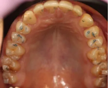
Fig. 1. Patient A.P. 24 years old. Occlusal contacts of the upper jaw registered with Bausch PROGRESS 100 µ
Fig. 3. Occlusal contacts of the upper jaw– two-stage registration
Fig. 2. Occlusal contacts of the lower jaw registered with Bausch PROGRESS 100 ì
Fig. 4. Occlusal contacts of the lower jaw – two-stage registration
RESULTS:
Fig. 5. Occlusal contacts of the upper jaw in CO
In contrast to Max, MIP presents contacts in a single frame of the record of maximum contact between teeth antagonists. Delta identifies differences between Max and MIP and shows areas of sliding as well as intermediate contacts that would otherwise be impossible to be registered.
Fig. 7. Occlusal scheme at MIP
Fig. 6. Occlusal contacts of the lower jaw in CO
For the purpose of correct interpretation of images we examine the results of computerized occlusal analysis during closure up to the maximum contact between teeth antagonists (maximal intercuspation - MIP). In the group of studied patients with bruxism and / or bruxomania it has been established an imbalance in the distribution of occlusal forces (in 89.3 % - 36 people there was a discrepancy in the distribution of forces in the lateral and in 85.7% - 34 people in the frontal areas of the dentition).
Data analysis shows that in bruxing patients an essential factor for preventing the spontaneous bilateral closure with balanced forces is the presence of interceptive contacts and sliding occurring from the first frame of the occlusion time until the MIP.
Interceptive contacts are displayed with the function Delta /Fig. 11/, which is equal to the difference between the maximum accumulated force MAX and MIP.
Max is the maximum closing force, registered on a series of frames in a movie /Fig. 9/. Max is a compilation of static (contacts in MIP) and dynamic (intermediate
Fig. 9. Occlusal scheme at Max.
Fig. 10. Patient S.P. 37 years old.: Occlusal contacts of the lower jaw at CO
Fig. 11. Occlusal scheme at Delta
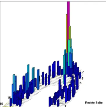
Fig. 12. Three-dimensional representation of intermediate contacts
Fig. 14. Three-dimensional representation of contacts in the Max
In our study patients with bruxism in neither case Max does not match to MIP and interceptive contacts are established in the way of closing.
It should be borne in mind that the clinical visualization of contacts with articulating paper along with contacts in CO always includes the contacts of Delta.
Fig. 8 and Fig. 10 present clinical registration of occlusal contacts in CO. Remarkable are the dense, large-area contacts on 14, 15, 24 and 25 which in daily clinical practice are perceived as strong contacts because of their large size and intensity. The three dimensional images of such contacts in the occlusal scheme in MIP, however, do not represent a dominance of these contacts /Fig. 13/. Fig.11 and Fig. 12 show that these are interceptive contacts that take place on the way from first contact up to the MIP, and they are not strong contacts.
DISCUSSION
The clinical problem when using the articulating paper for the registration of occlusal relationships is that based on the size and intensity of staining it was assessed whether a contact is “dense” or “light”. It is considered [4] that “those parts of the occlusal surface which are pressed together” in CO with the antagonist are “the dense” contacts. On the contrary - those which are “in a lightweight contact with the antagonist” are the “light” contacts. According to the teaching of Prosthetic Dentistry in the country “the dense contacts” with indicator paper are stained “intensively” and “the light are slightly marked.” This perceived in our country statement should not be taken as an axiom.
Our studies have shown that in many cases the relationship between the size of the contact and its force
force is measured with respect to the surface on which it acts and the broad contacts, even though intensively stained distribute the force over a larger area and as a result there is less concentration of the pressure. In contrast, small contacts concentrate the force upon small areas. It is known [8], that the smaller is the area of application of a force, the greater is the pressure. Computer analysis leads us to conclusion that in the daily practice in our country dentists probably read incorrectly the size of markings from the indicator paper, and interpret them reversely. Clinical occlusal analysis should be based on the understanding that the broad large contact means low pressure, whereas light, although seemed a little indication of the paper represents a high pressure which computerized occlusal analysis shows as a strong contact. Therefore, registration with articulating paper may serve as orientation about the location of contacts in CO. This method, however, could not be representative in terms of the area of contacts in CO and the forces applied in them. In this context, our studies confirmed the understanding of Quadeer et al. [19] that the size of markings with articulating paper should not be interpreted as a force applied in the contact, and should not be used to classify the contacts as strong and light.
Therefore, in patients with occlusal imbalances and unstable occlusion, such as patients with craniomandibular disorders, occlusal analysis can be considered to be objective and to be an indicator of subsequent therapeutic procedures only when performed by a clinical registering with articulating paper in combination with computerized occlusal analysis.
It should be taken into account that the registration with articulating paper does not provide data neither about the strength, nor for the timing of contacts or for their sequence.
The combination of computerized analysis of occlusion and its clinical registration with articulating paper provides an optimal opportunity to record and analyze all these parameters, allowing effective harmonization of occlusion in patients with functional pathology.
CONCLUSION
1. Droschl H,Permann I, Bantleon HP.Changes in occlusion and condylar positioning during retention with a gnathologic positioner. Eur J Orthod. 1989 Aug;11(3):221-227. [PubMed]
2. Egermark-Eriksson I, Ingervall B, Carlsson GE. The dependence of man-dibular dysfunction in children on func-tional and morphologic malocclusion. Am J Orthod. 1983 Feb;83(2):187-194. [PubMed]
3. Pirttiniemi P. Normal and in-creased functional asymmetries in the craniofacial area Acta. Odontol. Scand.
1998 Dec;56(6):342–345. [PubMed] 4. Sari S, Sonmez H. The relation-ship between occlusal factors and brux-ism in permanent and mixed dentition in Turkish children. J Clin Pediatr Dent.
2001 Spring;25(3):191–194. [PubMed] 5. Seligman DA, Pullinger AG: Assotiation of occlusal variables among refined TM patient diagnostic groups. J. Craniomandibular Disord. 1989 Fall; 3(4):227-236. [PubMed]
6. Filchev A. Anatomic and func-tional characterization of occlusal tooth contacts. Dissertation. Sofia. 1986 [In
Bulgarian]
7. Millstein P, Maya A. An evalua-tion of occlusal contact marking indica-tors. A descriptive quantitative method. J Am Dent Assoc. 2001 Sep;132(9): 1280-6. [PubMed] [CrossRef]
8. Peev T, Filchev A. Clinic of Pros-thetic Dentistry. Eco Print, Sofia. 2008. [In Bulgarian]
9. Kalachev J. Occlusal pressure and stress in the periodontium - analysis and guidelines for clinical use. Dissertation, Plovdiv, 2003. 41-48 [In Bulgarian]
10. Maness WL, Podoloff R. Distri-bution of occlusal contacts in maximum intercuspation. J Prosth Dent. 1989 Aug; 62(2): 238-42. [PubMed]
11. Tokumura K, Yamashita A. [Study on occlusal analysis by means of T-Scan system-its accuracy for measure-ment.] [in Japanese] Nippon Hotetsu Shika Gakkai Zasshi 1989 Nov; 33(5): 1037-1043. [PubMed]
12. Yamamura M, Takahashi A, Aoki H, Takeuchi N, Endo Y, Tamaki K, et al. [A study on display and accuracy of oc-clusal contacts by means of T-Scan Sys-tem.] [in Japanese] Kanagawa Shigaku REFERENCES:
Address for correspondence:
Assoc. Prof. Dr. Mariana Dimova, PhD
Department of Prosthetic Dentistry, Faculty of Dental medicine, Medical University Sofia.
1, St. G. Sofiiski Blvd., 1431 Sofia, Bulgaria e-mail: [email protected],
1990 Sep;25(2):236-241. [PubMed] 13. Tschernitschek H, Handel G, Gunay H. [T-scan - possibilities and limits of new occlusal diagnostic proce-dure.] [in German] Zahnarztl.Prax. 1990 Feb;41(2):54-56. [PubMed]
14. Scholz W, Pancherz H, Reichel R. [Review of T-SCAN systems for reg-istration of occlusal condition.] [in Ger-man] Zahnarztl.Prax. 1991 Jan 11; 42(1):6-9. [PubMed]
15. Waltz ME. The T-Scan system for occlusal registration. Gen Dent.
1991 Nov-Dec;39(6):451-454. [PubMed]
16. Filchev A, Ralev R. Propedeutics of prosthetic dentistry. Sofia, 2007. 120-125 [In Bulgarian]
17. Peev T. Cliniñ and treatment of occlusal tooth abrasion. Dissertation. Sofia. 1993. [In Bulgarian]
18. Quadeer S, Kerstein R, Kim RJ, Huh JB, Shin SW. Relationship be-tween articulation paper mark size and percentage of force measured with com-puterized occlusal analysis, J Adv Prosthodont. 2012 Feb,4(1):7-12. [PubMed] [CrossRef]
Acknowlegments
This research is related to the dissertation work of Assoc. Professor Mariana Dimova on “Current trends and gnathological prerequisites in the diagnosis and rehabilitation of craniomandibular disorders”, submitted for preliminary discussion at the Department of Prosthetic Dentistry, Faculty of Dental Medicine of Medical University - Sofia on 20. 05. 2013
The author is grateful to the Department of Prosthetic Dentistry at Faculty of Dental Medicine of Medical University - Plovdiv for the collegial approach and for the provided equipment in carrying out part of the research.