of
Wnt11
and
Ret
in Ureteric Tip Cells
Jason E. Cain1,2, Epshita Islam1, Fiona Haxho1, Lin Chen1, Darren Bridgewater1,2, Erica Nieuwenhuis1,3, Chi-Chung Hui1,3, Norman D. Rosenblum1,2,4,5,6*
1Program in Developmental and Stem Cell Biology, The Hospital for Sick Children, Toronto Medical Discovery Towers, Toronto, Ontario, Canada,2Division of Nephrology, The Hospital for Sick Children, Toronto, Ontario, Canada,3Department of Molecular and Medical Genetics, University of Toronto, Toronto, Ontario, Canada,4Department of Paediatrics, University of Toronto, Toronto, Ontario, Canada,5Department of Laboratory Medicine and Pathobiology, University of Toronto, Toronto, Ontario, Canada,
6Department of Physiology, University of Toronto, Toronto, Ontario, Canada
Abstract
Truncating GLI3 mutations in Pallister-Hall Syndrome with renal malformation suggests a requirement for Hedgehog signaling during renal development. HH-dependent signaling increases levels of GLI transcriptional activators and decreases processing of GLI3 to a shorter transcriptional repressor. Previously, we showed that Shh-deficiency interrupts early inductive events during renal development in a manner dependent on GLI3 repressor. Here we identify a novel function for GLI3 repressor in controlling nephron number. During renal morphogenesis, HH signaling activity, assayed by expression of
Ptc1-lacZ, is localized to ureteric cells of the medulla, but is undetectable in the cortex. Targeted inactivation ofSmo, the HH effector, in the ureteric cell lineage causes no detectable abnormality in renal morphogenesis. The functional significance of absent HH signaling activity in cortical ureteric cells was determined by targeted deletion ofPtc1, the SMO inhibitor, in the ureteric cell lineage.Ptc12/2UBmice demonstrate ectopicPtc1-lacZexpression in ureteric branch tips and renal hypoplasia
characterized by reduced kidney size and a paucity of mature and intermediate nephrogenic structures. Ureteric tip cells are remarkable for abnormal morphology and impaired expression of Ret and Wnt11, markers of tip cell differentiation. A finding of renal hypoplasia inGli32/2mice suggests a pathogenic role for reduced GLI3 repressor in thePtc12/2UBmice.
Indeed, constitutive expression of GLI3 repressor via theGli3D699allele inPtc12/2UBmice restores the normal pattern of HH
signaling, and expression of Ret and Wnt11 and rescued the renal phenotype. Thus, GLI3 repressor controls nephron number by regulating ureteric tip cell expression ofWnt11andRet.
Citation:Cain JE, Islam E, Haxho F, Chen L, Bridgewater D, et al. (2009) GLI3 Repressor Controls Nephron Number via Regulation ofWnt11andRetin Ureteric Tip Cells. PLoS ONE 4(10): e7313. doi:10.1371/journal.pone.0007313
Editor:Yihai Cao, Karolinska Institutet, Sweden
ReceivedJune 2, 2009;AcceptedSeptember 16, 2009;PublishedOctober 7, 2009
Copyright:ß2009 Cain et al. This is an open-access article distributed under the terms of the Creative Commons Attribution License, which permits unrestricted use, distribution, and reproduction in any medium, provided the original author and source are credited.
Funding:This research was supported by Operating Grants awarded by the Canadian Institutes of Health Research to Norman Rosenblum. Dr. Rosenblum is supported by a Canada Research Chair. The funders had no role in study design, data collection and analysis, decision to publish, or preparation of the manuscript.
Competing Interests:The authors have declared that no competing interests exist. * E-mail: [email protected]
Introduction
Development of the permanent mammalian kidney (the metanephros) is dependent on growth and branching of the ureteric bud and its daughter branches, a process termed renal branching morphogenesis. At the onset of this process, the ureteric bud elongates towards and invades the metanephric mesenchyme before undergoing spatial specification into ‘ureteric stalk’ and ‘ureteric tip’ domains. Reciprocal inductive interactions between the ureteric tip and surrounding metanephric mesenchyme results in division of the ureteric tip, forming the first of a series of ureteric branches, which ultimately constitute the mature collecting duct system. Simultaneously, each ureteric bud tip induces adjacent metanephric mesenchyme cells to undergo a mesenchyme-epithelial transformation and form the mesenchyme-epithelial components extending from the glomerulus to the distal tubule, a process known as nephrogenesis. The number of nephrons formed is directly related to the number of ureteric branches and their inductive capacity. Severe reductions in nephron number, characteristic of renal hypoplasia/dysplasia, are the leading cause of childhood renal failure. More subtle defects in nephron number
have been associated with the development of adult-onset essential hypertension and chronic renal failure [1,2,3,4,5].
The balance of GLI activator and repressor activities is critical during renal morphogenesis. Mutations that are predicted to generate a truncated protein similar in size to GLI3 repressor are observed in humans with Pallister-Hall Syndrome (PHS) and renal dysplasia [8]. The pathogenic role of constitutive GLI3 repressor activity during renal morphogenesis is further demonstrated by the renal dysplastic phenotype in mice engineered to express GLI3 repressor in a dominant manner [9] and in Shh-deficient mice [10]. Dysplastic kidney tissue inShh-deficient mice is characterized by sustained GLI3 repressor expression in the face of decreased levels of GLI activators (GLI1, GLI2 and full-length GLI3), resulting in a shift in the balance of GLI activators and GLI repressors in favor of repressor [10]. Remarkably, genetic elimination ofGli3in theShhnull background restores expression of GLI activators and normalizes renal morphogenesis [10]. The expression ofShhin ureteric cells suggests that it may control renal development via direct effects in the ureteric cell lineage. While conditional inactivation of Shh in ureteric cells results in renal hypoplasia, characterized by reduced kidney size and glomerular number [11], the dependency of this pathogenic phenotype onShh
signaling in ureteric cells is unknown.
Here we define the specific function of HH signaling in the ureteric cell lineage during murine kidney development, in genetic models of deficient or constitutively active signaling. HH signaling activity is specifically restricted to the ureteric cells of the medulla and ureter but is absent from the ureteric cell tips of the renal cortex. Genetic inactivation of Smo in the ureteric cell lineage exerted no deleterious effects on renal morphogenesis. In contrast, genetic inactivation of Ptc1 in the ureteric cell lineage caused ectopic HH signaling activity in ureteric tip cells, impaired ureteric tip cell-specific gene expression and renal hypoplasia. Genetic inactivation ofGli3alone, the primary GLI repressor, resulted in a similar phenotype suggesting a critical role for GLI3 repressor. Indeed, introduction of a constitutively active GLI3 repressor in a
Ptc1-deficienct background normalized the renal phenotype, restored the normal domain of HH signaling activity and rescued expression of genes specific to ureteric tip cells and required for their functions. We propose a model in which SHH-SMO signaling controls the spatial generation of GLI3 repressor, which is required in the cortical ureteric cells for ureteric tip cell-specific gene expression and cell function.
Results
Spatial Restriction of HH Signaling Activity to the Renal Medulla and Ureter
To begin to further investigate the role of SHH signaling during renal morphogenesis, we examined the expression ofPtc1
utilizing the Ptc1-lacZ reporter mouse [12]. Since Ptc1 is a downstream target of HH signaling, Ptc1-lacZ expression is indicative of the site of HH signaling activity [12,13,14]. In the
WT(Ptc1lacZ/+) kidney at E13.5,Ptc1-lacZis strongly localized to cells surrounding the presumptive ureter and the presumptive medullary stroma (Figure 1A,B), consistent with the pattern of
Ptc1mRNA expression [11].Ptc1-lacZis also weakly localized to the epithelium of the presumptive ureter and the distal or medullary collecting ducts (Figure 1A–C). Interestingly,Ptc1-lacZ
expression is not observed in any structures of the presumptive renal cortex, suggesting that HH signaling activity is restricted to the ureter and medullary regions of the developing kidney (Figure 1B,D). At a later stage (E18.5) of kidney development, a similar pattern of expression is maintained in the cells surrounding the ureter and medullary stroma (Figure S1A–C). However, at E18.5,Ptc1-lacZexpression is not observed in any
epithelial structures (Figure S1A–C). Taken together, Ptc1-lacZ
expression in both ureteric and metanephric mesenchyme-derived structures suggests a role for SHH function in both the ureteric bud and metanephric mesenchyme lineages of the early developing kidney but only in the presumptive ureter and medullary regions.
SHH-SMO-Dependent Signaling is not Required in the Ureteric Cell Lineage
We began to investigate the possible autocrine functions of SHH-SMO-dependent signaling by generating a loss of function model for HH signaling in the ureteric cell lineage.
Smoothened is required for the transduction of all HH signals. Similar toShhinactivation, inhibition of SMO with a steroidal alkaloid, cyclopamine, results in sustained GLI3 repressor in the absence of GLI activators [10]. Homozygous germline defi-ciency ofSmoin the mouse results in embryonic lethality prior to the commencement of metanephric development [15]. There-fore, we utilized Hoxb7creEGFP mice to generate a murine model in whichSmois genetically inactivated in the ureteric cell lineage [16,17], thereby eliminating SHH-SMO-dependant signaling.
Targeted deficiency ofSmoin the ureteric cell lineage did not adversely effect survival since mutants were recovered in the near expected Mendelian ratios (Table S1). To confirm thatSmo is abolished inSmo2/2UBmutants, we performed quantitative real-time PCR using ureteric bud tissue isolated at E11.5, a stage that closely follows metanephric induction. Interestingly,SmomRNA transcripts were decreased by,80% inSmo2/2UBureteric cells
compared toWT(WTvs.Smo2/2UB
: 0.6060.12 vs. 0.1260.08, p,0.05) (Figure 1E). Consistent with a loss ofSmo, analyses of
Ptc1-lacZ in Smo2/2UB
kidneys at E13.5 revealed a marked reduction in HH signaling activity in the ureteric cells of the ureter and medullary collecting ducts (Figure 1F,G). Examina-tion of the gross anatomical and histological features of newborn
Smo2/2UB
kidneys revealed no major differences compared to
WTkidneys (Figure 1H,I,L,M). Consistent with this observation, a more detailed analysis of nephrogenic and ureteric structures using immunofluorescence microscopy and RNAin situ hybrid-ization revealed that glomerulogenesis, nephrogenesis, nephron segmentation, ureteric branching morphogenesis and ureteric tip cell-specific gene expression was normal in Smo2/2UB kidneys (Figure S2). In order to determine if HH-SMO-dependant signaling is required in the mature kidney, we also examined
Smo2/2UB
kidneys at PN30. No histological abnormalities were detected in the Smo2/2UB
kidneys (Figure 1J,K,N,O). Taken together, these results demonstrate that HH-SMO-dependent signaling is not required in the ureteric cell lineage and suggest that SHH has no autocrine function [in the ureteric cells] during renal morphogenesis.
A Model of Cortical HH Signaling Activity in the Embryonic Kidney
Our results demonstrate that HH signaling activity is absent from the renal cortex. We determined the importance of this spatial restriction of HH signaling activity by generating a gain-of-function model in which HH signaling is ectopically activated in the cortical ureteric epithelium.
Patched1is a negative regulator of the HH signaling pathway. In the absence ofPtc1, repression of HH target genes is alleviated, even in the absence of SHH ligand. Homozygous germline deficiency of
Ptc1 in the mouse results in embryonic lethality prior to the commencement of metanephric development [12]. Therefore, we
utilizedHoxb7creEGFPmice to generate a murine model in which
Ptc1is genetically inactivated in the ureteric cell lineage [16,17] (refer to Methods). To confirm that inactivation ofPtc1results in a constitutively active HH signaling pathway in the ureteric cell lineage, we analyzed Ptc1-lacZ expression in Ptc12/2UB
kidneys. While Ptc1-lacZ expression is maintained in the cells surrounding the presumptive ureter and presumptive medullary stroma (Figure 2A,B), Ptc1-lacZ is markedly upregulated in the epithelium of the presumptive ureter and distal collecting ducts in
Ptc12/2UB
kidneys (Figure 1C vs. Figure 2C). Remarkably,Ptc1-lacZ
is ectopically expressed throughout the cortical or proximal collecting ducts and in the ureteric bud tips (Figure 2A,B,D), albeit in a mosaic pattern (Figure S1F). A similar pattern of upregulated
and ectopic ureteric bud epithelialPtc1-lacZ expression was also observed at a later stage (E18.5) of kidney development (Figure S1D-F). To confirm thatPtc1inactivation occurs by an early stage in renal morphogenesis, we assayed expression of Ptc1 mRNA, a surrogate marker of HH signaling activity, in ureteric bud tissue isolated from E11.5 kidney. Quantitative real-time PCR using primers designed for an undisrupted region of the mutant Ptc1
transcript demonstrated a 50-fold increase in Ptc1 mRNA transcripts in Ptc12/2UB
compared to WTureteric cells (WTvs.
Ptc12/2UB
: 2.2260.02 vs. 108.5166.47, p,0.001) (Figure S3H). Together, these results show that genetic elimination of PTC1 in the ureteric cell lineage leads to increased and ectopic HH signaling activity in the developing kidney.
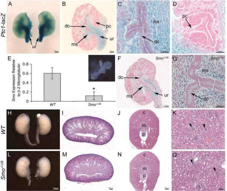
Figure 1.Ptc1-lacZexpression and site of HH signaling activity in the developing murine kidney.(A–D) Whole mount X-gal staining of kidney tissue isolated from E13.5Ptc1-lacZreporter mice reveals strong localization of HH signaling activity to the cells surrounding the presumptive ureter (ur), medullary stroma (ms) and weakly to the epithelium of the presumptive ureter and distal collecting ducts (dc). (D)Ptc1-lacZis not observed in any structures of the presumptive renal cortex. (E) Quantitative real-time PCR of isolated E11.5 ureteric buds (inset) demonstrates decreasedSmomRNA transcripts inSmo2/2UB
kidneys compared toWTlittermates (WTvs.Smo2/2UB
: 0.6060.12 vs. 0.1260.08, p,0.05). (F,G) Ptc1-lacZexpression inSmo2/2UB
kidneys at E13.5.Ptc1-lacZexpression is maintained in the cells surrounding the ureter and medullary stroma but is markedly reduced in the ureteric cells of the ureter and distal collecting ducts. (H,I,L,M) Macroscopic and histological analysis of newbornSmo2/2UB
kidneys is comparable toWTlittermates. (J,K,N,O) Analysis of matureSmo-deficient kidneys at PN30 demonstrates no histological abnormalities. c = cortex, m = medulla, p = papilla, pc = proximal collecting duct, arrowhead = mature glomerulus.
Figure 2. Renal hypoplasia in mice withPtc1-deficiency in the ureteric cell lineage.(A–D) InPtc1mutant kidneys (Ptc12/2UB), Ptc1-lacZ
expression is maintained in the cells surrounding the presumptive ureter (ur) and medullary stroma (ms). (A–C) HH signaling activity is upregulated in the epithelium of the presumptive ureter and distal collecting ducts (dc). (D) Ectopic HH signaling activity is observed in the proximal collecting ducts (pc) and weakly in ureteric bud tips. (E–L) Histological and immunofluorescence analysis ofWTandPtc12/2UB
kidneys at E18.5.Ptc1-deficient mice exhibit small kidneys, reduced density of medullary epithelial tubules (mt) and a paucity of mature glomeruli (g) and nephrogenic intermediate structures (n) and sparsity of the nephrogenic zone (arrows). (G,K) PAX2 (green) positive nephrogenic structures are reduced in thePtc12/2UB
kidneys. (H,L) LTL (green) positive proximal tubules are markedly reduced inPtc12/2UB
kidneys. (M) Kidney volume inPtc12/2UB
mutants is reduced by 45% in comparison to WTlittermates (p,0.01). (N) Ptc12/2UB
kidneys demonstrate a 45% reduction in mature glomerular number compared to WT
littermates (p,0.01). (O,P) Histological analysis also revealed a sparsity of the nephrogenic zone (arrows) and reduction in the number of nephrogenic structures inPtc12/2UB
compared toWTkidneys at E15.5. (Q,R) Analysis of ureteric branching morphogenesis inWTandPtc12/2UB
kidneys at E12.5. The number of ureteric branches was comparable betweenWTandPtc12/2UB
kidneys. However,Ptc12/2UB
kidneys exhibited abnormal ureteric tip morphology (arrowhead). (S–V) Whole mount GFP immunofluorescence of control and mutant kidneys. Kidney size and branching is mildly reduced at E13.5 (S,T) and is more pronounced at E15.5 (U,V).
doi:10.1371/journal.pone.0007313.g002
Ectopic HH Signaling Activity in the Proximal Epithelium Causes Renal Hypoplasia
Viable neonatal PTC1-deficient pups could not be recovered (Table S2). However, live Ptc12/2UB
mutant embryos were recovered in expected Mendelian ratios at all embryonic time points analyzed (Table S2). Macroscopic analyses of Ptc12/2UB
embryos revealed severe exencephaly, with 100% penetrance. Otherwise, Ptc12/2UB
embryos were indistinguishable fromWT
littermates (Figure S3). It is therefore likely that Ptc12/2UB
embryos reach term and die and/or are cannibalized immediately following birth.
Analysis of Ptc12/2UB
kidneys at E18.5 revealed renal hypoplasia, characterized by a 45% reduction in kidney volume (WTvs.Ptc12/2UB
: 163.67638.8761026mm3vs. 90.72624.056 1026
mm3, p,0.01) and reduced density of medullary epithelial tubules (Figure 2E,I,M). The extent of reduction in Ptc12/2UB
kidney size was variable, exhibiting a 1.7 fold range, but was 100% penetrant. Histological analyses of the renal cortex ofPtc12/2UB
kidneys revealed a paucity of mature glomeruli and nephrogenic intermediate structures in the nephrogenic zone (Figure 2F,J). Glomerular number in Ptc12/2UB
kidneys was decreased by 45% in comparison to WT littermates (WT vs. Ptc12/2UB
: 692.226116.35 vs. 374.25668.79, p,0.001) (Figure 2N). A deficiency in nephrogenic structures in Ptc12/2UB
kidneys was confirmed by examining the expression of PAX2, a marker of condensing mesenchyme and early nephrogenic structures (Figure 2G,K) and LTL, a marker of proximal tubules (Figure 2 H,L). A reduction in the number of intermediate nephrogenic structures was also observed inPtc12/2UB
kidneys by histology at E15.5 (Figure 2O,P). However, no difference in renal histology or the number of nephrogenic intermediate structures was observed between WT and Ptc12/2UB
kidneys at E13.5 suggesting that nephrogenesis is unaffected at that stage (Figure S4). Together, these results demonstrate that PTC1-deficiency leads to deficient nephrogenesis.
Ureteric branching is required for nephron formation. Since ureteric branch tips induce contiguous metanephric mesenchyme cells to engage in nephron formation, the number of ureteric branches is considered to be a critical determinant of the number of nephrons generated. To determine the effect ofPtc1-deficiency on early ureteric branching morphogenesis we quantified ureteric branching at E12.5 by whole mount immunofluorescence using Calbindin-D28K, a marker of ureteric bud epithelium. No significant difference in the number of ureteric branches was observed betweenWTandPtc12/2UB
kidneys (Figure 2Q,R and Figure S4F). However, we did observe abnormalities in the ureteric branch pattern. In contrast to WT kidneys, several irregularly shaped and dilated ureteric bud tips were observed in
Ptc12/2UB
kidneys (Figure 2Q,R). Analyses of ureteric branching morphogenesis a day later at E13.5 revealed a mild reduction in ureteric branch number in Ptc12/2UB
kidneys compared to controls (Figure 2S,T). At E15.5, this reduction was more even more pronounced (Figure 2U,V). Importantly, normal renal architecture is maintained inPtc12/2UB
kidneys. Taken together, these results demonstrate that ectopic HH signaling activity in proximal ureteric cells causes renal hypoplasia, characterized by marked defects in the formation of nephrogenic structures, reduced ureteric branching and a qualitative defect in early stage ureteric branch tips.
Effect of Ptc1-Deficiency on Proliferation and Apoptosis
Cell proliferation is crucial for ureteric branching morphogen-esis and nephrogenmorphogen-esis. Since SHH signaling controls renal cell proliferation [10] we investigated the possible contributions of
abnormal cell proliferation to the hypoplastic phenotype in PTC1-deficient kidneys at E13.5, a time point prior to the onset of the phenotype. Analysis of cell proliferation, using an in situ BrdU incorporation assay, revealed a 40% decrease in ureteric bud cell proliferation inPtc12/2UB
kidneys at E13.5 (WT vs.Ptc12/2UB
: 38.465.6 vs. 23.169.0, p = 0.05) (Figure 3A–C). In contrast, no significant difference in mesenchymal cell proliferation was detected (WT vs. Ptc12/2UB
: 2.6860.7 vs. 2.0560.6, p = 0.25) (Figure 3A,B,D).
Mesenchyme cell survival is critical to nephrogenesis. Moreover, elevated ureteric bud apoptosis has been implicated in the pathogenesis of renal hypoplasia [18]. Accordingly, we determined whether cell survival was impaired in PTC1-deficient kidneys at E13.5 (Figure S5). Analysis of apoptosis, using a TUNEL assay, revealed no significant difference in ureteric bud or metanephric mesenchyme cell death inPtc12/2UB
kidneys.
Taken together, these results suggest that enhanced and ectopic HH signaling activity in the ureteric cell lineage decreases ureteric cell proliferation but does not affect apoptosis.
Ectopic HH Signaling Activity in the Proximal Ureteric Epithelium Impairs Expression ofWnt11andRet in Ureteric Bud Tip Cells
Our findings of renal hypoplasia, abnormal ureteric tip morphology and decreased ureteric cell proliferation (inPtc12/2UB
kidneys) suggested that ectopic HH signaling activity in ureteric tips disrupts normal ureteric tip function.
Specification of ureteric bud tip cells distinct from ureteric stalk cells is essential for ureteric branching morphogenesis and nephrogenesis. Determination of ureteric tip cell fate is dependant on Gdnf/Ret signaling [19]. Furthermore, Gdnf/Ret signaling is required for the maintenance of Wnt11 expression, also in the ureteric bud tips cells [20,21]. Conversely,Wnt11promotesGdnf
expression in the surrounding metanephric mesenchyme suggesting thatGdnf,RetandWnt11participate in an autoregulatory feedback loop to regulate ureteric branching morphogenesis [20]. We examined the effect of ectopic HH signaling activity on ureteric tip cell-specific gene expression using in situ hybridization. In contrast toWTkidneys, expression ofRetandWnt11was markedly reduced in the majority of ureteric bud tips inPtc12/2UB
kidneys at E13.5 (Figure 3E–H). Consistent with the autoregulatory feedback loop, expression ofGdnf in the surrounding metanephric mesen-chyme was also markedly reduced in the Ptc12/2UB
kidneys (Figure 3I,J). The specificity of decreased Gdnf expression in
Ptc12/2UB
kidneys is demonstrated by normal expression ofOsr1,
Six2, and CITED1, each of which marks mesenchymal precursor cells [22,23,24,25,26]; Wnt4, a marker of pretubular aggregates [27]; andWnt9bwhich is required for the earliest inductive response in metanephric mesenchyme and acts upstream of Wnt4 [28] (Figure S6). To further investigate the ontogeny of abnormal ureteric tip cell gene expression inPtc12/2UB
mice, we assayedRet
and Wnt11 expression in isolated ureteric bud tissue using quantitative real-time PCR. At E11.5, a stage that immediately follows induction of the metanephric mesenchyme by the ureteric bud, expression ofRetwas comparable betweenWTandPtc12/2UB
ureteric cells (WT vs. Ptc12/2UB
: 1.6560.37 vs. 1.5060.61, p = 0.78) (Figure 3M). In contrast,Wnt11expression was reduced by,70% inPtc12/2UBureteric cells (WTvs.Ptc12/2UB: 1.7960.26
vs. 0.5160.27, p,0.05) (Figure 3N). Taken together, these results demonstrate that ectopic HH signaling activity in the proximal ureteric epithelium specifically impairs the expression ofWnt11and
Retin ureteric tip cells.
Given the impairment of tip cell gene expression, we investigated the possibility that HH signaling activity biases ureteric cells towards a distal cell fate. To determine whether Ptc12/2UB
tips cells had adopted characteristics of the ureteric stalk we performed DBA-lectin whole mount immunofluorescence microscopy onWT
andPtc12/2UB
kidneys at E13.5. DBA is a marker of the ureteric stalk but not the ureteric tip [29]. DBA expression was observed in ureteric tips inPtc12/2UB
kidneys in a mosaic pattern but rarely in
WTkidneys (Figure 3K,L and Figure S5E–H). The expression of
Wnt7b, another marker of ureteric stalk that is absent from the
ureteric tips [30], was comparable betweenWT and Ptc12/2UB
kidneys. Since ectopic HH signaling activity is also observed in proximal collecting ducts in mutant kidneys, we next investigated the possibility that HH signaling activity biases cortical collecting ducts towards a more medullary/distal cell fate. Expression of uroplakin III, a marker of the transitional epithelium of the ureter and renal pelvis, was unchanged in thePtc12/2UB
kidneys (Figure S7A–F). Similarly, the localization of aSMA, a marker of the smooth muscle population surrounding the ureter, was also unaltered (Figure S7G–L). Together, these results suggest that Figure 3. Ectopic HH signaling activity in the proximal epithelium impairs ureteric tip cell–specific gene expression.(A–D) Analysis of cell proliferation in E13.5 kidney tissue using in situ BrdU incorporation assay. BrdU (brown color) is decreased in ureteric cells inPtc12/2UB
kidney compared toWT. (C) Quantitative analysis of ureteric cell proliferation. Ureteric cell proliferation, quantitated as the percent of BrdU-positive ureteric cells, was decreased inPtc1-deficient kidneys (p = 0.05). (D) Quantitative analysis of mesenchymal cell proliferation. Mesenchymal cell proliferation, quantitated as the number of BrdU positive cells per mm2of renal tissue was comparable inWT andPtc12/2UB
kidneys. ub = ureteric bud, mes = metanephric mesenchyme. (E–H) RNAin situhybridization demonstrates expression ofRetandWnt11exclusively to the ureteric bud tips (arrowhead) inWTkidney tissue. In contrast, these mRNA transcripts are either absent or markedly reduced inPtc12/2UB
kidney tissue (arrowhead). (I,J) RNAin situhybridization demonstrates thatGdnfis markedly reduced in metanephric mesenchyme ofPtc12/2UB
kidneys (arrows). (K,L) DBA-lectin localizes predominantly to the ureteric stalk inWTkidneys and is excluded from the ureteric tips at E13.5. InPtc12/2UB
kidneys DBA-lectin is observed throughout the ureteric tips and ureteric stalks. (M,N) Quantitative real-time PCR of isolated WTandPtc12/2UB
ureteric buds at E11.5. (M)Ret
expression is similar betweenWTandPtc12/2UB
ureteric cells. (N)Wnt11expression is significantly decreased inPtc12/2UB
ureteric cells (p,0.05). doi:10.1371/journal.pone.0007313.g003
ectopic HH signaling activity in the proximal epithelium does not induce a distal ureteric bud cell fate. Furthermore, increased HH signaling activity in the distal epithelium (ureter and distal collecting ducts) has no deleterious effects on these structures.
Reduced Levels of GLI Repressor in the Renal Cortex Result in Renal Hypoplasia
The spatial restriction of HH signaling activity from the renal cortex during normal renal morphogenesis suggests that the cortex is a GLI repressor-dominant domain. We hypothesized that the deleterious effects of ectopic HH signaling in the proximal ureteric cells in Ptc12/2UB
kidneys is due to a reduction in local GLI repressor levels. We addressed this hypothesis by analyzing kidneys inGli3-deficient embryos.
GLI3 is the primary source of GLI repressor in mammalian cells [31]. Mice with homozygous deficiency inGli3die soon after birth or in late gestation [32]. We were able to recover viable Gli3 -deficient embryos as late as E18.5. Gli3-deficient embryos exhibited polysyndactyly and occasional exencephaly but were similar in size toWTlittermates (data not shown).Gli3+/XtJ
mice kidneys were indistinguishable from WT littermates (data not shown). Analysis of Gli3XtJ/XtJ kidneys at E18.5 revealed mild hypoplasia, characterized by a 15% decrease in kidney volume (p,0.05) and 15% reduction in glomerular number (p,0.05) compared toWTlittermates (Figure 4). Otherwise, glomerulogen-esis, nephron segmentation, and smooth muscle and urothelium differentiation was normal inGli3XtJ/XtJkidneys (Figure S8). These results are consistent with a functional role for GLI repressor in the renal cortex during renal morphogenesis.
GLI3 Repressor is Required for Ureteric Tip Cell Expression ofWnt11andRet
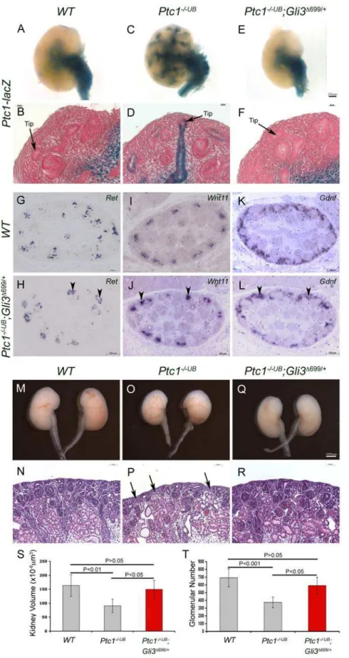
We further tested the contribution of GLI repressor in the proximal epithelium by reinstating GLI3 repressor levels in the
Ptc12/2UBmutants. We generated mice with bothPtc1-deficiency targeted to the ureteric cells and with one allele of WT Gli3
replaced with Gli3D699
, a constitutively active repressor form of GLI3. To determine what effect, if any, the reinstatement of GLI3 repressor had on HH signaling activity we analyzedPtc1-lacZin the Ptc12/2UB
;Gli3D699/+ kidneys. In the Ptc12/2UB
;Gli3D699/+ kidney at E13.5, Ptc1-lacZ expression is maintained in the cells surrounding the presumptive ureter and medullary stroma, consistent with the expression in WT and Ptc12/2UB
kidneys (Figure 5A,C,E). However, in contrast to ectopic Ptc1-lacZ
expression throughout the proximal ureteric cells in Ptc12/2UB
kidneys, the proximal collecting ducts and ureteric cell tips are devoid of Ptc1-lacZ expression in Ptc12/2UB;Gli3D699/+
kidneys (Figure 5A–F). These results suggest that reinstatement of GLI3 repressor inPtc12/2UB
kidneys, restores the normal pattern of HH signaling activity to the medullary and ureter domains and eliminates ectopic HH signaling activity in the renal cortex. Next we analyzed ureteric tip cell gene expression in Ptc12/2UB
; Gli3D699/+
kidneys. Remarkably, expression ofRetandWnt11was restored to comparable levels of that observed in WT and
Gli3D699/+
controls suggesting a rescue of ureteric tip cell differentiation (Figure 3E,H, Figure 5G,J, Figure S9). Further-more, consistent with a rescue ofRetandWnt11expression,Gdnf
expression was also restored by the reinstatement of GLI3 repressor (Figure 3I,J, Figure 5K,L, Figure S9). Moreover, macroscopic analysis of Ptc12/2UB
;Gli3D699/+
kidneys at E18.5 demonstrated a rescue in kidney size, comparable to that observed inWTlittermates (Figure 5M,O,Q). In fact, kidney volume was significantly restored from 55% in Ptc12/2UB kidneys alone, to
92% inPtc12/2UB
;Gli3D699/+
kidneys in comparison toWTkidney volume (WT vs. Ptc12/2UB;Gli3D699/+
, p.0.05; Ptc12/2UB vs.
Ptc12/2UB;Gli3D699/+
, p,0.05) (Figure 5S). Remarkably, histolog-ical analyses of the renal cortex also revealed a rescue in nephrogenesis in Ptc12/2UB;Gli3D699/+
kidneys (Figure 5N,P,R). In contrast to a 45% deficit in Ptc12/2UB kidneys, glomerular number inPtc12/2UB;Gli3D699/+
kidneys was significantly restored to 85% of that ofWT littermates (WTvs.Ptc12/2UB;Gli3D699/+
, p.0.05;Ptc12/2UB
vs.Ptc12/2UB;Gli3D699/+
, p,0.05) (Figure 5T). Taken together, these results demonstrate a requirement for GLI3 repressor for ureteric tip cell gene expression and function.
Discussion
Disruption of renal development in humans with Pallister-Hall Syndrome and truncating GLI3 mutations [33] and mice with elevated levels of GLI3 repressor [9,10] provides compelling evidence in favor of a critical role for GLI3-dependent signaling during mesenchymal-epithelial interactions during early stages of Figure 4. Loss of GLI repressor results in renal hypoplasia.(A–F) Analysis ofGli32/2kidneys E18.5. Histological analysis ofGli3deficient
kidneys revealed a reduction in kidney size (A,B) and a reduced density of the nephrogenic zone (C,D). (E) Kidney volume was reduced by 15% inGli3-deficient kidneys (p,0.05). (F) The number of mature glomeruli was reduced by 15% inGli3-deficient kidneys (p,0.05).
Figure 5. GLI3 repressor is required for ureteric tip cell-specific gene expression and function.(A–F) Normal of HH signaling activity is restored inPtc12/2UB;Gli3D699/+
kidneys at E13.5. In contrast toPtc12/2UB
kidneys,Ptc1-lacZexpression is absent from the cortical collecting ducts and ureteric bud tips inPtc12/2UB
;Gli3D699/+kidneys. (G–L) Expression ofRet,Wnt11andGdnfwas restored inPtc12/2UB
;Gli3D699/+kidneys at E13.5. Analysis
ofPtc12/2UB;Gli3D699/+
kidneys at E18.5 revealed a restoration in kidney size (M,O,Q) and density of the nephrogenic zone (N,P,R). Kidney volume (S) and glomerular number (T) is rescued inPtc12/2UB
;Gli3D699/+kidneys to comparable levels toWTlittermates.
doi:10.1371/journal.pone.0007313.g005
metanephric development. However, the functions of HH signaling during subsequent morphogenic events including nephrogenesis are unknown.
Here, we demonstrate that domains of GLI-dependent activator and repressor function are spatially patterned during renal morphogenesis. We investigated the functional significance of these domains in the ureteric cell lineage using genetic murine models of deficient or constitutively active HH signaling. Smo -deficiency targeted to the ureteric cell lineage does not disrupt kidney development, demonstrating HH-dependent GLI activa-tors are not required for ureteric cell function. The absence of HH signaling in the renal cortex ofWTmice suggests that the cortex is a zone of low GLI activator and high GLI repressor levels. We determined the importance of exclusion of HH signaling activity from the cortical collecting ducts in mice with Ptc1-deficiency targeted to the UB lineage. Absence of ureteric cellPtc1, a negative regulator of the HH signaling pathway, results in ectopic HH signaling activity in the cortical collecting ducts and ureteric bud tips. Ectopic HH signaling activity in the ureteric bud tips, which under normal circumstances is a domain of GLI repressor function, leads to decreased expression ofRetand Wnt11. These changes result in disruption of ureteric branching morphogenesis and nephrogenesis resulting in renal hypoplasia, likely due to impaired tip function. Remarkably, constitutive GLI3 repressor expression in thePtc12/2UB
background, restores ureteric tip cell-specific gene expression and normalizes renal morphogenesis, demonstrating a spatial requirement for GLI3 repressor in the proximal ureteric cells. Together, these results demonstrate a requirement for GLI3 repressor-dependent regulation of nephron number via ureteric tip cellWnt11- andRet-dependent functions.
Regulation of Ureteric Tip Cell Differentiation by GLI3 Repressor
We have established that loss of GLI3 repressor impairs ureteric tip cells by reducing Ret and Wnt11 expression. The precise mechanism by which GLI-dependent signaling may control Ret
andWnt11is unclear. Decreased expression ofWnt11precedes a decrease in Retexpression in the ureteric bud tips ofPtc12/2UB
kidneys. Wnt11 maintains Gdnf expression in the mesenchyme [20]. Thus, it is probable that reduced Gdnf expression in
Ptc12/2UB
mice is secondary to decreased Wnt11 expression in ureteric tip cells. Consistent with this,Wnt11null mice exhibit mild renal hypoplasia and a reduction inGdnf expression [20] almost identical to that observed inPtc12/2UB
mutants.
The mosaic expression ofPtc1-lacZexpression in the ureteric tips suggests that not all tip cells were exposed to ectopic HH signaling. This explains, in part, why Ret and Wnt11 expression is not completely abolished from the ureteric tips and why expression levels are variable among tips within the same kidney. It is tempting to speculate that the extent of ‘chimerism’ of a ureteric tip may influence the efficiency of its function. The ability of ‘mutant’ tips to confer some function inPtc12/2UB
kidneys could account for the mild phenotype since complete abolishment ofGdnf/Retsignaling would result in renal agenesis [34,35,36]. Impairment of tip cell gene expression has also been reported in mice with deletion ofb -catenin targeted to the ureteric cells [37]. Consistent with our findings,b-catenin-deficient mice demonstrate reduced expression of Ret,Wnt11and Gdnfand dilated ureteric tips. However, while early ureteric branching is normal in Ptc12/2UB
kidneys, it is arrested inb-catenin mutants resulting in severe renal dysplasia. Thus, it is likely that some residual inducing function is maintained in the ureteric tips inPtc12/2UB
mice during early stages of kidney development. However, the participation ofGdnf,RetandWnt11in an autoregulatory feedback loop to regulate ureteric branching
morphogenesis is likely to exacerbate the early decrement inWnt11,
Ret, and Gdnf contributing to the progressively more severe branching phenotype we observed inPtc12/2UB
mice.
Gli2andGli3can serve both redundant activator and repressor functions in a number of mammalian tissues [31,38,39]. Therefore, it is not surprising that the effect of Gli3-deficiency alone is less severe than that observed in conditional deletion of
Ptc1. Constitutive activity of the HH signaling pathway, such as that inPtc12/2UB
kidneys, is expected to inhibit the formation of bothGli2andGli3repressor isoforms. However, the capacity for the formation of Gli2repressor isoforms remains intact in Gli3 -deficient kidneys allowing for partial redundancy. Early lethality prior to the onset of metanephric development in the majority of
Gli2;Gli3 compound null mutants [31] limits the ability to investigate this functional redundancy in greater detail.
HH signaling is a known regulator of proliferation in mammalian tissues. Indeed, mutations inPtc1are associated with increased incidence of tumorigenesis [12]. In contrast, we observed a moderate decreased in ureteric cell proliferation in
Ptc1-deficient kidneys. Gdnf/Ret signaling has been shown to activate intracellular signaling pathways including the ERK MAP kinase pathway [40]. In the kidney, activation of this pathway leads to cellular events, including cell proliferation, cell survival and migration, and inhibition of this pathway results in reduced ureteric branching [41]. Therefore, reduced Ret and Gdnf
expression in Ptc12/2UB
kidneys could result in the observed reduction in proliferation. Consistent with this, the absence of a phenotype in Smo2/2UB kidneys suggests HH signaling is not required for ureteric cell proliferation.
Inactivating mutations inGli3andPtc1have been identified in humans with Greig Cephalopolysyndactyly Syndrome (GCPS) and Nevoid Basal Cell Carcinoma Syndrome (NBCCS, also known as Gorlin Syndrome) respectively [32,42]. Pertinent to the phenotype, mutations would lead to loss of GLI3 repressor and PTC1-deficiency respectively. Yet, currently no known association between human Gli3 inactivation or Ptc1 mutations and renal malformation exists. This is likely due to a number of reasons. Firstly, analysis of kidneys in patients with GCPS or NBCCS has not been performed. Our results provide a basis for analyses of kidney size in affected individuals. Secondly, the degree of haploinsufficiency in humans with Ptc1 mutations may be insufficient to result in a renal phenotype. Ptc1 heterozygous mutant mice exhibit phenotypes similar to those observed in humans with BNCCS [12], yet we did not observe any renal abnormalities in these mice. Furthermore,Ptc1homozygous null mice are embryonic lethal suggesting thatPtc1homozygous null mutations in humans are also incompatible with life [12]. Further, mutational analysis in humans exhibiting sporadic renal hypopla-sia for genes involved in HH signaling, includingGli3andPtc1, is also warranted, as done for other genes [43,44].
SHH Does Not Signal in a Autocrine Manner During Renal Morphogenesis
mesenchyme. In addition, recent in vitro and in vivodata in the pancreas, has suggested additional, non-canonical, mechanisms of GLI activation, downstream of SMO, via two HH-unrelated pathways, RAS and TGFb [45,46,47]. Whether non-canonical GLI activation occurs during renal morphogenesis remains unknown.
A Model of Spatial GLI Activator and Repressor Functional Domains
We propose a model whereby distinct SHH-dependent medullary GLI activator domain and cortical repressor domain functions are critical for normal renal ureteric patterning and function (Figure 6). It is likely these domains are established by a SHH gradient, emitted by the ureteric cells of the medullary collecting ducts. While SHH signaling is not required in the medullary collecting ducts themselves, the absence of signal in cortical ureteric cells, contributed to by diminishing SHH concentration and/or pathway inhibitors, is critical for ureteric tip cell gene expression required for ureteric branching and nephron induction. Identification of GLI3 repressor gene targets will provide novel insights into this novel mechanism of ureteric tip cell regulation and function. The presence of renal agenesis/ dysplasia in humans with Pallister-Hall syndrome and GLI3 repressor dominant murine models [9,10] suggests that a fine spatial and lineage-specific balance of GLI activator and GLI repressor must be maintained for normal renal morphogenesis. Furthermore, these results implicate misregulation of HH signaling as a possible underlying mechanism in unexplained human renal dysplasia.
Materials and Methods
Ethics Statement
Experiments using mice were approved in advance by the Animal Ethics Committee at The Hospital for Sick Children and
were carried out in accordance with the ‘Canadian Council of Animal Care.’
Mice
Hoxb7creEGFPmice [17] were mated toPtc1+/2,Ptc1+/lacZ
[12] or
Smo+/2 [15] mice to generate Hoxb7creEGFP;Ptc+/2, Hoxb7-creEGFP;Ptc+/lacZ
andHoxb7creEGFP;Smo+/2males. These males were mated to homozygous Ptc1 conditional (Ptc1neo/neo) [48] or Smo
conditional (Smoc/c
) [49] females to generateHoxb7creEGFP;Ptc12/neo
,
Hoxb7creEGFP;Ptc1lacZ/neo
andHoxb7creEGFP;Smo2/c
progeny in which
Ptc1or Smowas specifically removed from the UB lineage. These kidneys are referred to as ‘Ptc12/2UB
’ or ‘Smo2/2UB
’ in the text.
Hoxb7creEGFP;Ptc12/neo
, Hoxb7creEGFP;Ptc1lacZ/neo mutant kidneys were indistinguishable, displaying the same phenotype. Gli3D699/+ mice [9] were mated to Ptc1neo/neo mice to ultimately generate
Ptc1neo/neo;Gli3D699/+ mice. Hoxb7creEGFP;Ptc1+/2 or Hoxb7-creEGFP;Ptc1+/lacZ
males were mated toPtc1neo/neo;Gli3D699/+females to generate Hoxb7creEGFP;Ptc12/neo
;Gli3D699/+ or Hoxb7creEGFP;
Ptc1lacZ/neo;Gli3D699/+progeny, referred to as ‘Ptc12/2UB
;Gli3D699/+’ in the text. Gli3XtJ/+heterozygote mice [32] were intercrossed to generateGli3XtJ/XtJembryos.
Analysis of HH signaling activity was achieved by mating Ptc1-lacZmice [12] toHoxb7creEGFP mice orSmoc/cmice to generate
Hoxb7creEGFP;Ptc1+/lacZ
males orSmoc/c;Ptc1+/lacZ
females. Hoxb7-creEGFP;Ptc1+/lacZ
males were mated to Ptc1neo/neo females to generate Hoxb7creEGFP;Ptc1lacZ/neo (Ptc1lacZ/2UB) progeny that contain the Ptc1-lacZ reporter allele and in which Ptc1 is specifically removed from the UB lineage.Hoxb7creEGFP;Smo+/2 males were mated to Smoc/c;Ptc1+/lacZ
females to generate
Hoxb7creEGFP;Smo2/c;Ptc1+/lacZ
progeny that contain thePtc1-lacZ
reporter allele and in whichSmois eliminated from the UB lineage. PCR genotyping for each allele was performed as previously described [12,15,17,48,49].Gli3XtJ
heterozygote and homozygous mice were genotyped according to their characteristic limb phenotypes [32,50].
Figure 6. Model for HH signaling function in the ureteric epithelium.(A) During normal renal morphogenesis,Shhsecreted from the medullary collecting ducts (purple) establishes a gradient of HH signaling resulting in distinct medullary GLI activator (blue) and cortical GLI repressor domains. While HH signaling activity has no functional requirement in the ureteric cell lineage, the absence of HH signal, and thereby function of GLI3 repressor is critical for ureteric tip cell-specific gene expression and subsequent nephron induction (B). In the absence of cortical GLI repressor, such as inPtc12/2UB
mice, ureteric tip cell-specific gene expression is impaired resulting in reduced nephrogenesis and renal hypoplasia. doi:10.1371/journal.pone.0007313.g006
Embryonic day 0.5 (E0.5) was considered to be noon on the day of the plug. Littermates were used for all experiments in which normal and mutant embryos were compared.
b-galactosidase staining
Whole kidneys were briefly fixed in lacZ fix solution (25% gluteraldehyde, 100 nM EGTA, 1 M MgCl2, 0.1 M sodium phosphate) and rinsed in wash buffer (0.1 M sodium phosphate buffer, 2% nonidet-P40), 1 M MgCl2). Kidneys were then placed inlacZstaining solution (25 mg/ml X-gal, potassium ferrocyanide, potassium ferricyanide) at 37uC overnight in the dark. Once staining had occurred the reaction was terminated in wash buffer and post-fixed in 10% buffered formalin at 4uC. Whole kidneys were photographed using a Lieca EZ4D dissecting microscope and processed for embedding in paraffin wax and sectioned at 5mm. Sections were counterstained with eosin or nuclear fast red.
Histology and immunohistochemistry
Paraffin-embedded kidney sections were analyzed by histology after generating 4mm tissue sections and staining with haematox-ylin and eosin. Immunofluorescence was performed on formalin-fixed, paraffin-embedded kidney sections using anti-Pax2 (Cov-ance, Berkley, CA, 1:100 dilution), anti-pan cytokeratin (Sigma, St Louis, MO, 1:100 dilution), Lotus Tetragonolobus lectin (LTL) (Vector Laboratories, Burlingame, CA, 1:100 dilution), anti-NCAM (Sigma, 1:50 dilution), anti-WT1 (Santa Cruz, Santa Cruz, CA, 1:500 dilution), anti-a smooth muscle actin (aSMA) (Sigma, 1:500 dilution), anti-uroplakin III (Progen Biotechnik, Heidelberg, Germany, 1:10 dilution), anti-Cited1 (Labvision, 1:500 dilution). Alexa 488 goat anti-mouse and Alexa 568 goat anti-rabbit (Molecular Probes, Carlsbad, CA, 1:1000 dilution) were used as secondary antibodies. Whole mount immunofluore-sence was performed as described [51] with anti-Calbindin-D28K (Sigma, 1:200 dilution; secondary is Alexa 488 goat anti-mouse, Molecular Probes, 1:100 dilution) and Dolichos Biflorus Agglutin
(DBA)-lectin (Vector Laboratories, 1:200 dilution).
In situmRNA hybridization
Whole embryos were fixed in 4% PFA in PBS for 16 h at 4uC.In situ hybridization was performed on paraffin-embedded sections (4mm) using DIG-labeled cDNA probes encodingRet,Wnt11,Gdnf,
Wnt4,Six2, Wnt7b, Wnt9bandOsr1as previously described [52].
In situTUNEL assay, BrdU incorporation assay
Terminal deoxynucleotidyl transferase (TdT)-mediated dUTP nick end labeling (TUNEL) was performed using formalin fixed paraffin-embedded tissue sections as described in the manufactur-er’s instructions (Promega, Madison, WI). Cell proliferation was assayed in formalin fixed paraffin-embedded kidney tissue by incorporation of 5-bromo-2-deoxyuridine (BrdU, Roche Molecu-lar Biochemicals, Mannheim, Germany), as described [53]. Briefly, pregnant females received an intraperitoneal injection of BrdU (100 mg/g of body weight) 2 h prior to sacrifice. BrdU-positive cells were identified using anti-BrdU peroxidase-conju-gated antibody as described (Boehringer, Mannhiem). Immuno-reactivity was visualized using Aminoethyl Carbazole horseradish peroxidase chromogen/substrate solution (Zymed Laboratories, USA).
Ureteric bud isolation and real-time reverse transcriptase-PCR
Kidneys from E11.5WT, Smo2/2UB
andPtc1lacZ/2UB
mice were dissected and incubated in DMEM/Hams-F12 culture media
(GIBCO) containing 10% FCS and 0.2% Collagenase-B (Roche Diagnostics) for 10 min at 37uC. Kidneys were washed in ice-cold media containing 10% FCS and ureteric buds and metanephric mesenchyme were isolated by microdissection using 30 g needles. Ureteric buds were stored in RNAlater RNA stabilization reagent (Qiagen) and RNA was then isolated using the RNAqueous-Micro RNA Isolation kit (Ambion Inc.). cDNA was generated using First Strand cDNA Synthesis (Invitrogen) from total RNA. Real-time PCR reaction mix contained 1 ng of each cDNA sample, SYBR green PCR Mix (Applied Biosystems) and 300 nM of each primer to a total volume of 25ml. Primers forSmo(Exon 1),Ptc1(Exon 3),
RetandWnt11were designed using Primer 3 software and verified using the UCSC genome bioinformatics website (genome.ucs-c.edu). Real-time PCR Amplification was performed using the Applied Biosystems 7900 HT fast RT-PCR system. Relative levels of mRNA expression were determined using the standard curve method. Individual expression values were normalized by comparison tob-2 Microglobulin.
Calculation of kidney volume and the number of glomeruli
Kidney volume and glomerular number was measured according to Bertram et al. [54] with the following modifications: kidneys embedded in paraffin were exhaustively sectioned at 5mm, collected at 100mm intervals and stained with
Haematox-ylin and Eosin. The area of the tissue section was measured with AxioVision 4.6.3-SP1 (Zeiss) and multiplied by the section thickness. Total kidney volume is the sum of volumes for each section. Glomeruli were identified by the presence of both a podocyte layer and Bowman’s capsule.
Data analysis
Statistical analysis was performed using GraphPad Prism software (Version 3.01) (GraphPad Software Inc., San Diego, CA). Data were analyzed using a Student’st-test (two tailed). A probability of less than 0.05 was considered to indicate statistical significance. Values are given as means6SD or SEM.
Supporting Information
Figure S1 HH signaling activity in developing murine kidney.
Ptc1-lacZexpression and thereby HH signaling activity at E18.5. (A–C) In WT kidneys, Ptc1-lacZ is strongly localized cells surrounding the ureter (not shown) and the medullary stroma (ms). NoPtc1-lacZactivity is observed in the distal collecting ducts (dc) or any structures of the renal cortex. (D–F) In Ptc12/2UB
kidneys, in addition to strong localization ofPtc1-lacZto the cells surrounding the ureter and the medullary stroma, Ptc1-lacZ is ectopically expressed in the epithelium of the distal collecting ducts, proximal collecting ducts (pc) and in a mosaic pattern in the ureteric bud tips (tip). n = nephrogenic structure.
Found at: doi:10.1371/journal.pone.0007313.s001 (2.47 MB TIF)
Figure S2 HH signaling is not required in the ureteric cell lineage. (A–H) Immunofluorescence analysis of newborn Smo -deficient kidneys demonstrated no difference in podocyte differ-entiation (pod) (A,E), normal patterning of the nephrogenic zone (B,F)(green), and comparable densities of proximal tubules (C,G)(green) and collecting ducts (D,H)(red). (I,M) Ureteric branching morphogenesis is comparable betweenSmo2/2UB
and
Found at: doi:10.1371/journal.pone.0007313.s002 (4.05 MB TIF)
Figure S3 Exencephally inPtc1-deficient mutants. (A–F) Mac-roscopic analysis of Ptc1-deficient mice at all embryonic time points examined demonstrates severe exencephaly (arrowhead), with 100% penetrance. (G) No difference in body weights was detected betweenWTandPtc12/2UB
embryos (WTvs.Ptc12/2UB
: 1.49+0.13 vs. 1.57+0.13, p.0.05). (H) Quantitative real-time PCR of E11.5 isolated ureteric buds. Ptc1 mRNA transcripts are increased 50-fold inPtc12/2UB
ureteric cells (WT vs.Ptc12/2UB
: 2.23+0.2 vs. 108.51+6.47, p,0.001).
Found at: doi:10.1371/journal.pone.0007313.s003 (1.26 MB TIF)
Figure S4 Quantitation of Ptc1-deficient kidneys. (A,B) No histological differences were observed betweenWTandPtc12/2UB
kidneys at E13.5. (C,D) NCAM (red) positive nephrogenic structures where similar between WT and Ptc12/2UB
kidneys. (E) Quantitation of nephrogenesis at E13.5 demonstrates no significant difference in the number of NCAM positive nephro-genic structures inWTandPtc1-deficient kidneys. (F) Quantitation of ureteric branching morphogenesis at E12.5 demonstrates no significant difference in branch number in WT and Ptc12/2UB
kidneys. n = nephrogenic intermediate structure, ub = ureteric epithelium.
Found at: doi:10.1371/journal.pone.0007313.s004 (0.79 MB TIF)
Figure S5 Ptc1-deficiency does not effect metanephric cell survival. (A–D) Analysis of apoptosis in E13.5 kidney tissue using the TUNEL. TUNEL-positive cells (brown color) are rarely detected in the ureteric bud (ub) of WT orPtc12/2UB kidneys. There is no observable difference in TUNEL-positive cells in the mesenchyme (mes) betweenWTandPtc1-deficient kidneys. (C,D) Quantitative analysis of ureteric bud and mesenchymal apoptosis. (C) Ureteric cell apoptosis, quantitated as the percent of TUNEL-positive ureteric cells, was not significantly altered inPtc1-deficient kidneys (WTvs.Ptc12/2UB
: 0.32+0.13 vs 0.41+0.28, p = 0.78). (D) Mesenchymal cell apoptosis, quantitated as the number of TUNEL-positive cells per mm2 of renal tissue was comparable inWTand Ptc12/2UB
kidneys (WTvs.Ptc12/2UB
: 0.23+0.03 vs 0.19+0.04, p = 0.46). n = nephrogenic structure. (E–H) Whole mount Calbinidin-D28Kand DBA-lectin immunofluorescence at E13.5. Calbindin-D28Kis expressed in both ureteric stalks and tips in WT and Ptc12/2UB kidneys (E,F). DBA-lectin localizes predominantly to the ureteric stalk inWTkidneys and is excluded from the ureteric tips (G). In Ptc12/2UB
kidneys DBA-lectin is observed throughout the ureteric tips and ureteric stalks (H). (I,J)
Wnt7bis expressed in the ureteric stalks (arrow) but is absent from ureteric tips (arrowhead) in bothWTandPtc12/2UB
kidneys. Found at: doi:10.1371/journal.pone.0007313.s005 (2.29 MB TIF)
Figure S6 Ptc1-deficiency does not effect the nephron progenitor population. (A,D) RNAin situhybridization demonstrates normal expression of Ors1 and Six2 in the mesenchymal precursor
population of Ptc1-deficient kidneys. (E,F) Cited-1 immunofluo-rescence is comparable between WT and Ptc12/2UB
kidneys. (G,H) RNAin situhybridization demonstrates a reduced number of developing nephrogenic structures in Ptc12/2UB
kidneys but those present exhibit normal expression of Wnt4. (I,J) Wnt9b
expression is comparable betweenWTandPtc12/2UB
kidneys. Found at: doi:10.1371/journal.pone.0007313.s006 (3.66 MB TIF)
Figure S7 Ptc1-deficiency does not effect differentiation of the distal epithelium. (A–F) Uroplakin immunofluorescence (red color, arrows) demonstrates normal differentiation of the urothelium in the renal pelvis ofPtc1-deficient kidneys at E18.5. (C,F) Ectopic uroplakin-positive epithelium was not observed in the proximal epithelium. (G–L) Immunofluorescence for aSMA (red color, arrowheads) demonstrates normal smooth muscle differentiation surrounding the ureter and renal pelvis inPtc1-deficient kidneys. (I,L) Ectopic smooth muscle differentiation was not observed surrounding the proximal epithelium. rp = renal pelvis.
Found at: doi:10.1371/journal.pone.0007313.s007 (3.26 MB TIF)
Figure S8 Effect ofGli3inactivation of metanephric differentia-tion.(A–L) Immunofluorescence analysis of metanephric differen-tiation markers.Gli3-deficient kidneys demonstrated no difference in podocyte differentiation (aqua) (A,B), normal patterning of the nephrogenic zone (C,D)(green), comparable densities of proximal tubules (E,F)(green), comparable densities of collecting ducts (G,H)(red) and normal differentiation of smooth muscle (red) (I,J) and urothelium (red) (K,L).
Found at: doi:10.1371/journal.pone.0007313.s008 (3.80 MB TIF)
Figure S9 Early metanephric development inGli3Delta699/+mice is normal. RNA in situ hybridization demonstrates normal expression ofRet,Wnt11andGdnfinGli3Delta699/+
kidneys at E13.5. Found at: doi:10.1371/journal.pone.0007313.s009 (1.03 MB TIF)
Table S1 Mutant Mouse Frequency
Found at: doi:10.1371/journal.pone.0007313.s010 (0.04 MB DOC)
Table S2 Mutant Mouse Frequency
Found at: doi:10.1371/journal.pone.0007313.s011 (0.04 MB DOC)
Acknowledgments
We thank Ulrich Ruther (Dusseldorf, Germany) for permission to use the
Gli3D699/+
mice and Carlton Bates (Pittsburgh, USA) for providing the Hoxb7creEGFPmice.
Author Contributions
Conceived and designed the experiments: JEC NR. Performed the experiments: JEC EI FH DB. Analyzed the data: JEC NR. Contributed reagents/materials/analysis tools: LC EN CCH. Wrote the paper: JEC NR.
References
1. Brenner BM, Chertow GM (1994) Congenital oligonephropathy and the etiology of adult hypertension and progressive renal injury. American Journal of Kidney Diseases 23: 171–175.
2. Brenner BM, Garcia DL, Anderson S (1988) Glomeruli and blood pressure. Less of one, more the other? American Journal of Hypertension 1: 335– 347.
3. Hoy WE, Hughson MD, Singh GR, Douglas-Denton R, Bertram JF (2006) Reduced nephron number and glomerulomegaly in Australian Aborigines: a group at high risk for renal disease and hypertension. Kidney Int 70: 104–110. 4. Hughson MD, Douglas-Denton R, Bertram JF, Hoy WE (2006) Hypertension, glomerular number, and birth weight in African Americans and white subjects in the southeastern United States. Kidney Int 69: 671–678.
5. Keller G, Zimmer G, Mall G, Ritz E, Amann K (2003) Nephron number in patients with primary hypertension. N Engl J Med 348: 101–108.
6. Bai CB, Auerbach W, Lee JS, Stephen D, Joyner AL (2002) Gli2, but not Gli1, is required for initial Shh signaling and ectopic activation of the Shh pathway. Development 129: 4753–4761.
7. Park HL, Bai C, Platt KA, Matise MP, Beeghly A, et al. (2000) Mouse Gli1 mutants are viable but have defects in SHH signaling in combination with a Gli2 mutation. Development 127: 1593–1605.
8. Kang S, Graham JM, Jr., Olney AH, Biesecker LG (1997) GLI3 frameshift mutations cause autosomal dominant Pallister-Hall syndrome. Nat Genet 15: 266–268.
9. Bose J, Grotewold L, Ruther U (2002) Pallister-Hall syndrome phenotype in mice mutant for Gli3. Hum Mol Genet 11: 1129–1135.
10. Hu MC, Mo R, Bhella S, Wilson CW, Chuang PT, et al. (2006) GLI3-dependent transcriptional repression of Gli1, Gli2 and kidney patterning genes disrupts renal morphogenesis. Development 133: 569–578.
11. Yu J, Carroll TJ, McMahon AP (2002) Sonic hedgehog regulates proliferation and differentiation of mesenchymal cells in the mouse metanephric kidney. Development 129: 5301–5312.
12. Goodrich LV, Milenkovic L, Higgins KM, Scott MP (1997) Altered neural cell fates and medulloblastoma in mouse patched mutants. Science 277: 1109–1113. 13. Goodrich LV, Johnson RL, Milenkovic L, McMahon JA, Scott MP (1996) Conservation of the hedgehog/patched signaling pathway from flies to mice: induction of a mouse patched gene by Hedgehog. Genes Dev 10: 301–312. 14. Marigo V, Scott MP, Johnson RL, Goodrich LV, Tabin CJ (1996) Conservation
in hedgehog signaling: induction of a chicken patched homolog by Sonic hedgehog in the developing limb. Development 122: 1225–1233.
15. Zhang XM, Ramalho-Santos M, McMahon AP (2001) Smoothened mutants reveal redundant roles for Shh and Ihh signaling including regulation of L/R symmetry by the mouse node. Cell 106: 781–792.
16. Srinivas S, Goldberg MR, Watanabe T, D’Agati V, al-Awqati Q, et al. (1999) Expression of green fluorescent protein in the ureteric bud of transgenic mice: a new tool for the analysis of ureteric bud morphogenesis. Developmental Genetics 24: 241–251.
17. Zhao H, Kegg H, Grady S, Truong HT, Robinson ML, et al. (2004) Role of fibroblast growth factor receptors 1 and 2 in the ureteric bud. Dev Biol 276: 403–415.
18. Dziarmaga A, Clark P, Stayner C, Julien JP, Torban E, et al. (2003) Ureteric bud apoptosis and renal hypoplasia in transgenic PAX2-Bax fetal mice mimics the renal-coloboma syndrome. J Am Soc Nephrol 14: 2767–2774.
19. Shakya R, Watanabe T, Costantini F (2005) The role of GDNF/Ret signaling in ureteric bud cell fate and branching morphogenesis. Dev Cell 8: 65–74. 20. Majumdar A, Vainio S, Kispert A, McMahon J, McMahon AP (2003) Wnt11
and Ret/Gdnf pathways cooperate in regulating ureteric branching during metanephric kidney development. Development 130: 3175–3185.
21. Pepicelli CV, Kispert A, Rowitch DH, McMahon AP (1997) GDNF induces branching and increased cell proliferation in the ureter of the mouse. Dev Biol 192: 193–198.
22. Boyle S, Misfeldt A, Chandler KJ, Deal KK, Southard-Smith EM, et al. (2008) Fate mapping using Cited1-CreERT2 mice demonstrates that the cap mesenchyme contains self-renewing progenitor cells and gives rise exclusively to nephronic epithelia. Dev Biol 313: 234–245.
23. Kobayashi A, Valerius MT, Mugford JW, Carroll TJ, Self M, et al. (2008) Six2 defines and regulates a multipotent self-renewing nephron progenitor population throughout mammalian kidney development. Cell Stem Cell 3: 169–181. 24. Mugford JW, Sipila P, McMahon JA, McMahon AP (2008) Osr1 expression
demarcates a multi-potent population of intermediate mesoderm that undergoes progressive restriction to an Osr1-dependent nephron progenitor compartment within the mammalian kidney. Dev Biol 324: 88–98.
25. Mugford JW, Yu J, Kobayashi A, McMahon AP (2009) High-resolution gene expression analysis of the developing mouse kidney defines novel cellular compartments within the nephron progenitor population. Dev Biol 333: 312–323.
26. Self M, Lagutin OV, Bowling B, Hendrix J, Cai Y, et al. (2006) Six2 is required for suppression of nephrogenesis and progenitor renewal in the developing kidney. Embo J 25: 5214–5228.
27. Stark K, Vainio S, Vassileva G, McMahon AP (1994) Epithelial transformation of metanephric mesenchyme in the developing kidney regulated by Wnt-4. Nature 372: 679–683.
28. Carroll TJ, Park JS, Hayashi S, Majumdar A, McMahon AP (2005) Wnt9b plays a central role in the regulation of mesenchymal to epithelial transitions underlying organogenesis of the mammalian urogenital system. Dev Cell 9: 283–292.
29. Michael L, Sweeney DE, Davies JA (2007) The lectin Dolichos biflorus agglutinin is a sensitive indicator of branching morphogenetic activity in the developing mouse metanephric collecting duct system. J Anat 210: 89–97. 30. Yu J, Carroll TJ, Rajagopal J, Kobayashi A, Ren Q, et al. (2009) A
Wnt7b-dependent pathway regulates the orientation of epithelial cell division and establishes the cortico-medullary axis of the mammalian kidney. Development 136: 161–171.
31. Mo R, Freer AM, Zinyk DL, Crackower MA, Michaud J, et al. (1997) Specific and redundant functions of Gli2 and Gli3 zinc finger genes in skeletal patterning and development. Development 124: 113–123.
32. Hui CC, Joyner AL (1993) A mouse model of greig cephalopolysyndactyly syndrome: the extra-toesJ mutation contains an intragenic deletion of the Gli3 gene. Nat Genet 3: 241–246.
33. Hall JG, Pallister PD, Clarren SK, Beckwith JB, Wiglesworth FW, et al. (1980) Congenital hypothalamic hamartoblastoma, hypopituitarism, imperforate anus and postaxial polydactyly–a new syndrome? Part I: clinical, causal, and pathogenetic considerations. Am J Med Genet 7: 47–74.
34. Sanchez MP, Silos-Santiago I, Frisen J, He B, Lira SA, et al. (1996) Renal agenesis and the absence of enteric neurons in mice lacking GDNF. Nature 382: 70–73.
35. Schuchardt A, D’Agati V, Larsson-Blomberg L, Costantini F, Pachnis V (1994) Defects in the kidney and enteric nervous system of mice lacking the tyrosine kinase receptor Ret. Nature 367: 380–383.
36. Schuchardt A, D’Agati V, Pachnis V, Costantini F (1996) Renal agenesis and hypodysplasia in ret-k- mutant mice result from defects in ureteric bud development. Development 122: 1919–1929.
37. Bridgewater D, Cox B, Cain J, Lau A, Athaide V, et al. (2008) Canonical WNT/ beta-catenin signaling is required for ureteric branching. Dev Biol 317: 83–94. 38. Buttitta L, Mo R, Hui CC, Fan CM (2003) Interplays of Gli2 and Gli3 and their requirement in mediating Shh-dependent sclerotome induction. Development 130: 6233–6243.
39. Motoyama J, Milenkovic L, Iwama M, Shikata Y, Scott MP, et al. (2003) Differential requirement for Gli2 and Gli3 in ventral neural cell fate specification. Dev Biol 259: 150–161.
40. Jain S, Encinas M, Johnson EM, Jr., Milbrandt J (2006) Critical and distinct roles for key RET tyrosine docking sites in renal development. Genes Dev 20: 321–333.
41. Fisher CE, Michael L, Barnett MW, Davies JA (2001) Erk MAP kinase regulates branching morphogenesis in the developing mouse kidney. Development 128: 4329–4338.
42. Gorlin RJ (1987) Nevoid basal-cell carcinoma syndrome. Medicine (Baltimore) 66: 98–113.
43. Weber S, Taylor JC, Winyard P, Baker KF, Sullivan-Brown J, et al. (2008) SIX2 and BMP4 mutations associate with anomalous kidney development. J Am Soc Nephrol 19: 891–903.
44. Zhang Z, Quinlan J, Hoy W, Hughson MD, Lemire M, et al. (2008) A common RET variant is associated with reduced newborn kidney size and function. J Am Soc Nephrol 19: 2027–2034.
45. Dennler S, Andre J, Alexaki I, Li A, Magnaldo T, et al. (2007) Induction of sonic hedgehog mediators by transforming growth factor-beta: Smad3-dependent activation of Gli2 and Gli1 expression in vitro and in vivo. Cancer Res 67: 6981–6986.
46. Ji Z, Mei FC, Xie J, Cheng X (2007) Oncogenic KRAS activates hedgehog signaling pathway in pancreatic cancer cells. J Biol Chem 282: 14048–14055. 47. Pasca di Magliano M, Sekine S, Ermilov A, Ferris J, Dlugosz AA, et al. (2006)
Hedgehog/Ras interactions regulate early stages of pancreatic cancer. Genes Dev 20: 3161–3173.
48. Ellis T, Smyth I, Riley E, Graham S, Elliot K, et al. (2003) Patched 1 conditional null allele in mice. Genesis 36: 158–161.
49. Long F, Zhang XM, Karp S, Yang Y, McMahon AP (2001) Genetic manipulation of hedgehog signaling in the endochondral skeleton reveals a direct role in the regulation of chondrocyte proliferation. Development 128: 5099–5108.
50. Johnson DR (1967) Extra-toes: anew mutant gene causing multiple abnormal-ities in the mouse. J Embryol Exp Morphol 17: 543–581.
51. Cain JE, Nion T, Jeulin D, Bertram JF (2005) Exogenous BMP-4 amplifies asymmetric ureteric branching in the developing mouse kidney in vitro. Kidney Int 67: 420–431.
52. Ding Q, Motoyama J, Gasca S, Mo R, Sasaki H, et al. (1998) Diminished Sonic hedgehog signaling and lack of floor plate differentiation in Gli2 mutant mice. Development 125: 2533–2543.
53. Cano-Gauci DF, Song HH, Yang H, McKerlie C, Choo B, et al. (1999) Glypican-3-deficient mice exhibit developmental overgrowth and some of the abnormalities typical of Simpson-Golabi-Behmel syndrome. J Cell Biol 146: 255–264.