Outcome analysis of holmium laser and pneumatic lithotripsy
in the endoscopic management of lower ureteric calculus
in pediatric patients: a prospective study
_______________________________________________
Ankur Jhanwar
1, Ankur Bansal
1, Satyanarayan Sankhwar
1, Manoj Kumar
1, Gautam Kanodia
1, Gaurav
Prakash
11 King George Medical University, Lucknow, Uttar Pradesh, India
ABSTRACT
ARTICLE
INFO
______________________________________________________________ ______________________
Objective: To analyse outcomes of holmium laser and pneumatic lithotripsy in treat-ment of lower ureteric calculus in pediatric patients.
Materials and methods: Prospective study conducted between August 2013 and July 2015. Inclusion criteria were lower ureteric calculus with stone size ≤1.5cms. Exclusion criteria were other than lower ureteric calculus, stone size ≥1.5cms, congenital renal anomalies, previous ureteral stone surgery. Patients were divided into two groups. Group A underwent pneumatic and group B underwent laser lithotripsy procedure. Patient’s baseline demographic and peri-operative data were recorded and analysed. Post operatively X-ray/ultrasound KUB (Kidney, ureter and bladder) was performed to assess stone free status.
Results: A total of 76 patients who met the inclusion criteria to ureteroscopic in-tracorporeal lithotripsy were included. Group A and B included 38 patients in each. Mean age was 12.5±2.49 in Group A and 11.97±2.74 years in Group B respectively (p=0.38). Overall success rate was 94.73% in Group A and 100% in Group B, respec-tively (p=0.87).
Conclusion: Holmium Laser lithotripsy is as efficacious as pneumatic lithotripsy and can be used safely for the endoscopic management of lower ureteric calculus in pediatric patients. However, holmium laser requires more expertise and it is a costly alternative.
Keywords:
Lithotripsy, Laser; Ureteroscopy; Pediatrics
Int Braz J Urol. 2016; 42: 1178-82
_____________________
Submitted for publication: April 07, 2016
_____________________
Accepted after revision: May 10, 2016
_____________________
Published as Ahead of Print: August 11, 2016
INTRODUCTION
The incidence of stone disease in the pedia-tric age group is on the rise particularly in deve-loping nations like India. Management of ureteral stone in pediatric age group is often challenging. Open ureterolithotomy was the preferred treat-ment for ureteral stone before 1980’s (1). With the improvement in surgical skills and technological advancement of the endoscopic instruments, the management of ureteral stones in children is be-coming more similar to that in adults, and it has
Material and methods: This prospective study was conducted after obtaining ethical re-view board committee approval and an informed written consent was signed from all the included patients/parents/guardians in the department of Urology of a tertiary care teaching institute situa-ted in north India from Aug 2013 to July 2015. A total of 76 patients of 6-17 years of age met the inclusion criteria of having lower ureteric calculus (≤1.5cms) as evident by symptoms and radiolo-gical investigation (X-ray KUB/Ultrasound KUB/ Computed tomography of KUB (Kidney, ureter, and bladder region). Exclusion criteria were other than lower ureteric calculus, stone size ≥1.5cms, congenital renal anomalies, history of ureteral stone operation, active urinary tract infection, spi-nal deformities, bleeding diathesis.
Prior to intervention patients underwent complete physical examination, urine routine with culture and sensitivity, and blood investigation. The site and size of ureteral calculus was noted. Appropriate antibiotic was administered pre and post intervention. Patients were allocated into ei-ther group A or group B in 1:1 ratio. Group A pa-tients underwent pneumatic lithotripsy and group B underwent holmium laser lithotripsy. Patients were followed with X-ray KUB/USG KUB region and note of complaint if any. Operating time was calculated from insertion of pediatric cystoscope into meatus to the removal of ureteroscope out of meatus.
Technique
Both the ureteroscopic procedures were done under general anaesthesia in lithotomy po-sition. A rigid cystoscopy was performed to locate ureteric orifice and advancement of hydrophilic guidewire under fluoroscopic guidance into the renal pelvis or beyond the level of calculus. Ure-teral orifice was dilated with a balloon catheter (whenever indicated). A 6 to 7.5F semirigid ure-teroscope (Karl Storz) was used for ureteroscopic lithotripsy. Post operatively fluoroscopy was per-formed for reassessing any residual and position of double J stents. Ureteroscopy performed, urete-roscope placed distal to stone, the holmium laser fibre (365µm) pulse frequency: 8-10Hz, and power supply: 9.6-16W was used in group B and Swiss
lithoclast 2 device (Wolf) with 3F pneumatic pro-be was used in group A for lithotripsy. Stone was fragmented and retrieved and very tiny fragmen-tes were left for spontaneous passage. Double J stent was inserted in all patients. Foley catheter was placed post operatively. On postoperative day 2, stone-free state was checked with KUB films. Impacted calculus was defined as stone which did not change its position for at least 3 months.
Statistical analysis
The results are presented in mean±standard deviation (SD) and percentages. The unpaired t--test was used to compare two independent me-ans. The p-value <0.05 was considered statisti-cally significant. All the analysis was carried out using SPSS 16.0 versions (Chicago, Inc., USA).
RESULTS
Table 1 - Baseline characteristics of patients in both groups.
Parameters Group A (Pneumatic) Group B ( Laser) p value
No. of patients 38 38 1
Mean Age±SD 12.5±2.49 11.97±2.74 0.38
Male/Female ratio 34/4 32/6
Laterality (R/L) 26/12 28/10
Stone burden (in mm2) 8±3.09 8.2±3 0.77
Prior intervention Nil Nil NA
Impacted Calculus 2(5.2%) 3(7.89%) 1
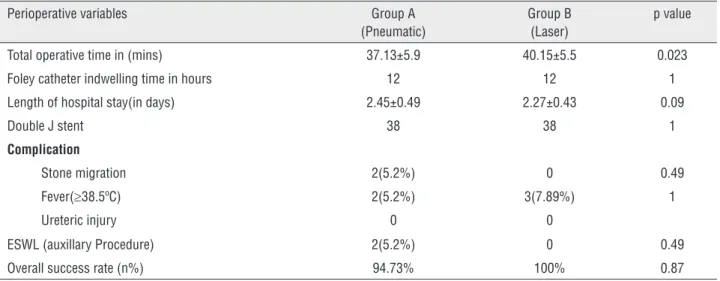
Table 2 - Perioperative clinical data of patients in both groups.
Perioperative variables Group A (Pneumatic)
Group B (Laser)
p value
Total operative time in (mins) 37.13±5.9 40.15±5.5 0.023
Foley catheter indwelling time in hours 12 12 1
Length of hospital stay(in days) 2.45±0.49 2.27±0.43 0.09
Double J stent 38 38 1
Complication
Stone migration 2(5.2%) 0 0.49
Fever(≥38.5ºC) 2(5.2%) 3(7.89%) 1
Ureteric injury 0 0
ESWL (auxillary Procedure) 2(5.2%) 0 0.49
Overall success rate (n%) 94.73% 100% 0.87
DISCUSSION
Lower ureteric calculus is not uncommon in pediatric age group. Different treatment moda-lities are available for the management of lower ureteric calculus for pediatric patients including ESWL (extracorporeal shock wave lithotripsy), ureteroscopy, percutaneous antegrade ureteros-copy, laparoscopic and open surgery (5). With the improvement in surgical skills and technical advancement in the working instruments (smal-ler calibre semi-rigid and flexible ureteroscopes), the management of lower ureteric calculus has changed from open surgery to minimal invasi-ve endoscopic lithotripsies (6). In the current era, ureteroscopy has become the preferred modality
of managing lower ureteric calculus with success rate approaching to 100% in both adults as well as in pediatric patients.
Young in 1912 was the first to perform ureteroscopy, inserted a cystoscope in a child with posterior urethral valve (7). Goodman in 1977 was the first to performed rigid ureteroscopy (8).
Different lithotriptors can be used for in-tracorporeal lithotripsy including electrohydraulic (EHL), ballistic (pneumatic), ultrasonic (US), laser (Ho: YAG). In the last few years lasers have been increasingly replacing others for intracorporeal li-thotripsy (9, 10).
of breaking all type of stone irrespective of their composition as compared to other lithotriptors and because of weaker shock waves there is lower risk of stone migration (11).
Pneumatic lithotripsy was first introduced into practice in 1992 in Switzerland (12). Advan-tage of pneumatic lithotripter when compared to other lithotriptors is its lower risk of perforating ureter and no thermal damage (13). Only concern with pneumatic lithotripter is stone migration, that ranges between 1.6% and 17.3% particularly with upper ureteral calculus (14, 15).
In the present study stone-free rate for lower ureteric calculus with holmium laser was 100% and 94.73% with pneumatic lithotripsy res-pectively (p=0.87). This study confirms previous reports in literature about efficacy of ureteros-copic Ho: YAG laser lithotripsy in treating distal ureteric stones (16, 17). Total operative time in this study was 40.15±5.55 min in laser lithotrip-sy while 37.13±5.94 min in pneumatic lithotriplithotrip-sy (p=0.023). In this study we observe 100% success rate with laser lithotripter and 94.73% with pneu-matic lithotripsy. Similarly, Salvado et al. (18) also reported 96% success rate of laser lithotripsy in the management of distal ureteral stone. Mano-har et al. (19) reported 84% success rate with laser lithotripsy. Our overall success rate for lower ure-teric calculus in pediatric age group approached 97% with the ureteroscopic procedure which was in accordance with the literature.
In our study we observed stone migration into the collecting system of two patients (5.2%) in group A patients (impacted calculus), which later was managed with ESWL (after 3 days), while none of our patients in group B experienced similar com-plication. Razzaghi et al. (2) reported higher inciden-ce of stone migration into renal collecting system with pneumatic lithotripter (17.9%) particularly with upper ureteric calculus and no such complication in the laser group. Salvado et al. (18) reported statis-tically insignificant difference of stone migration between the two modalities of lithotripsy. Similarly, Manohar et al. (19) did not observed any statisti-cally significant difference of stone migration rates between pneumatic and laser lithotripsy groups. This is because of the improvement in surgical skills and technological advancement.
In this study, we retrogradely inserted Double J stent in all patients. In our belief ureteral stenting with double J stent prevents postoperati-ve sepsis, and ureteral mucosal edema, although there was no clear precise indication for ureteral stenting such as ureteral injury or perforation. Ho-wever, there are several prospective randomized controlled trails comparing stented versus non stented ureteroscopic lithotripsy and they reported similar outcomes (20). In the present study, length of hospital stay was comparable and we did not found any statistically significant difference tween the two groups (p=0.09). This might be be-cause none of our patients in the study had suffe-red any major complication related to procedure. Some studies in the literature reported complica-tion rate of ureteroscopy between 9-25%. Howe-ver, incidence of major complication is less than 0.1% n (5). Stone analysis in our study showed ammonium acid urate and uric acid stones the predominant variety. The major advantage with the holmium laser lithotripsy when compared with pneumatic lithotripter is that laser lithotripter will fragment any stone irrespective of composition.
CONCLUSIONS
Both holmium laser and pneumatic litho-tripsy are equally efficacious and can be used safely for the endoscopic management of lower ureteric calculus in pediatric patients. However, holmium laser requires more expertise and it is a costly alternative and comparatively more advan-tageous in impacted calculus.
CONFLICT OF INTEREST
None declared.
REFERENCES
1. Khaladkar S, Modi J, Bhansali M, Dobhada S, Patankar S. Which is the best option to treat large (>1.5 cm) midureteric calculi? J Laparoendosc Adv Surg Tech A. 2009;19:501-4. 2. Razzaghi MR, Razi A, Mazloomfard MM, Golmohammadi
3. Breda A, Ogunyemi O, Leppert JT, Schulam PG. Flexible ureteroscopy and laser lithotripsy for multiple unilateral intrarenal stones. Eur Urol. 2009;55:1190-6.
4. Grasso M, Conlin M, Bagley D. Retrograde ureteropyeloscopic treatment of 2 cm. or greater upper urinary tract and minor Staghorn calculi. J Urol. 1998;160:346-51.
5. Türk C, Knoll T, Petrik A, Sarica K, Skolarikos A, Straub M, Seirz C. EAU Guidelines on Urolithiasis. European Association of Urology 2013.
6. Schock J, Barsky RI, Pietras JR. Urolithiasis update: clinical experience with the Swiss LithoClast. J Am Osteopath Assoc. 2001;101:437-40.
7. Young HH, Mckay RW. Congenital valvular obstruction of the prostatic urethra. Surg. Gynecol Obstet 1929;48:509. 8. Goodman TM. Ureteroscopy with pediatric cystoscope in
adults. Urology. 1977;9:394.
9. Breda A, Ogunyemi O, Leppert JT, Schulam PG. Flexible ureteroscopy and laser lithotripsy for multiple unilateral intrarenal stones. Eur Urol. 2009;55:1190-6.
10. Bader MJ, Eisner B, Porpiglia F, Preminger GM, Tiselius HG. Contemporary management of ureteral stones. Eur Urol. 2012;61:764-72.
11. Zarrabi A, Gross AJ. The evolution of lasers in urology. Ther Adv Urol. 2011;3:81-9.
12. Denstedt JD, Eberwein PM, Singh RR. The Swiss Lithoclast: a new device for intracorporeal lithotripsy. J Urol. 1992;148:1088-90.
13. Piergiovanni M, Desgrandchamps F, Cochand-Priollet B, Janssen T, Colomer S, Teillac P, et al. Ureteral and bladder lesions after ballistic, ultrasonic, electrohydraulic, or laser lithotripsy. J Endourol. 1994;8:293-9.
14. Murthy PV, Rao HS, Meherwade S, Rao PV, Srivastava A, Sasidharan K. Ureteroscopic lithotripsy using mini-endoscope and Swiss lithoclast: experience in 147 cases. J Endourol. 1997;11:327-30.
15. Menezes P, Kumar PV, Timoney AG. A randomized trial comparing lithoclast with an electrokinetic lithotripter in the management of ureteric stones. BJU Int. 2000;85:22-5. 16. Tawfiek ER, Bagley DH. Management of upper urinary tract
calculi with ureteroscopic techniques. Urology. 1999;53:25-31. 17. Leijte JA, Oddens JR, Lock TM. Holmium laser lithotripsy
for ureteral calculi: predictive factors for complications and success. J Endourol. 2008;22:257-60.
18. Salvadó JA, Mandujano R, Saez I, Saavedra A, Dell’oro A, Dominguez J, et al. Ureteroscopic lithotripsy for distal ureteral calculi: comparative evaluation of three different lithotriptors. J Endourol. 2012;26:343-6.
19. Manohar T, Ganpule A, Desai M. Comparative evaluation of Swiss LithoClast 2 and holmium:YAG laser lithotripsy for impacted upper-ureteral stones. J Endourol. 2008;22:443-6. 20. Denstedt JD, Wollin TA, Sofer M, Nott L, Weir M, D’A Honey
RJ. A prospective randomized controlled trial comparing nonstented versus stented ureteroscopic lithotripsy. J Urol. 2001;165:1419-22.
_______________________ Correspondence address: