Weak Interactions between
Salmonella
enterica
FlhB and Other Flagellar Export
Apparatus Proteins Govern Type III Secretion
Dynamics
Jonathan L. McMurry1*, Tohru Minamino2, Yukio Furukawa2, Joshua W. Francis1¤a,
Stephanie A. Hill1¤b, Katy A. Helms1¤c, Keiichi Namba2
1Department of Molecular & Cellular Biology, Kennesaw State University, Kennesaw, Georgia, United States of America,2Graduate School of Frontier Biosciences, Osaka University, Osaka, Japan
¤a Current address: Department of Biochemistry, Emory University School of Medicine, Atlanta, Georgia, United States of America
¤b Current address: Medical College of Georgia, Georgia Regents University, Augusta, Georgia, United States of America
¤c Current address: Mercer University School of Medicine, Macon, Georgia, United States of America
Abstract
The bacterial flagellum contains its own type III secretion apparatus that coordinates protein export with assembly at the distal end. While many interactions among export apparatus proteins have been reported, few have been examined with respect to the differential affini-ties and dynamic relationships that must govern the mechanism of export. FlhB, an integral membrane protein, plays critical roles in both export and the substrate specificity switching that occurs upon hook completion. Reported herein is the quantitative characterization of interactions between the cytoplasmic domain of FlhB (FlhBC) and other export apparatus
proteins including FliK, FlhACand FliI. FliK and FlhACbound with micromolar affinity. KDfor
FliI binding in the absence of ATP was 84 nM. ATP-induced oligomerization of FliI induced kinetic changes, stimulating fast-on, fast-off binding and lowering affinity. Full length FlhB purified under solubilizing, nondenaturing conditions formed a stable dimer via its trans-membrane domain and stably bound FliH. Together, the present results support the previ-ously hypothesized central role of FlhB and elucidate the dynamics of protein-protein interactions in type III secretion.
Introduction
The bacterial flagellum is a proton-driven rotary nanomachine responsible for motility in many species [1,2,3]. Most proteins that comprise a flagellum reside beyond the cytoplasmic membrane and must be secreted. Secretion occurs via a specialized type III secretion system (T3SS or“export apparatus”[4,5]). Utilizing protonmotive force [6,7,8], the apparatus
a11111
OPEN ACCESS
Citation:McMurry JL, Minamino T, Furukawa Y, Francis JW, Hill SA, Helms KA, et al. (2015) Weak Interactions betweenSalmonella entericaFlhB and Other Flagellar Export Apparatus Proteins Govern Type III Secretion Dynamics. PLoS ONE 10(8): e0134884. doi:10.1371/journal.pone.0134884
Editor:Michael Hensel, University of Osnabrueck, GERMANY
Received:April 22, 2015
Accepted:July 14, 2015
Published:August 5, 2015
Copyright:© 2015 McMurry et al. This is an open access article distributed under the terms of the
Creative Commons Attribution License, which permits unrestricted use, distribution, and reproduction in any medium, provided the original author and source are credited.
Data Availability Statement:All relevant data are within the paper and its Supporting Information Files.
Funding:This work was supported by NIH grant GM080701 (to J.L.M.) and KSU College of Science & Mathematics Mentor-Protege Awards (to J.W.F. and S.A.H.). The funders had no role in study design, data collection and analysis, decision to publish, or preparation of the manuscript.
translocates flagellar proteins across the cytoplasmic membrane into the central channel within the growing flagellum through which they transit to their final location [9]. Homologous T3SSs effect many modes of bacterial pathogenesis using needle-like structures that closely resemble flagella [10].
The core flagellar T3SS consists of three soluble proteins (FliH, FliI and FliJ) and six integral membrane proteins (FlhA, FlhB, FliO, FliP, FliQ and FliR) that are housed within the mem-brane-supramembrane ring of the basal body. Like most of the other proteins, FlhB is necessary for secretion and is critical for the substrate specificity switching that occurs upon hook com-pletion as the export apparatus shifts from rod and hook-type proteins to filament-type pro-teins [4,11]. FlhB undergoes asparagine-mediated autohydrolysis at N269-P270 [12,13]. Mutations in FlhB that slow or abolish this cleavage give rise to dramatically altered flagellar structures due to defects in switching [14]. FliK is one of the rod- and hook-type substrates rec-ognized via the flagellar T3SS during hook assembly [15]. A specific interaction of FlhB with FliK is central to the switch, in which the T3SS stops exporting rod and hook-type proteins and begins exporting filament-type proteins [16,17] via a mechanism modeled as a“temporal tape measure”in which FliK interacts with both FlhB and hook proteins [18,19]. The interaction between FliK and FlhB is thought to vary as a function of hook length, though the details of how remain unknown [20]. In addition to FliK, FlhB has been reported to bind FliH, FliI, FliJ and perhaps the cytoplasmic domain of FlhA (FlhAC) [21], though the veracity and
conse-quences of these interactions are largely unknown. Full-length FlhA and FlhB exhibited no binding to each other in affinity blots [22].
While a great deal of work has described apparatus proteins with respect to requirements for secretion, qualitative interactions and structure, understanding of dynamic interactions has lagged. Kinetic relationships are fertile ground for exploration and characterizing them will provide a better understanding of T3S and transmembrane transport in general. Much of what is known about interactions among export proteins, substrates and chaperones derives from copurification and affinity blotting experiments that have limitations such as requirements for attainment of equilibrium and that only high affinity interactions can be observed. Using a type of optical biosensing, biolayer interferometry (BLI)[23], and analytical ultracentrifugation, the present study was able to address oligomerization of full-length, membrane integrated FlhB as well as the complex kinetic interactions of FlhB and its cytoplasmic domain with other T3S apparatus proteins.
Similar to surface plasmon resonance (SPR), BLI allows real-time measurement of protein-protein interactions and determination of kinetic and affinity constants [24]. Ligand proteins are tethered to fiber optic sensors and dipped into analyte-containing buffers to measure asso-ciation. Dissociation is monitored after movement to buffer without analyte. Instrument response, measured in nanometers of shift of the interference pattern of white light caused by analyte-induced changes in the distance between two reflecting surfaces over time, yields asso-ciation and dissoasso-ciation rate information. Fits of raw data to kinetic models allow assignment of rate and affinity constants. In the case of simple binding, fits to single exponentials allow determination of kofffrom the dissociation phase since reassociation is negligible due to
dilu-tion of dissociated analyte. Fitting the associadilu-tion phase yields observed rate constants (kobs),
from which koncan be extracted given analyte concentration and koff.
We were able to characterize the mostly weak, complex interactions of FlhBCwith FliK,
FlhACand FliI. Provision of ATP to FliI dramatically altered binding, weakening affinity. FlhB
Materials and Methods
Overexpression and purification
Plasmids used in this study are shown inS1 Table. His-tagged variants of the soluble export proteins and the cytoplasmic domains of FlhA and FlhB (“FlhAC”and“FlhBC”) were overpro-duced and purified. Overnight cultures ofE.coliBL21DE3(pLysS) cells harboring plasmids encoding His-tagged proteins were subcultured and grown in Luria broth at 30°C to an OD600
~ 0.4. Expression was induced by addition of 0.2 mM IPTG, after which growth was continued for four hours. Cells were harvested by centrifugation and pellets were frozen at -80°C until use.
All purification steps were performed on ice or at 4°C. Pellets from 1 L cultures were thawed and resuspended in 25 ml lysis buffer (50 mM Tris pH 8.0, 500 mM NaCl, 10 mM imidazole, 0.1% Tween-20 and 200μg ml-1lysozyme). Resuspended cells were passed through a French press at 20,000 psi and then subjected to centrifugation for 20 min at 10,000 x G at 4°C. The resulting clarified supernatant was transferred to a tube containing 1 ml of equilibrated Talon (BD Biosciences) immobilized metal affinity chromatography (IMAC) resin.
Batch binding was allowed to proceed with gentle agitation for 20 min after which the resin was pelleted by brief centrifugation and washed twice with 20 ml wash buffer (50 mM Tris pH 8.0, 500 mM NaCl, 25 mM imidazole, 0.1% Tween-20). The resin was transferred to a column and washed with an additional 10 ml. Elution was achieved by addition of elution buffer (wash buffer with 250 mM imidazole). Proteins were exchanged into HBS-T (10 mM HEPES, pH 7.4, 150 mM NaCl. 0.05% Tween 20) by gel filtration and used immediately, or glycerol was added to 10% and proteins were snap frozen in liquid nitrogen and stored at -80°C until use. Concen-trations were determined by Bradford assay [25] using BSA as standard.
Full-length FlhB was overproduced and purified under nondenaturing conditions from sol-ubilized crude membrane fractions as described for FlhA [22]. For AUC studies the uncleava-ble variant of full-length FlhB, FlhB(N269A), the method of Fleming et al. [26] was modified as follows: cells overexpressing FlhB(N269A) were resuspended in 10 mM phosphate buffer pH 8.0, 500 mM NaCl, 20% glycerol, 10 mMβ-mercaptoethanol, 10 mM imidazole and lysed by sonication. Lysates were centrifuged at 10,000 x g to pellet unbroken cells. Supernatants were ultracentrifuged at 100,000 x g to pellet membranes. The crude membrane fraction was resus-pended in lysis buffer (same as above but with 1% Thesit), homogenized and stirred at 4°C for 1 hour. After centrifugation at 100,000 x g for 45 min, the supernatant was retained as solubi-lized membrane fraction and subjected to IMAC to purify the FlhB(N269A). Wash and elution buffers were the same as the lysis except containing 20mM and 250 mM imidazole, respec-tively. To exchange the Thesit for E8C5, a detergent with the same partial specific volume as
water and hence amenable to analytical ultracentrifugation, purified FlhB(N269A), ~25 ml, was diluted in 1 L dilution buffer (10 mM phosphate buffer, pH 8.0, 1% Thesit, 20% glycerol, 20mMβ-mercaptoethanol and then loaded onto a 1 mL SP Sepharose column. The column was washed with 50 ml of 10 mM phosphate buffer, 33 mM C8E5, 10 mM NaCl at 1 ml min-1.
FlhB(N269A) was eluted in 1 ml fractions in the phosphate/C8E5buffer with 500 mM NaCl.
Optical biosensing
biotin to protein for 30 min at room temperature followed by separation of protein from free biotin by repeated passage over a desalting column. After loading ligands onto SA sensors, a baseline was established in buffer prior to association at varying analyte concentrations. Disso-ciation was subsequently measured in buffer only. All phases were done in HBS-T, except the full-length FlhB experiment, in which the Tween was replaced with 1% Triton X-100. Raw data were analyzed with GraphPad Prism.
For numerical simulation of FliK-FlhBCbinding, a conformational change model (A + BÐ
ABÐAB) was made in which A is analyte (FlhBC), B is ligand, AB is the bound complex and
AB
is a conformationally altered state. Rate constants k1and k2govern association and
disso-ciation of the free proteins and k3and k4describe the shift to and from the AB
state, respec-tively. The set of differential equations used for the simulations were:
dAB=dt¼k1ABþk4AB k2AB k3AB
dA=dt¼k2AB k1AB
dAB=dt¼k3AB k4AB
Data were plotted as fractions of maximal binding (Bmax), which was iteratively determined. A
15% correction factor to account for differences between the signal produced by ABrelative to AB was included in the simulations.
Analytical ultracentrifugation
Sedimentation equilibrium ultracentrifugation was performed using a Beckman Optima XL-A ultracentrifuge and an AnTi 60 rotor essentially as described [27], except that the buffer con-tained 33 mM C8E5. Scans were collected at 280 nm with a spacing of 0.001 cm in the step
mode with twenty averages per step. Three scans were superimposed prior to analysis with Optima XL-A/XLI version 4.0 (Beckman).
Results
Biosensing Survey
To examine dynamic interactions between FlhBCand other apparatus proteins, FlhBCwas
used as analyte versus each of the other apparatus proteins as ligand (Fig 1A). A starkly
Fig 1. FlhBCkinetic survey.Purified T3S proteins were biotinylated and used as ligands. All analyte concentrations were 1μM. Association and
dissociation phases were 180 seconds in all cases. A, FlhBCanalyte binding to ligands FlhAC(red), FlhBC(blue), FliH (green), FliI (orange), FliJ (magenta)
and FliK (black). B, nonspecific binding control of analyte apparatus proteins binding to sensor-tethered biotinylated BSA. Analyte colors are the same as ligand colors in A. C, Analyte apparatus proteins binding to ligand FlhBC. Colors are the same as in B.
different shift profile from nonspecific control binding (Fig 1B, black trace) was observed for FliK. Smaller differences were noted for all other export proteins, e.g. a small amplitude fast on state for FlhAC, indicating some interaction with FlhBC. Observed binding was complex and
nonspecific binding (NSB) as evidenced by response to BSA as ligand was in many cases signif-icant. FliJ, FliH and FlhBCligands also exhibited binding different from BSA, but were resistant
to further analysis due to NSB, low signal and other reasons. Biotinylated FlhBCwas tethered
to SA sensors and screened for binding versus analyte soluble export apparatus proteins at 1μM (Fig 1C). Differences in FliI binding were noted. FliK-FlhBC, FlhAC-FlhBCand FlhBC
-FliI interactions were selected for further kinetic characterization.
Kinetic characterizations
We previously reported a KDof 3.2μM for FliK-FlhBCbinding determined by steady state
analysis of SPR data [13]. Delving further into the complexity to better understand the kinet-ics, BLI sensorgrams were collected for a concentration course ranging from 0 to 5μM FlhBC.
As shown inFig 2A and 2B, association and dissociation phases could be fit by two exponen-tials, i.e. parallel events, but there was no global solution that yielded constants that fit two independent states. Instead, numerical simulations were performed using differential equa-tions constructed from a conformational change model. Simulaequa-tions of association-then-dissociation are shown for 5, 4, 3, 2 and 1μM inFig 2C–2G. Parameters for constants used in the simulations are shown inTable 1and include slow transitions to and from the AB
state. Plotting k1determined from simulations, which is equivalent to the observed rate constant
(kobs) for initial binding in that it also accounts for dissociation occurring during the
associa-tion phase, vs. analyte concentraassocia-tion (Fig 2H) yielded konof 5.5 x 10⁴M−1s−1. Combination
with a koffof 0.44 s−1gave a KDof 8.0μM for the initial binding event, consistent with our
ear-lier study. Supporting the conformational change model is the observation that the amplitude of the slow-off state in the dissociation phase varied proportionately with the length of the association phase (S2 Fig). It should be noted that amplitude variations between full kinetic characterizations and theFig 1survey are likely a function of different specific binding activi-ties of different preparations for both ligand and analyte. All concentration courses in this experiment were done with dilutions of the same preparation. We also note that the overall KDdetermined by steady state analysis, i.e. including the slow states, for FliK-FlhBCinFig 2is
2.1μMS3 Fig), almost identical to that of the preparations used in the earlier report despite very different amplitudes.
Ligand FlhAC-analyte FlhBCbinding also exhibited complexity and did not fit global
one-state association-then-dissociation models. Single exponentials did fit the association phase (Fig 3A). Global two-state exponentials could fit dissociation with koffs of 0.13 s−1and 4.7 x
10−3s−1(Fig 3B). Saturation analysis (Fig 3C) yielded a K
Dof 1.1μM. Plotting kobsvs
[FlhBC](Fig 3D) led to an estimate of konof 8.5 x 10⁴M−1s−1and thus a nominal one-state
koffof 0.09 s−1, though caution should accompany interpretation of these values (see
Discussion).
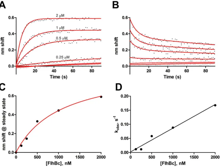
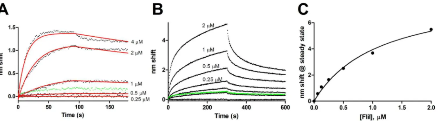
FlhB binds FliI via its cytoplasmic domain. As shown inFig 4, ligand FlhBCbound FliI both
in the absence and presence of Mg2+-ATP. For FliI without ATP (Fig 4A), fits to a global one-state model yielded a KDof 84 nM with a konof 1.8 x 10⁴M−1s−1and koffof 1.5 x 10⁻³s−1.
Pre-incubation of analyte FliI with an excess of ATP resulted in near elimination of nonspecific binding and more complex kinetics; substantial fast-on and fast off-states are seen relative to the no ATP sample. Data do not fit two-state models, perhaps reflecting additional states induced by oligomerization of FliI (seeDiscussion). KDdetermined from steady state analysis
Fig 2. Simulation of FliK-FlhBCbinding.A and B, association and dissociation phases. Concentrations of FlhBCwere 5, 4, 3, 2, 1, 0.5, 0.25, and 0μM. The
0.5 and 0.25μM traces are unlabeled. Fits to individual two-state exponentials are shown as red lines. C-G, Simulations of the 5, 4, 3, 2 and 1μM data with a conformational change model using global parameters for koffand konand kofffor transition to the conformationally altered state (Table 2). H, Apparent konvs
[FlhBC] to determine the global kon(= slope).
Full-length FlhB interactions
FlhBC-FlhBCinteractions (Fig 1) were at best minimally observable, consistent with earlier
studies that found questionable or no interaction [12,21]. We report here purification of solubi-lized FlhB under non-denaturing conditions using a procedure modified from a prior method used to purify FlhA [22]. The uncleavable but export competent N269A variant [14] was used to assure retention of the carboxyl-terminal subdomain in the solubilizing conditions used Table 1. Parameters determined by simulation of FliK-FlhBCbinding.
[FlhBC],μM 5 4 3 2 1
k1(s-1) 0.25 0.25 0.2 0.1 0.05
k2(s-1) 0.44 0.44 0.44 0.44 0.44
k3(s-1) 0.0085 0.0085 0.0085 0.0085 0.0085
k4(s-1) 0.008 0.008 0.008 0.008 0.008
Bmax 1.7549 1.7549 1.7549 1.7549 1.7549
doi:10.1371/journal.pone.0134884.t001
Fig 3. FlhAC-FlhBCbinding.Ligand FlhACwas exposed to 2, 1, 0.5, 0.25 and 0.125μM FlhBC. A, association with fits to a one-state model B, dissociation
with fits to a global two-state model C, steady state analysis. D, kobsvs. [FlhBC] to estimate kinetic constants, R2= 0.98.
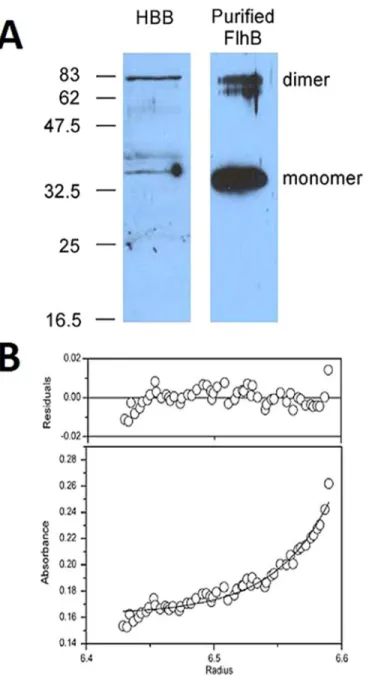
(though later purification of wild-type FlhB from pMM9, which complements aflhBnull, showed that the subdomain consisting of residues 270–383 is retained (S1 Fig)). Anti-FlhB immunoblots of hook-basal bodies (HBBs) prepared from SJW880 [28] under conditions in which the C ring and export apparatus proteins are retained [29] (gift from Noreen R. Francis) demonstrated significant SDS-stable dimerization, as did purified FlhB(N269A) (Fig 5A). Full-length FlhB(N269A), solubilized in the neutrally buoyant, nondenaturing detergent C8E5,
formed a stable dimer in a sedimentation equilibrium ultracenrifugation experiment (Fig 5B). Fits to a single species model produced a molecular weight of 84.1 kDa, consistent with a FlhB dimer. The tagged monomer is ~42.3 kDa.
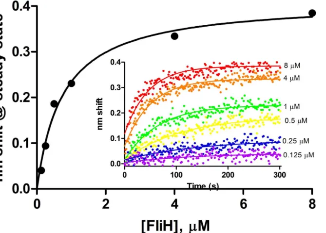
Purified wild-type FlhB exhibited specific binding to FliH as ligand. Dissociation anomalies perhaps due to detergent effects prevented kinetic analysis. KDdetermined from steady state
analysis (Fig 6) was 0.8μM. The 2μM sample was excluded from analysis due to anomalous readings from that channel, though its inclusion would render a KDof 0.9μM with a
concomi-tant reduction in R2from 0.99 to 0.82. Further experiments with other analytes were precluded by instability of the FlhB preparations; we hope to examine them in future studies.
In summary, the present results assign rate and affinity constants to binding of FlhB to sev-eral apparatus proteins and provide mechanistic clues for T3S. They are consistent with a con-formational change in FlhB upon FliK binding and ATP-induced kinetic alterations in FlhB-FliI interactions as well as weak FlhB-FlhA cytoplasmic domain interactions and FliH interactions with full length FlhB.
Fig 4. FlhBC-FliI binding.FlhBCwas used as ligand to examine binding of FliI in the absence and presence of ATP. A, Binding of 4, 2, 1, 0.5 and 0.25μM FliI
with fits to a global one-state association-then-dissociation model shown in red. The green points are 4μM FliI exposed to a sensor without FlhBC, e.g. NSB.
B, FlhBC-FliI binding with FliI preequilibrated with 5 mM ATP and 5 mM MgCl2added to all phases. FliI concentrations were 2, 1, 0.5, 0.25, 0.13, 0.062 and
0μM. Green points represent the 2 uM sample binding to sensor without ligand. C, Steady state analydis of FliI with ATP from panel B, KD= 1.1μM.
doi:10.1371/journal.pone.0134884.g004
Table 2. Kinetic constants determined from BLI experiments for binding to FlhBC.
Analyte KD kon(M−1s−1) koff(s−1)
FlhAC 1.0μM 8.5 x 10⁴ 0.085
FliI (-ATP) 84 nM 1.8 x 10⁴ 1.5 x 10⁻³
FliI (+ATP) 1.1μM ND ND
FliK 8.0μM 5.5 x 10⁴ 0.44
ND, not determined. Constants are expressed with respect to the monomer concentrations.
Discussion
Perhaps the best information about protein-protein interactions in the flagellar T3SS originates from intergenic suppression studies, e.g. [16,30]. However, much of the current conception of these interactions is based on qualitative equilibrium methods such as immunoaffinity blotting and copurification that can effectively identify only strong interactions [12,21,22,31]. These methods can also make interpretation difficult due to NSB. NSB was certainly a challenge in the present study and may be an inherent consequence of examining pairwise interactions Fig 5. Full-length FlhB forms a dimer.A, anti-FlhB immunoblot of hook-basal body preparation (HBB) and purified FlhB(N269A). Approximate locations of molecular weight standards in kDa are shown at left. B, sedimentation equilibrium analytical centrifugation. A fit is shown to a single-species model, the molecular weight of which is 84.1 kDa (monomer of tagged FlhB(N269A) = 42.3 kDa).
between proteins whose normal environment is within a membrane-integrated macromolecu-lar complex. We were able to measure NSB via the proxy of BSA binding, performing full anal-yses only when it was not a substantial fraction of total binding.
BLI of course has interpretative limitations as well. Since sensors are coated in ligand, sen-sors without ligands are not true references in that they can present a surface that differs in electrostatic and other properties. Use of BSA as a non-related ligand presents similar concerns, though binding of export proteins to both BSA and sensors without ligand was similar (data not shown). Ligand activity and presentation upon biotinylation could explain failure to observe binding on reversal of ligand-analyte pairings.
Interestingly, significant binding was not observed with several interactions previously reported. While present results cannot rule out interactions not observed, it is possible that at lower concentrations than those used in affinity blotting, FlhB engages in a different set of interactions. For example, ligand FlhBCbound only FliI and perhaps FlhBC, exhibiting
essen-tially no affinity for other apparatus proteins even though positive in affinity blotting [21]. Another possibility is that interactions may be biologically relevant even though they associate slowly but are stable once formed. The weaker interactions (FliK-FlhBC, FlhBC-FliI (with
ATP), and FlhAC-FlhBC) are the ones more likely to undergo dynamic changes during export
as they exhibit significant off rates.
All of the characterized interactions exhibited more than simple one-state binding. Whether the secondary events are biologically relevant is an open question. We suspect the initial, fast Fig 6. Steady state analysis of FliH binding to full length wild-type FlhB.Association phases from which steady state amplitudes were determined are shown in the inset. FliH concentrations ranged from 0.125 to 8μM.
but low affinity events are the relevant ones and that the slower on and off events may be due to aggregation, surface-associated denaturation or other biologically irrelevant events. Surface-associated denaturation in BLI has been observed for calmodulin-nitric oxide synthase [32] andHelicobacter pyloriUreE-UreG binding [33]. A potential example in this study is the slow-off FlhAC-FlhBCdissociation phase (Fig 3B).
Kinetic and affinity constants determined with varying degrees of veracity are shown in
Table 2.
FliK-FlhB
Cinteractions
Though known to interact for many years, binding between FliK and FlhBCwas first directly
observed using biosensing [13]. The one-state with conformational change model described herein fits observed FlhB-FliK binding. Affinity for the initial binding and dissociation is 8μM, in good agreement with the previous report, which was determined by saturation binding and reflects both states. The fast-on, fast-off initial binding is consistent with intergenic suppression data and failure to observe interactions by equilibrium methods. Slow transitions to and from the conformationally altered state (AB
, seeResults) render it a minor event, but one sufficient to explain the complexity observed. Whether it is a biologically relevant state and whether it changes in the presence of other proteins, e.g. substrates, or structural changes brought about by hook completion, remains a subject for further investigation, though conformational flexi-bility appears to be important for FlhB function [34]. Alterations in the dynamics of the con-formational change would also be consistent with the temporal tape measure model.
FlhA
C-FlhB
Cinteractions
Compared to the NSB indicated by FlhBCbinding to BSA sensors (Fig 1B), FlhAC-FlhBC
bind-ing exhibited fast on and fast off components (Fig 3). The most likely interpretation is that these proteins possess weak affinity for one another and that the slow-off state represents an irreversible, biologically irrelevant state. Conversely, it may signal a conformationally changed, high affinity state, but qualitative evidence suggesting weaker [21] or undetectable [22] binding supports the former interpretation. FlhAC-FlhBCinteractions are depicted as gating the
mem-brane pore, e.g. [35]. Weak binding in the absence of the transmembrane domains may be exemplary of this.
FlhB-FliI Binding
ATP induces hexamerization in FliI [36,37]. The present results show that in addition to induc-ing oligomerization, ATP alters FliI interactions with FlhBC. Presumably monomeric FliI
with-out ATP shows tight binding to FlhBCwith relatively low NSB (Fig 4A). Addition of an excess
of ATP resulted in substantially lower affinity (Fig 4C) but faster association and dissociation and additional complexity consistent with the hypothesis that FliI6undergoes repeated binding
and release events in delivering export-competent substrates to the export gate [38] and recent observations of FliI turnover in the basal body [39].
FlhB dimerization and interaction with FliH
Ferris et al. extensively searched for FlhBC-FlhBCinteractions, finding none using equilibrium
methods [12]. Our BLI data, too, indicate very little interaction between the cytoplasmic domains, though there may be some low affinity binding. Indeed, one interpretation of the complexity observed in BLI of FliK-FlhBCbinding was oligomerization of FlhBC[13].
stable dimer in detergent micelles. Additional evidence from the HBBs suggests that FlhB forms dimersin vivoas well. The high affinity of the transmembrane domain-containing FlhB and the extremely low affinity (if present at all) of the cytoplasmic domains for themselves may hint at the dynamics of FlhB; the proximity of cytoplasmic domains forced by dimerization of the transmembrane domains may facilitate otherwise weak binding that may undergo cycles of association and dissociation as secretion occurs.
FliH exhibited significant binding only to full-length FlhB (Fig 6). Two possibilities suggest themselves: the binding site may reside at least partly within the transmembrane domain of FlhB; or FliH may be active with respect to FlhB binding in detergent micelles, which can be considered unsurprising as it partitions with the membrane even in the absence of basal bodies [40]. Other apparatus proteins were not investigated in this study with respect to binding the full-length FlhB due to the difficulty of the purification and the loss of FlhB binding activity over time. We hope to characterize these events in a future study.
The present results expand knowledge of the dynamic interactions of FlhB with other export apparatus proteins and assign rate an affinity constants to them. In short, FlhB stably dimerizes and stably binds FliH; FlhBCbinds FliK and FlhACwith micromolar affinity and complex
kinetics. Interactions with FliI shift upon addition of ATP, lowering affinity but increasing the rates of association and dissociation. The complexities observed underlie the mechanism of T3S. How interactions change when they are more than pairwise, asin vivo, is an active area of investigation.
Supporting Information
S1 Fig. Purification of full length FlhB.A, Coomassie stained SDS-PAGE of samples taken during purification. Lanes are: 1, uninduced cells; 2, induced cells; 3, crude lysate; 4, 1st low-speed supernatant (clarified lysate); 5, 1sthigh-speed supernatant; 6, 1sthigh-speed pellet (crude membranes); 7, solubilization (overnight); 8, second high-speed pellet; 9, solubilized sample (load); 10, flow-through 11; pooled first wash; 12, final wash; E1-5, eluted protein frac-tions 1–5. B, Immunoblotting analysis of uninduced (U), induced (I) and purified (E2, diluted 10x), with positions of FlhBTM+CNand FlhBCCdenoted with arrowheads at right. Note that
anti-His only responds to FlhBTM+CNas the His-tag is amino-terminal, anti-FLAG is
overex-posed and anti-FlhB is more reactive to FlhBCC, as has been noted previously (31).
(JPG)
S2 Fig. Slow-off state increases in amplitude as a function of association time.Ligand FliK was exposed to analyte 2μM FlhBCfor various times of association after which dissociation
was monitored for 300 s. Time of association was 10 s (green), 30 s (yellow), 60 s (orange), 180 s (blue), and 900 s (brown).
(TIF)
S3 Fig. Slow-off state increases in amplitude as a function of association time.Ligand FliK was exposed to analyte 2μM FlhBCfor various times of association after which dissociation
was monitored for 300 s. Time of association was 10 s (green), 30 s (yellow), 60 s (orange), 180 s (blue), and 900 s (brown).
(TIF)
S1 Table. Plasmids used in this study.
Acknowledgments
We are grateful to John C. Salerno for helpful comments and assistance with the simulations. This work was supported by NIH R15 GM080701.
Author Contributions
Conceived and designed the experiments: JLM TM KN. Performed the experiments: JLM YF JWF SAH KAH. Analyzed the data: JLM TM YF KN JWF SAH KAH. Contributed reagents/ materials/analysis tools: JLM TM JWF SAH KAH KN. Wrote the paper: JLM TM KN JWF SAH KH.
References
1. Zhao X, Norris SJ, Liu J (2014) Molecular architecture of the bacterial flagellar motor in cells. Biochem-istry 53: 4323–4333. doi:10.1021/bi500059yPMID:24697492
2. Terashima H, Kojima S, Homma M (2008) Flagellar motility in bacteria structure and function of flagellar motor. Int Rev Cell Mol Biol 270: 39–85. doi:10.1016/S1937-6448(08)01402-0PMID:19081534
3. Minamino T, Imada K, Namba K (2008) Molecular motors of the bacterial flagella. Curr Opin Struct Biol 18: 693–701. doi:10.1016/j.sbi.2008.09.006PMID:18848888
4. Minamino T (2014) Protein export through the bacterial flagellar type III export pathway. Biochim Bio-phys Acta 1843: 1642–1648. doi:10.1016/j.bbamcr.2013.09.005PMID:24064315
5. Evans LDB, Hughes C, Fraser GM (2014) Building a flagellum outside the bacterial cell. Trends Micro-biol 10: 566–572.
6. Minamino T, Namba K (2008) Distinct roles of the flagellar ATPase and proton motive force in bacterial flagellar protein export. Nature (London) 451: 485–488.
7. Paul K, Erhardt M, Hirano T, Blair DF, Hughes KT (2008) Energy source of flagellar type III secretion. Nature (London) 451: 489–492.
8. Minamino T, Morimoto YV, Hara N, Namba K (2011) An energy transduction mechanism used in bacte-rial flagellar type III protein export. Nat Commun 2: 475. doi:10.1038/ncomms1488PMID:21934659
9. Evans LDB, Poulter S, Terentjev T, Hughes C, Fraser GM (2013) A chain mechanism for flagellar growth. Nature 504: 287–290. doi:10.1038/nature12682PMID:24213633
10. Burkinshaw BJ, Strydnaka NCJ (2014) Assembly and structure of he T3SS. Biochim Biophys Acta 1843: 1649–1663. doi:10.1016/j.bbamcr.2014.01.035PMID:24512838
11. Ferris HU, Minamino T (2006) Flipping the switch: bringing order to flagellar assembly. Trends in Micro-biol 14: 519–525.
12. Ferris HU, Furukawa Y, Minamino T, Kroetz MB, Kihara M, Namba K, et al. (2005) FlhB regulates ordered export of flagellar components via autocleavage mechanism. J Biol Chem 280.
13. Morris DP, Roush ED, Thompson JW, Moseley MA, Murphy JW, McMurry JL (2010) Kinetic characteri-zation ofSalmonellaFliK-FlhB interactions demonstrates complexity of the type III secretion substrate-specificity switch. Biochemistry 49: 6386–6393. doi:10.1021/bi100487pPMID:20586476
14. Fraser GM, Hirano T, Ferris HU, Devgan LL, Kihara M, Macnab RM (2003) Substrate specificity of type III flagellar protein export inSalmonellais controlled by subdomain interactions in FlhB. Mol Microbiol 48: 1043–1057. PMID:12753195
15. Minamino T, González-Pedrajo B, Yamaguchi K, Aizawa S-I, Macnab RM (1999) FliK, the protein responsible for flagellar hook length control inSalmonella, is exported during hook assembly. Mol Microbiol 34: 295–304. PMID:10564473
16. Williams AW, Yamaguchi S, Togashi F, Aizawa S-I, Kawagishi I, Macnab RM (1996) Mutations infliK
andflhBaffecting flagellar hook and filament assembly inSalmonella typhimurium. J Bacteriol 178: 2960–2970. PMID:8631688
17. Hirano T, Minamino T, Namba K, Macnab RM (2003) Substrate specificity classes and the recognition signal forSalmonellatype III flagellar export. J Bacteriol 185: 2485–2492. PMID:12670972
18. Moriya N, Minamino T, Hughes KT, Macnab RM, Namba K (2006) The type III flagellar export specificity switch is dependent on FliK ruler and a molecular clock. J Mol Biol 359: 466–477. PMID:16630628
19. Erhardt M, Singer HM, Wee DH, Keener JP, Hughes KT (2011) An infrequent molecular ruler controls flagellar hook length in Salmonella enterica. EMBO J 30: 2948–2961. doi:10.1038/emboj.2011.185
20. Uchida K, Aizawa S (2014) The flagellar soluble protein FliK determines the minimal length of the hook inSalmonella entericaSerovar Typhimuriu. J Bact 196: 1753–1758. doi:10.1128/JB.00050-14PMID: 24563036
21. Minamino T, Macnab RM (2000) Interactions among components of theSalmonellaflagellar export apparatus and its substrates. Mol Microbiol 35: 1052–1064. PMID:10712687
22. McMurry JL, Van Arnam JS, Kihara M, Macnab RM (2004) Analysis of the cytoplasmic domains of Sal-monellaFlhA and interactions with components of the flagellar export machinery. J Bacteriol 186: 7586–7592. PMID:15516571
23. Concepcion J, Witte K, Wartchow C, Choo S, Yao D, Persson H, et al. (2009) Label-free detection of biomolecular interactions using biolayer interferometry for kinetic characterization. Comb Chem High Throughput Screen 12: 791–800. PMID:19758119
24. Abdiche Y, Malashock D, Pinkerton A, Pons J (2008) Determining kinetics and affinities of protein inter-actions using a parallel real-time label-free biosensor, the Octet. Anal Biochem 377: 209–217. doi:10. 1016/j.ab.2008.03.035PMID:18405656
25. Bradford MM (1976) A rapid and sensitive method for the quantitation of microgram quantities of protein using the principle of protein-dye binding. Anal Biochem 72: 248–254. PMID:942051
26. Fleming KG, Ackerman AL, Engelman DM (1997) The effect of point mutations on the free energy of transmembraneα-helix dimerization. J Mol Biol 272: 266–275. PMID:9299353
27. Minamino T, Saijo-Hamano Y, Furukawa Y, Gonzalez-Pedrajo B, Macnab RM, Namba K (2004) Domain organization and function of Salmonella FliK, a flagellar hook-length control protein. J Mol Biol 341: 491–502. PMID:15276839
28. Patterson-Delafield J, Martinez RJ, Stocker BAD, Yamaguchi S (1973) A newflagene inSalmonella typhimurium—flaR—and its mutant phenotype—superhooks. Arch Mikrobiol 90: 107–120. PMID: 4575053
29. Francis NR, Sosinsky GE, Thomas D, DeRosier DJ (1994) Isolation, characterization and structure of bacterial flagellar motors containing the switch complex. J Mol Biol 235: 1261–1270. PMID:8308888
30. Kihara M, Minamino T, Yamaguchi S, Macnab RM (2001) Intergenic suppression between the flagellar MS ring protein FliF ofSalmonellaand FlhA, a membrane component of its export apparatus. J Bacter-iol 183: 1655–1662. PMID:11160096
31. Minamino T, Macnab RM (2000) Domain structure ofSalmonellaFlhB, a flagellar export component responsible for substrate-specificity switching. J Bacteriol 182: 4906–4914. PMID:10940035
32. McMurry JL, Chrestensen CA, Scott IM, Lee EW, Rahn AM, Johansen AM, et al. (2011) Rate, affinity and calcium dependence of CaM binding to eNOS and nNOS: effects of phosphorylation. FEBS J 278: 4943–4954. doi:10.1111/j.1742-4658.2011.08395.xPMID:22004458
33. Benoit SL, McMurry JL, Hill SA, Maier RJ (2012)Helicobacter pylorihdrogenase accessory protein HypA and urase accessory protein UreG compete for UreE recognition".Biochim Biophys Acta1820: 1519–1525.
34. Meshcheryakov VA, Barker CS, Kostyukova AS, Samatey FA (2013) Function of FlhB, a membrane potein impocated in the bacterial flagellar type III secretion system. PLoS One 8: e68384. doi:10.1371/ journal.pone.0068384PMID:23874605
35. Minamino T, Kinoshita M, Hara N, Takeuchi S, Hida A, Koya S, et al. (2012) Interaction of a bacterial fla-gellar chaperone FlgN with FlhA is required for efficient export of its cognate substrates. Mol Microbiol 83: 775–788. doi:10.1111/j.1365-2958.2011.07964.xPMID:22233518
36. Claret L, Calder SR, Higgins M, Hughes C (2003) Oligomerization and activation of the FliI ATPase cen-tral to bacterial flagellum assembly. Mol Microbiol 48: 1349–1355. PMID:12787361
37. Kazetani K, Minamino T, Miyata T, Kato T, Namba K (2009) ATP-induced hexamerization facilitates bacterial flagellar protein export. Biochem Biophys Res Comm 388: 323–327. doi:10.1016/j.bbrc. 2009.08.004PMID:19665005
38. Erhardt M, Namba K, Hughes KT (2010) Bacterial nanomachines: the flagellum and type III injectisome. Cold Spring Harb Perspect Biol 2: a000299. doi:10.1101/cshperspect.a000299PMID:20926516
39. Bai F, Morimoto YV, Yoshimura SDJ, Hara N, Kami-ike N, Namba K, et al. (2014) Assembly dynamics and the roles of FliI ATPase of the bacterial flagellar export apparatus. Sci Rep 4: 6528. doi:10.1038/ srep06528PMID:25284201