elegans
Marco Boccitto1,2*, Todd Lamitina3, Robert G. Kalb1,2,4
1Department of Pediatrics, Division of Neurology, Children’s Hospital of Philadelphia Research Institute, Philadelphia, Pennsylvania, United States of America, 2Department of Neuroscience, University of Pennsylvania School of Medicine, Philadelphia, Pennsylvania, United States of America,3Department of Physiology, University of Pennsylvania, Philadelphia, Pennsylvania, United States of America,4Department of Neurology, University of Pennsylvania School of Medicine, Philadelphia, Pennsylvania, United States of America
Abstract
The DAF-2 Insulin/IGF-1 signaling (IIS) pathway is a strong modifier ofCaenorhabditis eleganslongevity and healthspan. As aging is the greatest risk factor for developing neurodegenerative diseases such as Amyotrophic Lateral Sclerosis (ALS), we were interested in determining if DAF-2 signaling modifies disease pathology in mutant superoxide dismutase 1 (SOD1) expressingC. elegans. Worms with pan-neuronal G85R SOD1 expression demonstrate significantly impaired locomotion as compared to WT SOD1 expressing controls and they develop insoluble SOD1 aggregates. Reductions in DAF-2 signaling, either through a hypomorphic allele or neuronally targeted RNAi, decreases the abundance of aggregated SOD1 and results in improved locomotion in a DAF-16 dependant manner. These results suggest that manipulation of the DAF-2 Insulin/IGF-1 signaling pathway may have therapeutic potential for the treatment of ALS.
Citation: Boccitto M, Lamitina T, Kalb RG (2012) Daf-2 Signaling Modifies Mutant SOD1 Toxicity in C. elegans. PLoS ONE 7(3): e33494. doi:10.1371/ journal.pone.0033494
Editor:Mikhail V. Blagosklonny, Roswell Park Cancer Institute, United States of America
ReceivedJanuary 19, 2012;AcceptedFebruary 14, 2012;PublishedMarch 20, 2012
Copyright:ß2012 Boccitto et al. This is an open-access article distributed under the terms of the Creative Commons Attribution License, which permits unrestricted use, distribution, and reproduction in any medium, provided the original author and source are credited.
Funding:This work was supported by grants from the National Institutes of Health R21NS060754 and RO1NS052325. The funders had no role in study design, data collection and analysis, decision to publish, or preparation of the manuscript.
Competing Interests:TL is an academic editor for PLoS ONE. This does not alter the authors’ adherence to all the PLoS ONE policies on sharing data and materials.
* E-mail: [email protected]
Introduction
ALS is an adult onset neurodegenerative disease characterized by progressive weakness, due to dysfunction and eventual death of motor neurons. The majority of cases of ALS are sporadic but single gene mutations have been described that lead to inherited versions of the disease. These genes include SOD1, TAR DNA binding protein (TDP43), fused in sarcoma, progranulin, ubiquilin 2 and a hexanucleotide repeat expansion of a noncoding region in C9ORF72 [1,2,3,4,5,6,7]. Expression of some of these mutant proteins in model organisms has been used to successfully model ALS pathology.
Point mutations in SOD1 (e.g., G85R) are an example of a genetic cause of familial ALS that has been successfully modeled in transgenic mice and nematodes [8]. The G85R point mutation causes a toxic gain-of-function, which in mice leads to ubiquiti-nated SOD1 aggregates and motor neuron death [9]. C. elegans expressing human G85R SOD1 in the nervous system accumulate SOD1 aggregates and demonstrate reduced mobility compared to WT SOD1 expressing worms [10]. The availability of numerous loss-of-function mutants affecting highly conserved signaling pathways makeC. elegansan ideal system in which to explore the relationship between such pathways and SOD1 aggregation and toxicity in anin vivosetting.
Aging is the greatest risk factor for the development of ALS. The Insulin/IGF-1 signaling (IIS) pathway is a robust modifier of longevity and aging in C. elegans [11]. Loss of function of the Insulin/Insulin-like growth factor receptor, DAF-2, promotes longevity via signaling cascades mediated by inhibition of the
Results
Decreased crawling speed ofG85Rworms is ameliorated by decreased IIS signaling
We began by monitoring the average crawling speed ofG85R, G85R;daf-2(e1370), G85R;daf-16(mgDf50), daf-2(e1370) and daf-16(mgDf50) worms on a bacterial lawn (OP50) at 96, 120, 144 and 168 hours after depositing eggs onto plates with bacteria (figure 1 A). Group differences were observed at 96, 120 and 144 hrs post egg drop (96, 120 and 144 hrs: F(4,25)= 22.96,
F(4,25)= 11.08, F(4,28)= 12.57respectively by single factor ANOVA p,0.01 at all timepoints). At 96, 120 and 144 hrs after growth initiation, the G85R;daf-2(e1370) worms crawled approximately twice as fast asG85Rworms (p,0.05 at 96, 120 and 144 hrs by Tukey’s post-hoc) while at the 168 hr time point no significant difference was observed. Although not statistically significant, G85R;daf-16(mgDf50)worms tended to perform worse thanG85R worms at all time points. These results suggest that reduced IIS ameliorates the toxic effects of mutant SOD1, and while this benefit is maintained for 144 hrs of life, it can not be sustained
after this point. The fact thatG85Rworms that are also null for DAF-16 tend to perform worse thanG85R worms suggests that part of the worm’s endogenous response to proteotoxic insults, such as mutant SOD1, may include activation of DAF-16.
In order to control for any potential variation in locomotion and/or behavioral differences in worms with thedaf-2(e1370)or daf-16(mgDf50) mutations, we examined the average crawling speed of worms carrying these mutations on a non-SOD1 background. There were no statistically significant differences between these two groups in locomotory activity at any time point observed (by Tukey’s post-hoc), suggesting that these mutations are modulating the toxicity ofG85Ras opposed to altering other aspects of behavior or locomotion in general.
Decreased swim speed ofG85Rworms is ameliorated by decreased IIS signaling
While the crawling assay allowed us to identify an improvement in the locomotory activity ofG85Rworms when they were on the daf-2(e1370) background, the inability to control for behavior
Figure 1.A) Videos of worms crawling on OP50 at the indicated times were taken and used to calculate worm speed using the parallel worm tracker software. B) Average swim speed normalized to size was calculated at the indicated times using the parallel worm tracker. C) Swim speed normalized to size was calculated forTDP43andTDP43;daf-2(e1370)worms 72 hrs post egg drop.
during a given observation window made speed measurements variable. In order to reduce variability we examined swimming worms, a context which elicits continual movement.
We compared the following worm strains in the swimming assay: WT SOD1, G85R, G85R;daf-2(e1370), G85R;daf-16(mgDf50), and G85R;daf-2(e1370);daf-16(mgDf50) (figure 1 B). Between group differences were detected at all timepoints by single factor ANOVA (72, 96, 120, 144, and 168 hrs: F(4,25)= 15.53, F(4,26)= 14.22,
F(4,25)= 8.67, F(4,25)= 10.82, F(4,25)= 9.56p,0.01 at all timepoints).
WT SOD1worms were significantly faster thanG85Rworms at all timepoints (p,0.01 by Tukey’s post-hoc).G85R;daf-2(e1370)worms were also significantly faster than G85R worms at all timepoints (p,0.05 by Tukey’s post-hoc) and had mobility equivalent toWT SOD1worms at all time points (no significant difference by Tukey’s post-hoc). Ablation of daf-16 in the G85R;daf-2(e1370) worms eliminated the observed rescue effect of daf-2(e1370) as no statistically significant difference was observed betweenG85Rand G85R;daf-2(e1370);daf-16(mgDf50) worms (by Tukey’s post-hoc). These observations provide further data in support of the hypothesis that thedaf-2(e1370)background is strongly protective against the toxicity of G85R SOD1 as assessed by locomotory function and that this protection isdaf-16dependent.
There was a trend forG85R;daf-16(mgDf50)worms to perform worse than either G85R or (G85R;daf-2(e1370);daf-16(mgDf50) worms although it was only significant at 96 hrs (p,0.05 by Tukey’s post-hoc). The observation that G85R;daf-16(mgDf50) worms tended to perform worse than G85R;daf-2(e1370);daf-16(mgDf50) worms suggests that part of the beneficial effect of daf-2(e1370)may bedaf-16independent. As in the crawling assay, the fact thatG85R;daf-16(mgDf50)worms tended to perform worse than G85R worms also suggests a potential role ofdaf-16 in the worm’s endogenous response to proteotoxic insults. Like the crawling assay, the swimming assay further supports the hypothesis that reduced IIS activity reducesG85R SODtoxicity.
In order to determine the specificity ofdaf-2(e1370)on SOD1 toxicity we tested the effects ofdaf-2(e1370)on worms expressing TDP-43 in the nervous system (Psnb-1::hTDP-43) (Figure 1 C). Like the mutant SOD1 expressing worms, transgenic expression of TDP-43 in the C.elegansnervous system causes locomotory defects and protein aggregation [24]. In a comparison of swim speed of TDP-43worms versusTDP-43;daf-2(e1370)worms, we found daf-2(e1370)improvedTDP-43induced swimming deficit (p,0.01 by t-test).
Improvement of theG85Rphenotype is dependent on decreased IIS in the nervous system
daf-2(RNAi) has previously been described to mimic the longevity/healthspan promoting effects of the daf-2(e1370) hypo-morphic allele and RNAi todaf-16has been shown to abrogate the benefits of loss of function of daf-2 [25]. We next compared locomotion ofG85Rworms feddaf-2, daf-16or empty vector (EV) RNAi. Unexpectedly, feeding neither daf-2 nordaf-16 RNAi to G85Rworms had a significant effect on locomotion (Figure 2 D,E). One possible explanation for these differences is the variable effectiveness of RNAi in the worm nervous system.
The resistance of worm neurons to RNAi can be mitigated by transgenic over expression of SID-1 in the nervous system [26]. SID-1 allows for passive cellular uptake of double stranded RNA, therefore increasing a cells response to RNAi. SID-1 is not normally expressed in neurons, therefore one way to selectively increase the nervous systems response to RNAi is through transgenic expression of SID-1 in the nervous system of worms that are null forsid-1in peripheral tissues. In order to maximize the efficacy of RNAi in the nervous system ofG85R worms we
generated Psnb-1::G85R::YFP; sid-1(pk3321)[Punc119::sid-1];Pmyo-6::mcherry worms, hereafter referred to as G85R;sid-1. G85R and G85R;sid-1 worms were fed G85R-YFP or empty vectro (EV) RNAi.G85R;sid-1 worms fed G85R-YFPRNAi showed decrease YFP intensity in the nervous system and a significant (p,0.05 by t-test) increase in locomotory activity compared to G85R;sid-1 worms fed EV RNAi (Figure 2 A–B). FeedingG85Rworms G85R-YFPRNAi had no effect on fluorescence intensity or locomotory activity (Figure 2 A and data not shown). These observations lead to two important conclusions 1) they confirm the increased efficacy of neuronal RNAi in the sid-1 nervous system expressing sid-1(pk3321)[Punc119::sid-1];Pmyo-6::mcherrybackground and 2) dem-onstrate that the locomotory phenotype in these worms is likely to be due to mutant G85R SOD1 expression and not integration of the transgene into a critical locus.
To better understand the role of IIS in the nervous system on G85R toxicity we compared the following groups: RNAi todaf-2, daf-16and EV fed toG85Rand G85R;sid-1worms. WhileG85R worms fed daf-2 RNAi showed no significant improvement in mobility compared toG85Rworms fed EV,G85R;sid-1worms fed daf-2RNAi had significantly improved mobility compared to all other groups (ANOVA F(3,16)= 13.66 p,0.01 and p,0.01 by Tukey’s post-hoc) (Figure 2 E). This disparity between the effects onG85RversusG85R;sid1worms suggests the need for decreased IIS activity in the nervous system in this model to rescue locomotory activity.
We also compared locomotion ofG85R;daf-2(e1370)worms fed daf-16 RNAi or EV. Feeding G85R;daf-2(e1370) worms daf-16 RNAi abrogated a significant (p,.05 by t-test) amount of the daf-2(e1370)induced rescue of locomotory function as compared to feeding EV RNAi (figure 2 C). It is interesting that the daf-16 (RNAi) appears to diminish daf-2(e1370) rescue on a non-Punc119::sid1;sid1(pk3321) background suggesting it is working outside of the nervous system. This raises the possibility that although reduced IIS is required in the nervous system to rescue locomotory activity in this model, there are daf-16 dependent effects in non-nervous system tissues.
IIS activity modulates the solubility of SOD1
were interested in further characterizing SOD1 abundance at various ages.
Total SOD1 burden does not correlate with locomotor activity
While no differences in YFP intensity were evident by eye when observing the different worm strains, we wished to explore the total burden of SOD1 at various times more closely. In order to do this we used the COPAS worm sorter to measure YFP intensity and time-of-flight (TOF, i.e., length) in individual animals from a
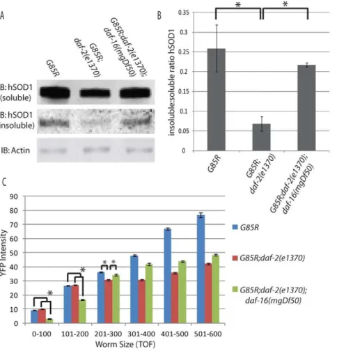
mixed population of approximately ten thousand worms from the following groups: G85R, G85R;daf-2(e1370) and G85R;daf-2(e1370);daf-16(mgDf50) (figure 3C). Using TOF as a proxy for age, we were unable to identify a direct correlation between total SOD1 burden and locomotory activity. The data were binned into TOF measurements of 100 units and significant differences between groups were observed in all bins by ANOVA (0–100 TOF F(2,11369)= 881.69, 101–200 TOF F(2,7209)= 250.90, 201– 300 TOFF(2,4880)= 48.1, 301–400 TOFF(2,3075)= 243.23, 401– 500 TOF F(2,1435)= 411.05, 501–600 TOF F(2,708)= 277.65). In
Figure 2.A) YFP signal was imaged in G85R or G85R;unc119p::sid1;sid1(pk3321)worms fed empty vector (EV) or G85R:YFP RNAi in order to demonstrate the efficacy of RNAi in neurons on theunc119p::sid1;sid1(pk3321)background B–E) Average speed normalized to size of swimming worms fed bacteria expressing the indicated RNAi.
young worms( 0–200 TOF) SOD1 intensity was equivalent between G85R and G85R;daf-2(e1370) worms (no significant differences between groups by Scheffe’s post-hoc), yet significant locomotory differences were observed between these groups at all ages assessed using the swimming assay. Conversely, young G85R;daf-2(e1370);daf-16(mgDf50) worms have significantly less YFP intensity thanG85RandG85R;daf-2(e1370) worms (p,0.05 by Scheffe’s post-hoc), yet they perform worse than G85R;daf-2(e1370)worms and equivalent toG85Rworms in our locomotory assays. Looking at older worms (300–600 TOF), G85R worms have a total SOD1 burden that significantly exceeds that of G85R;daf-2(e1370);daf-16(mgDf50) worms (p,0.05 by Scheffe’s post-hoc in 301–400, 401–500 and 501–600 bins) yet they show similar locomotory activity in the swim test. This is unlikely to simply be a floor-type effect as G85R;daf-16(mgDf50) worms perform worse in the swimming assay than both G85R and G85R;daf-2(e1370);daf-16(mgDf50) worms, suggesting that there is room for decline in the locomotory phenotype. Taken together these results suggest that overall SOD1 burden is not responsible for the locomotory differences observed between these groups, but rather suggest that the locomotory differences result from changes in how SOD1 toxicity is handled in these various backgrounds.
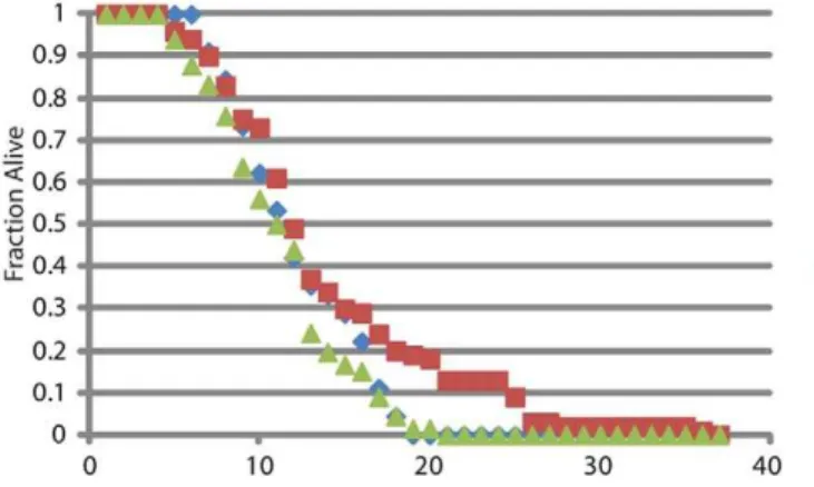
IIS effects on longevity in the SOD1 background The toxicity of mutant SOD1 expression in the C. elegans nervous system was previously reported to have a negative effect on lifespan [10]. In order to determine whether decreaseddaf-2
signaling in this background has a beneficial effect on lifespan, similar to its effect on locomotion, longevity was monitored inWT SOD1, G85R;daf-2(e1370) and G85R;daf-2(e1370);daf-16(mgDf50) worms (figure 4).WT SOD1andG85R;daf-2(e1370);daf-16(mgDf50) worms had similar lifespans, while theG85R;daf-2(e1370)worms had a modest but statistically significant increase in lifespan (p,0.05 by Mann-Whitney analysis). Interestingly, there is no correlation between health and lifespan as assayed by locomotory activity.
Discussion
Aging is a common risk factor for many neurodegenerative diseases [28]. The IIS pathway is a well characterized genetic modifier of aging inC. elegans[11] and we demonstrate here that alterations in this pathway can robustly modify the toxicity of mutant SOD1 as assessed by mobility, protein aggregation and longevity. We find that reduceddaf-2activity has a beneficial effect on the toxicity ofG85R SOD1and that this beneficial effect is daf-16 dependent. While aging in the worm has previously been shown to be coordinated at the organismal level, we found a requirement for IIS to be reduced in the nervous system in order to modify SOD1 toxicity. Although the genetic modifiers of aging are less well characterized in humans, these data suggest that IIS and pathways which beneficially modify lifespan/healthspan in humans may be potential targets for therapeutic intervention in ALS.
Figure 3.A) Representative western blot from three experiments looking at soluble vs insoluble SOD1 B) Quantification of SOD1 insoluble : soluble ratio C) COPAS data showing average YFP intensity for bins of various worm TOF.
Although their exact role in disease pathology is not entirely understood, aggregated proteins are associated with numerous neurodegenerative diseases, including ALS [29,30]. As aging occurs, the total burden of aggregated proteins increases, suggesting a diminished capacity for proper folding and/or degradation of aggregation prone proteins with age [31]. In this study we demonstrate that a LOF mutation ofdaf-2diminishes the amount of insoluble SOD1; an effect that might be due to an increased capacity for clearance/folding in these worms. Similar observations have been made regarding the ability of modifica-tions of IIS to modulate the solubility and toxicity of other disease related proteins [12,24,32]. This is likely due to the ability of decreased IIS to induce expression of chaperones such as the HSP family of proteins [19]. An RNAi screen performed on these worms identified chaperones as the most highly represented functional class of proteins found to negatively modify SOD1 aggregation in this model [10]. Increased capacity for folding/ clearance in worms with reduced IIS is likely to contribute to decreasing the toxcicity of mutant SOD1 in this model. We can not rule out the contribution of other changes in metabolism, lipid biogenesis, and free radical scavenger expression which have also been linked to increased lifespan and stress resistance due to decreased IIS [18,21,22].
Our data from the COPAS support both the concept that: 1) the daf-2(e1370) background can help reduce buildup of insoluble protein and 2) the ratio of soluble to insoluble G85R SOD1, rather than its total abundance, is associated with toxicity in this model. Using TOF as an approximation for age, it appears thatG85R worms accumulate less G85R SOD1 on the daf-2(e1370) as opposed to wild type background over time. This suggests an increased capacity for clearance of SOD1 over time in the daf-2(e1370) background. The abundance of SOD1 in G85R;daf-2(e1370);daf-16(mgDf50) worms is less than both G85R and G85R;daf-2(e1370)in young animals yet their mobility is reduced compared toG85R;daf-2(e1370)and equivalent toG85Rat all time points assessed. Although the relative abundance of SOD1 varies between these strains over time, their relative locomotory activity remains constant, suggesting that it is not total SOD1 abundance that dictates toxicity.
Previous work with a worm model of Ab toxicity has demonstrated that the daf-2(e1370) allele is protective against Ab1–42 aggregates in two distinct ways. Firstdaf-2(e1370)led to
activation ofhsf-1 which resulted in breakdown of Ab1–42fibrils.
Seconddaf-2(e1370)led to activation ofdaf-16which increased the abundance of Ab1–42 in high molecular weight aggregates. It is
possible that both activities diminish Ab1–42toxicity by removing
Ab1–42from the putatively toxic fibril pool [23]. If these
observations can be generalized, they could account for the lack of correlation between total SOD1 abundance and locomotory deficits in these worms.G85R;daf-2(e1370);daf-16(mgDf50) worms may not show the robust increase in SOD1 over time seen in the G85R worms because they are not accumulating the large aggregates of protein. These worms still suffer from SOD1 toxicity due to high levels of insoluble SOD1, but may never reach the same YFP intensity asG85Rworms due to a lack of high molecular weight aggregation facilitated bydaf-16. Activation ofhsf-1in the G85R;daf-2(e1370);daf-16(mgDf50) worms may also explain their increased mobility compared to G85R;daf-16(mgDf50) worms in the motility assay.
Using worms with neuronal expression of SID1, we demon-strate that the beneficial effect of reduced daf-2 is likely to be mediated by decreased IIS signaling in the nervous system. This contrasts with its effects on longevity, where daf-2 RNAi (on a background lacking pk3321to enhance neuronal RNAi) mimics the lifespan extending effects of the daf-2(e1370) allele. In this setting, daf-2 RNAi does not influence gene expression in the nervous system [33].G85R;daf-2(e1370) worms fed daf-16 RNAi should have normal levels of DAF-16 in their nervous system and it would be activated due to thedaf-2(e1370)background. If the beneficial effect of decreased daf-2 activity was completely mediated by the nervous system, then these worms should have comparable locomotory function toG85R;daf-2(e1370)worms fed empty vector. We find these worms to have an intermediate phenotype with a significant reduction in locomotory activity as compared to worms on empty vector RNAi plates. This suggests that part of the beneficial effect ofdaf-2(e1370)might be mediated through non-neuronal tissue(s). Alternatively, daf-16(RNAi) may partially, although not completely, reduce daf-16 expression in both neuronal and non-neuronal cells. Taken together, these findings suggest that although decreased IIS is required in the in the nervous system in order to have a beneficial effect on SOD1 toxicity, some of the downstream actions of reduced neuronal IIS may be functioning in the periphery.
Our results demonstrate the strong capacity of the IIS pathway to modulate G85R proteotoxicity. One possible mechanism of action for this beneficial effect is through the ability of this pathway to increase the cellular capacity to prevent toxic insoluble protein accumulation. Interestingly this beneficial activity of IIS may not be completely cell autonomous but may be in part a manifestation of alterations in cellular aging coordinated at the organismal level. This pathway may represent a possible therapeutic target for proteotoxic diseases cause by insoluble proteins.
Figure 4. Longevity analysis, percent alive per day.
Materials and Methods
Worm Strains and Handling
C. eleganswere cultured under standard conditions at 20uC and fed theE. colistrain OP50 [34]. The following worm strains were used: daf-16(mgDf50) and daf-2(e1370) were obtained from the CGC. Psnb-1::hTDP-43;Pmtl-2::GFP and daf-2(e1370); Psnb-1::hTDP-43;Pmtl-2::GFPwere a generous gift of Chris Link. Psnb-1::G85R SOD1::YFPandPsnb-1::WT SOD1::YFPwere a generous gift from Arthur Horwich and Jiou Wang. Punc119::sid1;-sid1(pk3321) was a generous gift from Martin Chalfie. Double and triple worms were generated by standard genetic crosses and verified by PCR or fluorescence expression.
Locomotory Assay
Video recordings of worms were made using the image acquisition tool in Matlab 2009b. These videos were then analyzed using the parallel worm tracker software (downloaded from the Goodman lab http://wormsense.stanford.edu/tracker/). For monitoring locomotion on OP50 a 1 minute video was recorded from each plate at the center of the lawn of OP50. At 50 worms were analyzed for each genotype at each timepoint. The number of worms was greater than 30 for each genotype at each timepoint. For the swimming assay worms were suspended in a pool of M9 and their swimming was recorded for 30 seconds. At least 30 worms were analyzed per group per timepoint. Statistics were performed using the average of each video as an n of 1 for speed or size/speed.
Feeding RNAi
HT115 bacteria containing the indicated genes in the L4440 vector were grown overnight at 37uC in 50 ug/ml ampicillin. They were then seeded on to NGM plates supplemented with 12.5 ug/ml tetracycline and 4 mM IPTG and allowed to grow overnight at room temperature. Gravid adults were allowed to drop eggs on the RNAi plates for 2 hrs and were then removed. Plates were then kept at 20uC until they were assayed.
Imaging
Worms were immobilized in 25 mM levamisole on agar pad slides and then coverslipped. Images were acquired at a constant intensity on a confocal microscope using a 40 uM Zstack.
Soluble vs Insoluble Protein Assay
Approximately 100 ul of packed worms were lysed via sonication in 300 ul RIPA buffer (150 mM NaCl, 50 mM Tris pH 8.0, 1 mM EGTA, 5 mM EDTA, 1% NP40, 0.5% Sodium Deoxycholate, 0.1% SDS) with complete protease inhibitor cocktail. A soft spin of 800 g for 5 min was performed to remove unlysed worms and large debris from the lysate. The supernatant was then spun at 99,0006g for 30 min @ 4uC. The supernatant was kept as the soluble fraction. The pellet was sonicated again in RIPA as a wash step to ensure removal of all soluble protein. It was centrifuged again at 99,0006g for 30 min @ 4uC. The pellet was then solublized in 50 ul urea buffer (40 mM Tris, 7 M urea, 2 M thiourea, 1% CHAPS). Equal volumes of sample were run on SDS-page gels under reducing conditions and probed with anti SOD1 antibody (Cell Signaling #2770) and anti actin (Sigma). Westerns were visualized using the Odyssey system and quantified using ImageJ.
COPAS
COPAS was used to sort and collect fluorescence intensity from 10,000 worms from each group as described in [35].
Longevity Assay
An egg drop was performed on NGM plates with OP50. The lifespan assay was carried out at 20uC and worms were transferred to fresh NGM OP50 plates as necessary in order to avoid starving the animals. Each day worms were counted. FUDR was not included in this assay so during the active reproductive period of the worms they were transferred to a new plate each day.
Acknowledgments
We would like to thank Dr. David Raizen and Maria Lim for their valuable comments and assistance withC. eleganstechniques on this project. We would like to thank Martin Chalfie, Arthur Horwich, Chris Link, and Jiou Wang for generously sharing worms strains.
Author Contributions
Conceived and designed the experiments: MB BK TL. Performed the experiments: MB. Analyzed the data: MB. Contributed reagents/ materials/analysis tools: TL. Wrote the paper: MB.
References
1. Renton AE, Majounie E, Waite A, Simon-Sanchez J, Rollinson S, et al. (2011) A hexanucleotide repeat expansion in C9ORF72 is the cause of chromosome 9p21-linked ALS-FTD. Neuron 72: 257–268.
2. DeJesus-Hernandez M, Mackenzie IR, Boeve BF, Boxer AL, Baker M, et al. (2011) Expanded GGGGCC hexanucleotide repeat in noncoding region of C9ORF72 causes chromosome 9p-linked FTD and ALS. Neuron 72: 245–256. 3. Deng HX, Chen W, Hong ST, Boycott KM, Gorrie GH, et al. (2011) Mutations in UBQLN2 cause dominant X-linked juvenile and adult-onset ALS and ALS/ dementia. Nature 477: 211–215.
4. Vance C, Rogelj B, Hortobagyi T, De Vos KJ, Nishimura AL, et al. (2009) Mutations in FUS, an RNA processing protein, cause familial amyotrophic lateral sclerosis type 6. Science 323: 1208–1211.
5. Schymick JC, Yang Y, Andersen PM, Vonsattel JP, Greenway M, et al. (2007) Progranulin mutations and amyotrophic lateral sclerosis or amyotrophic lateral sclerosis-frontotemporal dementia phenotypes. J Neurol Neurosurg Psychiatry 78: 754–756.
6. Van Deerlin VM, Leverenz JB, Bekris LM, Bird TD, Yuan W, et al. (2008) TARDBP mutations in amyotrophic lateral sclerosis with TDP-43 neuropa-thology: a genetic and histopathological analysis. Lancet Neurol 7: 409–416. 7. Rosen DR, Siddique T, Patterson D, Figlewicz DA, Sapp P, et al. (1993)
Mutations in Cu/Zn superoxide dismutase gene are associated with familial amyotrophic lateral sclerosis. Nature 362: 59–62.
8. Chattopadhyay M, Valentine JS (2009) Aggregation of copper-zinc superoxide dismutase in familial and sporadic ALS. Antioxid Redox Signal 11: 1603– 1614.
9. Reaume AG, Elliott JL, Hoffman EK, Kowall NW, Ferrante RJ, et al. (1996) Motor neurons in Cu/Zn superoxide dismutase-deficient mice develop normally but exhibit enhanced cell death after axonal injury. Nat Genet 13: 43–47.
10. Wang J, Farr GW, Hall DH, Li F, Furtak K, et al. (2009) An ALS-linked mutant SOD1 produces a locomotor defect associated with aggregation and synaptic dysfunction when expressed in neurons of Caenorhabditis elegans. PLoS Genet 5: e1000350.
11. Kenyon C, Chang J, Gensch E, Rudner A, Tabtiang R (1993) A C. elegans mutant that lives twice as long as wild type. Nature 366: 461–464.
12. Hsu AL, Murphy CT, Kenyon C (2003) Regulation of aging and age-related disease by DAF-16 and heat-shock factor. Science 300: 1142–1145. 13. Lin K, Hsin H, Libina N, Kenyon C (2001) Regulation of the Caenorhabditis
elegans longevity protein DAF-16 by insulin/IGF-1 and germline signaling. Nat Genet 28: 139–145.
14. Williams TW, Dumas KJ, Hu PJ (2010) EAK proteins: novel conserved regulators of C. elegans lifespan. Aging (Albany NY) 2: 742–747.
15. Guarente L, Kenyon C (2000) Genetic pathways that regulate ageing in model organisms. Nature 408: 255–262.
16. Lin K, Dorman JB, Rodan A, Kenyon C (1997) daf-16: An HNF-3/forkhead family member that can function to double the life-span of Caenorhabditis elegans. Science 278: 1319–1322.
18. Zhou KI, Pincus Z, Slack FJ (2011) Longevity and stress in Caenorhabditis elegans. Aging (Albany NY) 3: 733–753.
19. Murphy CT, McCarroll SA, Bargmann CI, Fraser A, Kamath RS, et al. (2003) Genes that act downstream of DAF-16 to influence the lifespan of Caenorhabditis elegans. Nature 424: 277–283.
20. Artal-Sanz M, Tavernarakis N (2009) Prohibitin couples diapause signalling to mitochondrial metabolism during ageing in C. elegans. Nature 461: 793–797. 21. Shmookler Reis RJ, Xu L, Lee H, Chae M, Thaden JJ, et al. (2011) Modulation
of lipid biosynthesis contributes to stress resistance and longevity of C. elegans mutants. Aging (Albany NY) 3: 125–147.
22. Artal-Sanz M, Tavernarakis N (2010) Opposing function of mitochondrial prohibitin in aging. Aging (Albany NY) 2: 1004–1011.
23. Cohen E, Bieschke J, Perciavalle RM, Kelly JW, Dillin A (2006) Opposing activities protect against age-onset proteotoxicity. Science 313: 1604–1610. 24. Zhang T, Mullane PC, Periz G, Wang J (2011) TDP-43 neurotoxicity and
protein aggregation modulated by heat shock factor and insulin/IGF-1 signaling. Hum Mol Genet 20: 1952–1965.
25. Dillin A, Crawford DK, Kenyon C (2002) Timing requirements for insulin/IGF-1 signaling in C. elegans. Science 298: 830–834.
26. Calixto A, Chelur D, Topalidou I, Chen X, Chalfie M (2010) Enhanced neuronal RNAi in C. elegans using SID-1. Nat Methods 7: 554–559.
27. David DC, Ollikainen N, Trinidad JC, Cary MP, Burlingame AL, et al. (2010) Widespread protein aggregation as an inherent part of aging in C. elegans. PLoS Biol 8: e1000450.
28. Kenyon CJ (2010) The genetics of ageing. Nature 464: 504–512.
29. Rubinsztein DC (2006) The roles of intracellular protein-degradation pathways in neurodegeneration. Nature 443: 780–786.
30. Ticozzi N, Ratti A, Silani V (2010) Protein aggregation and defective RNA metabolism as mechanisms for motor neuron damage. CNS Neurol Disord Drug Targets 9: 285–296.
31. Morimoto RI, Cuervo AM (2009) Protein homeostasis and aging: taking care of proteins from the cradle to the grave. J Gerontol A Biol Sci Med Sci 64: 167–170.
32. Morley JF, Brignull HR, Weyers JJ, Morimoto RI (2002) The threshold for polyglutamine-expansion protein aggregation and cellular toxicity is dynamic and influenced by aging in Caenorhabditis elegans. Proc Natl Acad Sci U S A 99: 10417–10422.
33. Apfeld J, Kenyon C (1998) Cell nonautonomy of C. elegans daf-2 function in the regulation of diapause and life span. Cell 95: 199–210.