Listeria monocytogenes
Infection in Macrophages
Induces Vacuolar-Dependent Host miRNA Response
Anna K. D. Schnitger1, Alzbeta Machova1, Roman Ulrich Mueller2, Ariadne Androulidaki3, Bernhard Schermer2, Manolis Pasparakis3, Martin Kro¨nke1, Nikoletta Papadopoulou1*
1Institute for Medical Microbiology, Immunology and Hygiene, University of Cologne, Cologne, Germany,2Department of Medicine, University of Cologne, Cologne, Germany,3Institute of Genetics, University of Cologne, Cologne, Germany
Abstract
Listeria monocytogenesis a Gram-positive facultative intracellular pathogen, causing serious illness in immunocompromised individuals and pregnant women. Upon detection by macrophages, which are key players of the innate immune response against infection, L. monocytogenes induces specific host cell responses which need to be tightly controlled at transcriptional and post-transcriptional levels. Here, we ask whether and how host miRNAs, which represent an important mechanism of post-transcriptional regulation in a wide array of biological processes, are altered by a model pathogen upon live infection of murine bone marrow derived macrophages. We first report that L. monocytogenes subverts the host genome-wide miRNA profile of macrophagesin vitro. Specifically, we show that miR-155, miR-146a, miR-125a-3p/5p and miR-149 were amongst the most significantly regulated miRNAs in infected macrophages. Strikingly, these miRNAs were highly upregulated upon infection with the Listeriolysin-deficientL. monocytogenesmutantDhly, that cannot escape from the phagosome thus representing a vacuolar-contained infection. The vacuolar miRNA response was significantly reduced in macrophages deficient for MyD88. In addition, miR-146a and miR-125a-3p/5p were regulated at transcriptional levels upon infection, and miR-125a-3p/5p were found to be TLR2 responsive. Furthermore, miR-155 transactivation in infection was regulated by NF-kB p65, while miR-146a and miR-125a-3p/5p expression was unaffected in p65-deficient primary macrophages upon L. monocytogenes infection. Our results demonstrate that L. monocytogenes promotes significant changes in the miRNA expression profile in macrophages, and reveal a vacuolar-dependent miRNA signature, listeriolysin-independent and MyD88-dependent. These miRNAs are predicted to target immune genes and are therefore most likely involved in regulation of the macrophage innate immune response against infection at post-transcriptional levels.
Citation:Schnitger AKD, Machova A, Mueller RU, Androulidaki A, Schermer B, et al. (2011)Listeria monocytogenesInfection in Macrophages Induces Vacuolar-Dependent Host miRNA Response. PLoS ONE 6(11): e27435. doi:10.1371/journal.pone.0027435
Editor:Robin Charles May, University of Birmingham, United Kingdom
ReceivedMay 20, 2011;AcceptedOctober 17, 2011;PublishedNovember 17, 2011
Copyright:ß2011 Schnitger et al. This is an open-access article distributed under the terms of the Creative Commons Attribution License, which permits unrestricted use, distribution, and reproduction in any medium, provided the original author and source are credited.
Funding:This work was supported by funds from the University of Cologne. The funders had no role in study design, data collection and analysis, decision to publish, or preparation of the manuscript.
Competing Interests:The authors have declared that no competing interests exist.
* E-mail: [email protected]
Introduction
Listeria monocytogenesis a Gram positive facultative intracellular bacterium that has been used as a model pathogen to study host-pathogen interactions [1]. This opportunistic food-borne host-pathogen causes serious illness to immunocompromised individuals, preg-nant women and the developing foetus [2]. To establish an infection in a host organism, the bacterium must first overcome the barriers of the innate immune system. Macrophages are professional phagocytes which provide a first line of innate immune defence against invading pathogens. Detection of L. monocytogenes by macrophages at the cell surface, within phago-somes or the cytosol triggers distinct host cell transcriptional responses via pattern recognition receptors (PRR) [3–4]. Signalling via the so-called vacuolar/Toll-like receptor (TLR) - and the cytosolic/nuclear oligomerisation domain (NOD)-like receptor (NLR) - dependent pathways lead to mitogen-activated protein kinase (MAPK), nuclear factor kappa B (NF-kB) and Interferon Regulatory Factor-3 (IRF3) activation [5]. As a consequence, numerous proinflammatory mediators and other molecules are expressed that further instruct elicitation of antigen-specific acquired immunity and clearance of infection.
Recognition of PAMPs by PRRs of immune cells results in expression of distinct subsets of miRNAs. PRR-triggered miRNAs are thought to target and negatively regulate activated signalling cascade components. In particular, it has been proposed that miR-146a and miR-155 negatively regulate lipopolysaccharide (LPS)-induced TLR4 signalling in myeloid cells with NF-kB transcrip-tion factor involved in regulating miR-146a expression [15–16]. These miRNAs target protein-coding genes involved in receptor-induced signalling such as TNF receptor-associated factor 6 (TRAF6), IL-1 receptor-associated kinase (IRAK) 1 and IRAK2 by miR-146a [15,17], or Fas-associated death domain (FADD) and IkB kinase epsilon (IKKe) by miR-155 [18,19]. In addition, let-7e and miR-155 were found to target TLR4 and SOCS1, respectively, in response to LPS, regulated by Akt1 in macro-phages [20]. In infection, miR-155 negatively regulates the epithelial cell response upon infection with Gram-negative bacterium Helicobacter pylori [19], while in macrophages infected with another Gram-negative bacteriumFrancisella tularensis, miR-155 expression resulted in enhanced pro-inflammatory response [21]. Similarly, infections with the protozoan parasite Cryptosporid-ium parvumin epithelial cells [22] or detection of heat-killedCandida albicans by macrophages [23], lead to alterations of host miRNA profile, most likely involved in immunoregulation. Furthermore, analysis of the host miRNA profile upon Salmonella Typhimurium
infection also identified the let-7 family of miRNAs as regulators of the inflammatory response in both epithelial cells and macro-phages [24]. More recently, it was shown that miR-29 targets interferon (IFN)-cproduction by natural killer (NK) cells, CD4+T cells and CD8+T cells during systemic infection with intracellular bacteria in mice [25]. However, it is unclear whether infection with any Gram-positive intracellular bacteria can subvert the genome-wide miRNA profile of macrophages.
To address this question, we employed the model pathogenL. monocytogenes and first determined whether the host genome-wide miRNA profile is altered upon infection of primary murine macrophages. We found that miR-155, miR-146a, miR-125a-3p/ 5p and miR-149 were amongst the most significantly regulated miRNAs in infected macrophages. To precisely dissect at which stage of infection and via which host pathways Listeria may promote induction of these miRNAs, we used genetically modified
L. monocytogenesstrains, to independently activate the extracellular/ vacuolar response, and compare it to wild typeListeriathat gain access to the cytosol and thus elicits both vacuolar and cytosolic immune responses in macrophages. To accurately identify the host pathways potentially involved in regulation of Listeria-induced miRNAs, we directly compared primary macrophages deficient for MyD88 (MyD882/2), which transmits most TLR signals, or NF-kB p65 (p65MYEL
KO), which transcriptionally regulates the inflammatory response uponListeriainfection, to wild type cells. We have identified miR-155, miR-146a, miR-125a-3p/5p and miR-149 as highly responsive elements of the vacuolar host response, differentially regulated by inflammatory mediators in macrophages.
Results
L. monocytogenesinfection in primary macrophages induces significant host miRNA expression
To determine the impact of L. monocytogenes infection in the global miRNA profile of macrophages, we performed parallel profiling of 585 miRNAs using TaqMan Rodent miRNA Arrays A and B (v2.0) on total RNA from bone marrow derived macrophages (BMDM) infected for 6 h with a calculated multiplicity of infection (MOI) 10 compared to non-infected cells,
from three biological replicates. Using the Applied Biosystems (ABI) RQ Manager 1.2 and DataAssist v2.0 software for analyses, we comprehensively identified 385 miRNAs that were expressed in our samples, of which 13 were significantly ($1.5 fold, P
value#0.05) up-regulated at 6 h post-infection (pi; Table 1 and Figure S1). Interestingly,Listeria-induced miRNAs included miR-155, miR-146a and miR-147, which are known modulators of the inflammatory response upon TLR ligation in macrophages [15– 16,26]. Additionally, two recently described miRNAs with unknown function in infected macrophages, namely miR-125a-3p and miR-125a-5p, and a newly described miRNA, namely miR-149, were significantly up-regulated in Listeria-infected BMDMs. Surprisingly, no miRNAs were found to be significantly down-regulated in infected compared to non-infected macrophag-es (Figure S1).
To validate the accuracy of our array results and study further the regulation of these miRNAs upon infection, we generated four new sets of experiments, each including: two timepoints of infection with two types of bacteria (MOI 10) in two different sorts of BMDMs. These samples were subsequently used to quantify miRNA expression by real time quantitative PCR (RT-qPCR) using Taqman assays. The results from these experiments validate that miR-155 and miR-125a-3p were significantly upregulated at 6 h pi by approximately 5-fold (Figure 1A–B). These two miRNAs represent the highest regulated among the fiveListeria-induced miRNAs. At 6 h pi miR-146a, miR-125a-5p and miR-149 were significantly induced by 1.5–2.0 folds (Figure 1C–E). Interestingly, miR-125a-3p and miR-149 were also significantly up-regulated as early as 3 h pi (Figure 1B and 1E). Taken together, our miRNA-array and RT-qPCR data clearly demonstrate thatL. monocytogenessubverts the host miRNA profile, causing specific miRNAs to be upregulated early upon infection in BMDMs. These results provide the first miRNA signature of a Gram-positive intracellular pathogen in murine primary macrophages.
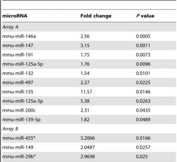
Table 1.Significant miRNA expression uponL. monocytogenesinfection in Bone Marrow Derived Macrophages.
microRNA Fold change Pvalue
Array A
mmu-miR-146a 2.56 0.0005
mmu-miR-147 3.15 0.0011
mmu-miR-191 1.75 0.0073
mmu-miR-125a-5p 1.76 0.0096
mmu-miR-132 1.54 0.0101
mmu-miR-497 2.27 0.0225
mmu-miR-155 11.57 0.0146
mmu-miR-125a-3p 5.38 0.0263
mmu-miR-200c 2.31 0.0435
mmu-miR-139-5p 1.82 0.0489
Array B
mmu-miR-455* 3.2066 0.0166
mmu-miR-149 2.0487 0.0257
mmu-miR-29b* 2.9698 0.025
Genome-wide profiling was performed by Taqman Rodent miRNA Arrays A and B v2.0 (ABI, Life Technologies).
Figure 1. Regulation ofL. monocytogenes-induced miRNAs in macrophages.Bone marrow derived macrophages (BMDMs) from wild type (WT; n = 4) and MyD882/2(n = 4) mice were infected (MOI 10) withL. monocytogenes(Lm) and the LLO-deficient mutant
Dhly(LmDhly) for 3 h and 6 h. Total RNA was extracted and the expression levels of miR-155 (A), miR-146a (B), miR-125a-3p (C), miR-125a-5p (D) and miR-149 (E) were quantified by RT-qPCR using TaqMan miRNA assays. Data was normalized to the endogenous control sno202 and fold changes of miRNA expression relative to the non-infected control of its own genotype was calculated by the 22DDCTmethod. Data represents the mean
6SEM from three to four biological replicates. Statistical significance of miRNA expression between infected and non infected WT BMDMs was determined by the Studentt -test; *P,0.05; **P,0.01; ***P,0.001. Statistical significance of miRNA induction in infection between WT and MyD882/2BMDMs was determined by two-way ANOVA test;#P,0.05,##P,0.01,###P,0.001.
doi:10.1371/journal.pone.0027435.g001
L. monocytogenesinduced miRNA expression is LLO-independent
Upon uptake by macrophages,L. monocytogenesis initially found within the phagosomal vacuole before escaping into the cytosol where it replicates. In parallel, a distinct set of genes is transcriptionally activated upon vacuolarL. monocytogenesinfection, important for intracellular bacteria sensing [4]. Destruction of the phagosomal membrane by L. monocytogenesis essential for in vivo
bacterial virulence and is mediated by the pore-forming haemolysin Listeriolysin-O (LLO), encoded by thehlygene [27]. Therefore, deletion of the gene encoding LLO (Dhly) renders this mutant unable to enter the host cell cytosol, avirulent and low in immunogenicity [28].
In our study, we employed this mutant (Dhly-Lm) as a tool to investigate compartment-specific miRNA-induction in BMDMs at 3 h and 6 h pi, by which timepoints wild type Lm are already replicating in the host cytosol, in contrast toDhly-Lmwhich remain confined in the phagosome (Figure S2). The intracellular growth of both types of bacteria in macrophages is illustrated in Figure S3. Strikingly, quantitative analyses of 155, 146a, miR-125a-3p, miR-125a-5p and miR-149 revealed that all five miRNAs were significantly upregulated by the vacuole-contained bacteria at both timepoints (Figure 1A–E). Our findings suggest that these miRNAs are induced in macrophages not only upon wild type/cytosolic Listeria infection but also upon vacuolar infection i.e. independent of bacterial virulence and access to the host cytosol.
Vacuolar miRNA response to infection is MyD88-dependent
Downstream signalling of all TLRs (except of TLR3), IL-1 and IL-18 receptors depends on the adaptor molecule MyD88 [29]. In macrophages, the so-called vacuolar transcriptional response upon
L. monocytogenes infection is MyD88-dependent, while escape of bacteria in the cytosol induces a MyD88-independent/cytosolic transcriptional response [3–4,30]. To accurately demonstrate which type of host response regulates theL. monocytogenes-induced miRNA expression in primary macrophages, and further analyse the strong effect of Dhly-Lm infection on miRNA induction, we quantified changes in miRNA expression upon infection with wild typeLmorDhly-Lmfor 3 h and 6 h in MyD882/2compared to wild type (WT) BMDMs. Interestingly, expression of miR-155, miR-125a-3p, miR-146a and miR-149 was significantly reduced in MyD882/2 compared to WT BMDMs upon infection with
Dhly-Lm(Figure 1A–C and 1E, respectively). The effect of MyD88 ablation in miR-125a-5p induction was moderate (Figure 1D). These findings suggest that L. monocytogenes promotes a predom-inant vacuolar miRNA response that is MyD88-dependent.
We additionally analysed BMDM culture supernatants from the same infections for tumour necrosis factor (TNF) production, a gene that represents induction of the vacuolar response upon
Listeria infection in macrophages [4]. In agreement to the published data, TNF production was induced upon wild typeLm
andDhly-Lminfection in WT BMDMs and completely abolished in MyD882/2BMDM (Figure S4), while bacterial uptake in cells of both genotypes was comparable (Figure S5).
L. monocytogenesinfection promotes upregulation of miRNA genes at transcriptional level
MiR-125a-3p and miR-125a-5p represent two opposing strands of the same primary transcript (pri-miR; Figure S6). Nevertheless,
Listeria-induced expression levels of the two mature miRNAs varied between the two strands by 4.5-folds (Figure 1B and 1D)
suggesting differential regulation upon infection. Regulation of mature miRNA expression can occur on the level of transcription as well as post-transcriptionally [15–16,31]. To determine whether
L. monocytogenes regulates miRNA-125a-3p/5p expression in BMDMs at transcriptional level, we analysed the induction of pri-miR-125a, in parallel to pri-miR-146a, upon 3 h and 6 h pi with wild typeLmandDhly-Lm.
We found that pri-miR-125a and pri-miR-146a were both upregulated upon cytosolic and vacuolar infections, while pri-miR-125a induction was significantly augmented upon Dhly-Lm
infection (Figure 2). Furthermore, we compared miRNA tran-scriptional regulation upon infection of MyD882/2 and WT
BMDMs and found that pri-miR-125a upregulation was signifi-cantly decreased in infected MyD882/2 BMDM (Figure 2A) suggesting that MyD88 signalling is also required for the upregulation of the primary transcript upon infection, most likely upon immediate recognition ofListeriaPAMPs extracellularly or in the phagosomal vacuole. In contrast, pri-miR-146a induction was unaffected in MyD882/2BMDMs compared to WT cells infected by either wild typeLmorDhly-Lm(Figure 2B). Our findings suggest thatListeriainfection in BMDMs regulates both miR-125a-3p/5p and miR-146a at transcriptional levels, the first in a vacuolar/ MyD88-dependent manner and the latter in a MyD88-indepen-dent manner.
TLR2-mediates miR-125a-3p and miR-125a-5p upregulation in macrophages
Upon infection,L. monocytogenes-derived lipoteichonic acids [32] and lipoproteins [33] are recognised by membrane-bound TLR2 receptors which transmit their signals via MyD88 [34]. Activation of TLRs in immune cells results in expression of distinct subsets of miRNAs, including miR-146a [15] and miR-155 [16]. To investigate the response of miR-125a-3p and miR-125a-5p upon receptor-specific activation, we stimulated BMDMs with the TLR2 synthetic ligand Pam3CSK4. Interestingly, our results showed that miR-125a-3p and miR-125a-5p were significantly induced upon Pam3CSK4 treatment in BMDMs, providing the first evidence to support involvement of the TLR2 pathway in the induction of these two miRNAs (Figure 3A–B). Recently, miR-125a-3p and miRNA-125a-5p were reported to be moderately upregulated upon TLR4 ligation by LPS, a major component of the cell wall of Gram-negative bacteria, in BMDMs subjected to microarray analyses (P values of 0.095 and 0.066 respectively) [23]. Unlike this study, we used ultra-pure LPS to prevent residual protein impurities influencing the induction attributed to TLR4-signalling, and found that miR-125a-3p and miR-125a-5p were both significantly upregulated upon specific TLR4 ligation (P,0.01 andP,0.001, respectively) similarly to miR-146a which served as positive control (Figure 3A–C). Most importantly, we compared TLR2- and TLR4-mediated upregulation of miR-125a-3p, miR-125a-5p and miR-146a in BMDMs from WT and MyD882/2mice, and could show that TLR2-mediated upregula-tion of all three miRNAs by Pam3CSK4 was significantly decreased in MyD882/2 cells and thus was MyD88-dependent (Figure 3A–C). Compared to WT cells, MyD882/2 BMDMs
and miR-146a primary transcripts in WT BMDMs, while induction of pri-miR-125a was MyD88-dependent (Figure 3D– E). These findings support that miR-125a-3p and miR-125a-5p are new members of the group of miRNAs that are induced upon TLR/MyD88-signalling and underline a role in the vacuolar response of macrophage to infection.
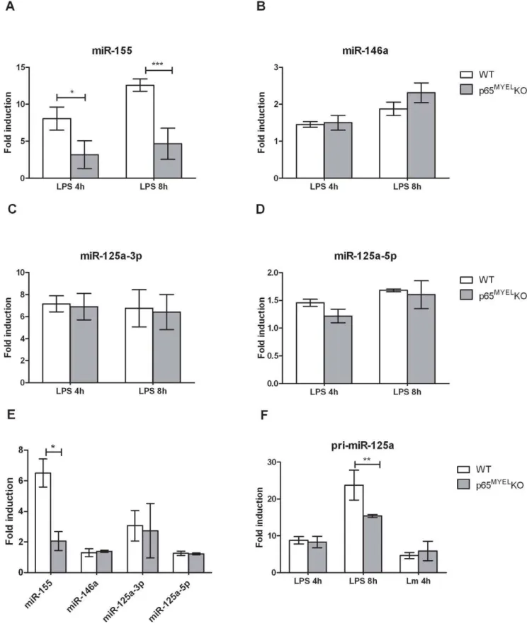
NF-kB p65 regulates miR-155, but not miR-125a-3p/5p and miR-146a, expression in response toL.
monocytogenesor LPS
A potential mechanism for selectively regulating miRNA transcript levels upon infection is via nuclear transcription factors [15,22,35]. Activation of NF-kB transcription factors downstream of TLRs is crucial in coordinating the transcriptional response to infection, and various NF-kB subunit knock-out mice show increased susceptibility to L. monocytogenes infection [36]. Few studies report that miR-155 and miR-146a have active NF-kB binding sites in their promoter elements [15,37], or their induction depends on signalling via the IkB kinase (IKK) complex [21–23]. We hypothesised that NF-kB p65 is involved in regulating transcription of L. monocytogenes-induced miRNAs, and to accu-rately demonstrate its direct role we compared BMDMs from mice in which p65 is genetically ablated (p65MYELKO) to WT cells upon mild (MOI 1) infection with L. monocytogenes for 4 h or exposure to LPS for 4 h and 8 h. To corroborate the absence of functional p65 we first assessed the response of TNF, a p65-regulated gene. As expected, p65MYEL KO BMDMs showed reduced TNF production upon infection or LPS treatment compared to WT cells (Figure S7). Similarly, miR-155, which possesses two active NF-kB binding sites in the BIC/miR-155 promoter region, was significantly reduced in p65MYEL KO BMDMs compared to WT cells upon LPS treatment and L. monocytogenes infection (Figure 4A and 4E), suggesting the involvement of the canonical NF-kB pathway, and specifically of the p65 subunit. Surprisingly, miR-146a expression which was shown to be NF-kB p65 dependent upon LPS treatment in mouse
embryonic fibroblasts [38] was unaffected in p65MYEL KO compared to WT BMDMs treated with LPS or infected withL. monocytogenes (Figure 4B and 4E). Similarly, miR-125a-3p and miRNA-125a-5p which are predicted to derive from exon1 of a non-coding RNA transcript on chromosome 17 (Ensembl: Ncrna 00085-003) that has several putative NF-kB binding sites in the pri-miR-125a promoter region, was unaffected by genetic ablation of p65 in infected or LPS treated BMDMs (Figure 4C–E). Furthermore, we specifically investigated the pri-miR-125a kinetics and found that only at 8 h after LPS exposure, its expression was significantly reduced in p65MYELKO compared to WT BMDMs (Figure 4F). Based onin silicoanalyses of the 1.5 kb region upstream of the pri-miR-125a transcriptional start site for transcription factor binding sites, using the Genomatix MatIn-spector software package, we found that apart of the two putative NF-kB p65 sites, there are also putative NF-kB p50 and IRF binding sites present in the promoter region (Figure S6). These results suggest that regulation ofL. monocytogenes-induced pri-miR-125a may be propelled by NF-kB p50 or another transcription factor, which is also likely for miR-146a, while miR-155 transactivation uponL. monocytogenesinfection and LPS treatment in BMDMs is predominately NF-kB p65 dependent.
Discussion
It is well established that miRNAs are important players of the post-transcriptional mechanism that regulates gene expression in many biological processes, including cell proliferation, differenti-ation, immunity and tumorigenesis. Host-pathogen interactions must be tightly controlled at all levels to prevent excessive inflammation and spread of infection. It is increasingly appreciated that miRNAs are important immunoregulators; however our knowledge on the role of miRNAs upon bacterial infection is far from comprehensive. Infection of macrophages with one of the best studied Gram positive intracellular pathogensL. monocytogenes
initiates a sequence of responses at transcriptional and post-transcriptional levels which are vital for host survival. In this
Figure 2.L. monocytogenes-induced transcriptional regulation of miR-146a and miR-125a.Bone marrow derived macrophages (BMDMs) were infected (MOI 10) withL. monocytogenes(Lm) and the LLO-deficient mutantDhly(LmDhly) for 3 h and 6 h. Total RNA was extracted and the expression levels of primary transcript (pri)-miR-125a (A) and pri-miR-146a (B) were quantified by RT-qPCR using TaqMan assays. Data was normalized to the endogenous control gene HPRT1 and fold changes of miRNA induction in infected compared to control cells of each genotype were calculated by the 22DDCTmethod. Data represents the mean
6SEM from three biological replicates. Statistical significance of miRNA expression between infected and non infected WT BMDMs was determined by the Studentt-test; *P,0.05, **P,0.01, ***P,0.001. Statistical significance of miRNA induction in infection between WT and MyD882/2BMDMs was determined by two-way ANOVA test;#P
,0.05,##P,0.01,###P,0.001. doi:10.1371/journal.pone.0027435.g002
model, we first explored whether miRNAs are part of the anti-listerial immune response of macrophages. Our study reveals the
Listeria-induced miRNA signature at 6 h pi in primary macro-phages. Specifically, infection induced significant upregulation of 13 miRNAs, including miR-155, miR-146a, miR-125a-3p/5p and miR-149. To our knowledge, this is the first comprehensive profile
of miRNA expression upon Gram-positive bacterial infection. Notably, at 6 h pi, we did not detect significantly downregulated miRNAs in infected BMDMs compared to non-infected control cells. To establish profound down-regulation of miRNAs, longer timepoints may need to be explored [22,24–25] though in that case, the contribution of cytokines and interferons produced by
Figure 3. Regulation of miR-125a-3p/5p and miR-146a expression upon TLR2 and TLR4 stimulation. Wild type (WT; n = 3) and MyD882/2(n = 2) mouse bone marrow derived macrophages (BMDMs) were treated with 1
mg/ml ultra-pure LPS or 2mg/ml Pam3CSK4 for 6 h. Total
RNA was extracted and miRNA expression levels were quantified by RT-qPCR using TaqMan miRNA assays. MiR-125a-3p (A), 125a-5p (B) and miR-146a (C) expression in MyD882/2BMDMs. Pri-miR-125a (D) and pri-miR-146a (E) expression upon TLR stimulation in WT and MyD882/2BMDMs. Data was normalized to sno202 or HPRT1, endogenous controls for miR or pri-miR expression, respectively, and fold changes were calculated by the 22DDCTmethod and expressed relative to non-infected control of each genotype. Data represents the mean
6SEM of at least two biological replicates. Statistical significance of miR/pri-miR induction was determined by the Studentt-test compared to non infected control (*P,0.05, **P,0.01, ***P,0.001) or two-way ANOVA between WT and KO cells (#P,0.05,##P,0.01,###P,0.001).
Figure 4. Regulation of miR-125a-3p/5p, miR-146a and miR-155 expression in macrophages by NF-kB p65.Wild type (WT) and p65MYEL
KO BMDMs were treated with 100 ng/ml ultra-pure LPS for 4 h and 8 h (A–D, F) or infected (MOI 1) withL. monocytogenes(Lm) for 4 h (E–F). Total RNA was extracted and the expression levels of miR-125a-3p, miR-125a-5p, miR-146a and miR-155 were quantified by TaqMan miRNA assays. Data was normalized to sno202 or HPRT1, endogenous controls for miR or pri-miR expression, respectively, and fold changes were calculated by the 22DDCTmethod and expressed relative to the mock-treated/mock-infected control for each timepoint/condition in each genotype. Data represents the mean6SEM of at least two independent experiments, including minimum two mice from each genotype. Statistical significance of miR/pri-miR induction between WT and p65MYELKO BMDMs was determined by two way ANOVA; *P,0.05, **P,0.01, ***P,0.001.
doi:10.1371/journal.pone.0027435.g004
infected macrophages should also be considered when attributing the direct and indirect effects of bacterial infection on the host miRNA profile [16].
Our aim was to investigate potential miRNA induction linked to PRR activation which is vital for innate immune defence against intracellular pathogens.
To validate our array data, we performed quantitative RT-PCR and confirmed that miR-155, miR-146a, miR-125a-3p/5p and miR-149 were significantly increased in macrophages upon early sensing of live bacteria, suggesting that these miRNA genes may be implicated in the inflammatory immune response. MiR-149 has not been characterised before in macrophages upon TLR activation, while miR-125a-3p/5p were only recently reported to be upregulated upon LPS treatment or heat-killed C. albicans
infection, in monocytes [39] and macrophages [23], respectively. MiR-155 and miR-146a are induced in different cell types upon viral, parasitic, fungal or Gram-negative bacterial infections as well as upon specific TLR ligation and are thought to modulate the inflammatory response [15–24,37].
To determine how L. monocytogenes infection promotes host miRNA induction, we differentiated the phases of infection to vacuolar or cytosolic which are characterized by distinct transcriptional profiles in macrophages [3–4]. To accomplish this, we employed two different types ofL. monocytogenesEGDe bacteria, the wild type and its isogenic mutantDhly[28] which is confined to the phagosome and therefore does not induce cytosolic immune signalling. Interestingly, like wild type infection, vacuolar/Dhly-Lm
infection caused significant upregulation of all five miRNAs, suggesting that the haemolytic action of LLO and therefore the cytosolic surveillance pathways are not implicated in the miRNA response of macrophages to infection. To investigate whether this response is regulated via the TLR signalling pathway and establish the effect of vacuolar infection to the host miRNA response, we compared miRNA induction between WT and MyD882/2 BMDMs, upon wt-Lm and Dhly-Lm infections. Our data shows that miR-155, miR-125a-3p, miR-146a and miR-149 response is MyD88-dependent upon Dhly-Lm infection, suggesting that these miRNAs are among the early-response genes expressed in macrophages upon phagosomal detection of Listeria PAMPs. These data also provide the first evidence on 149 and miR-125a-3p regulation by MyD88.
From all Listeria-induced miRNAs, we were particularly attracted to miRNA-125a-3p and miRNA-125a-5p, because they represent the two strands of the same miRNA duplex, and unlike their trend of induction upon LPS treatment or heat-killed C. albicans infection [23], upregulation uponListeriainfection varied significantly between the two miRNA strands. Listeria promotes miR-125a gene expression upon MyD88 signalling at the transcriptional level which was comparable to the induction and regulation of the mature miR-125a-3p. In contrast, miR-125a-5p expression during bothwt-andDhly-Lminfections was moderately affected by MyD88 deletion in BMDMs. Based on the mechanism proposed by Schwarz et al. [13], miRNA 3p and 5p strand accumulation depends on free energy properties of miRNA duplexes, and can be developmentally regulated in specific cell lineages [40]. It is tempting to speculate that microbial infection may also influence the mechanisms that regulate mature miRNA abundance and strand stability in activated macrophages, providing an additional element to the proposed mechanism, perhaps depending on miRNA target abundance and the state of the infected cell. Furthermore, recent evidence on miRNA-125a-3p/5p in lung carcinoma suggests that the two strands exhibit opposing functions in regulating invasive and metastatic capabil-ities of lung cancer cells [41]. Although their role in infection is still
unresolved, we clearly show that 125a-3p/5p, alongside miR-155 and miR-146a, are part of the early innate immune response of macrophages toListeriainfection. The majority of the targets for miR-155 and miR-146a are still unknown [15,17–19,21,42–44], while the targets for miR-125a-3p/5p and miR-149 have not yet been explored. We carried out computational analyses by
TargetScanMouse 5.1software as previously described [22,45], and identified a variety of immune-related putative mRNA targets of all fiveL. monocytogenes-induced miRNAs (Table S1). MiR-125a-3p and miR-125a-5p potentially target the interleukin-1 receptor 1 (IL-1 R1), and the IL-6 receptor (IL-6 R), respectively. MiR-125a-5p shares the same 59-UTR sequence (seed region) with miR-125b and may therefore target the same mRNAs. In murine macrophages, miR-125b was shown to be induced by LPS and to target TNFa[18] and in H69 epithelial cells it was upregulated uponC. parvuminfection via NF-kB [22]. However, miR-125b was not upregulated in our arrays, which is not surprising since the two miRNAs derive from transcripts located on different chromosomal locations, and are probably regulated differently in BMDMs infected withL. monocytogenescompared to other types of infection. The recognition ofListeriaPAMPs by TLR2 in macrophages is well established [32–34]. MiR-155 and miR-146a have been shown to be induced upon ligation of TLR2 and TLR4 [15–16]. In contrast, the role of TLR2 on miR-125a-3p/5p induction and regulation has not been recognised. We have demonstrated that miR-125a-3p/5p were upregulated upon TLR2 activation, which turned out to be MyD88-dependent. The TLR2-MyD88-depen-dent induction of miR-125a-3p/5p was found to be regulated at transcriptional levels. Furthermore, miR-125a-3p/5p were signif-icantly upregulated upon LPS stimulation, although the require-ment of MyD88 at both mature and primary-transcript levels was less evident, possibly due to involvement of TLR4-TRIF-mediated signalling, that is also induced upon LPS treatment in BMDMs [38]. Recent evidence suggests that miR-125a-5p is involved in the atherosclerotic response of monocytes/macrophages since its expression was induced by ox-LDL stimulation of THP-1 cells while its down-regulation lead to reduced lipid uptake and inflammatory cytokine secretion, including IL-6, TNF- a, and TGF-b[46]. Therefore, miR-125a gene is likely to have a broad role in modulating the inflammatory response of myeloid cells.
A potential mechanism for selectively regulating miRNA transcript levels upon immune stimuli is via nuclear transcription factors [22,35,47]. Promoter binding by NF-kB or pharmacolog-ical inhibition of IKK signalling was used previously to show regulation of miR-146a and miR-155 expression upon Gram-negative bacterial infections or LPS stimulation in several independent studies [15,21,23,37–38]. Similarly, duringC. parvum
infection in epithelial cells several miRNA genes were activated upon NF-kB p65 binding to their promoter regions [22]. NF-kB p65 is a member of the NF-kB family of transcription factors, crucial for induction of proinflammatory gene transcription upon
Listeriainfection [36]. We used NF-kB p65 deficient BMDMs to investigate miR-125a-3p/5p regulation upon L. monocytogenes
infection or LPS treatment, in parallel to 146a and miR-155. Unlike miR-155, miR-125a-3p/5p and miR-146a expression were unaffected in p65MYEL KO compared to WT BMDMs
transcription in specific conditions and types of infection. Similarly, we observed increased TNF production upon wtand
Dhly-Lm infections, which coincides with increased miR-155 expression, both of which were regulated by NF-kB p65. Perhaps
Listeria-induced miR-155 is involved in regulating TNFa mRNA stability in BMDMs. Finally, infection with the Gram-negative bacteriumFrancisella novicida, but notFrancisella tularensis, induced miR-155 expression in human monocytes [21]. Similarly to F. novicida,L. monocytogenesDhlycannot escape from the phagosome in the cytoplasm of the host cell, unlike the virulent strainF. tularensis
which led to a significantly lower miR-155 expression. Thus, it is possible that miR-155 is a common element of the host innate immune response to phagosome-confined bacteria following activation of TLR2/MyD88 signalling.
In summary, our study reveals the first genome-wide Listeria -induced miRNA profile in primary murine macrophages. Our findings show significant upregulation of specific miRNAs upon infection. Most importantly, expression of these miRNAs was augmented during the vacuolar host response, in a MyD88-dependent manner, and differentially regulated by NF-kB p65, suggesting that they are part of the early innate immune response of macrophages to bacterial infection. The Listeria-induced miRNA signature is composed of the well established in immune regulation miR-155 and mR-146a, as well as the newly detected miR-149 and the miR-125a-3p/5p duplex, all of which are predicted to target important immune-related genes. Furthermore, this study signifies the miRNA host response upon Gram positive intracellular bacterial infection in macrophages providing new aspects of regulation in host-pathogen interactions, at post-transcriptional levels.
Materials and Methods
Ethics Statement
All animal procedures were conducted in accordance with European (EU directive 86/609/EEC), national (TierSchG), and institutional guidelines and protocols of the University of Cologne, and were approved by local governmental authorities (Landesamt fu¨r Natur, Umwelt und Verbraucherschutz Nordrhein-Westfalen) under the licenses 8.87-50.10.45.08.219 and 8.87-50.10.37.09.242.
BMDM primary cell culture
BMDMs were isolated from the femurs and tibias of WT and KO C57BL/6 mice, 8–12 weeks old. After red blood cell lysis, cells were plated in RPMI supplemented with 10% FCS, 15% L929-conditioned medium as a source of the macrophage colony-stimulating factor (M-CSF), 100 units/ml penicillin and strepto-mycin, 2 mM glutamine, 1 mM sodium glutamate and HEPES. Cells were cultured for 7–8 days in bacterial dishes in a humidified incubator with 5% CO2at 37uC and differentiated BMDMs were
used for experiments on day 8–10. Over 95% of these cells were F4/80 and CD11b double positive as determined by FACS analyses (Figure S8).
Reagents
Ultra-pure LPS fromE. coliserotype EH100 (Ra) TLR-grade was obtained from Alexis Biochemicals and Pam3CSK4 was purchased from Invivogen. TNF production was measured in the culture supernatants using an ELISA kit (R&D Systems). TRITC-conjugated Phalloidin and goat anti-rabbit IgG FITC conjugates were purchased from SIGMA; polyclonal antibody against L. monocytogenes was purchased from Acris. Mmu-miRNA detection assays were obtained from Applied Biosystems (ABI, Life Technol-ogies): sno202 (assay ID: TM 001232); miR-125a-3p (assay ID: TM
002199); miR-125a-5p (assay ID: TM 002198); miR-146a (assay ID: TM 000468); miR-149 (assay ID: TM 002255); miR-155 (assay ID: TM 002571); pri-mir-146a (Mm03306349); pri-mir-125a (Mm03306233); HPRT1(Mm01545399_m1).
Mice
All mice were kept under specific pathogen-free conditions. WT C57BL/6 mice were purchased from Charles River. MyD882/2 mice, on a C57BL/6 genetic background, were previously described by Adachi et al., 1998 [48]. The C57BL/6 Mx1Cre mice were crossed with p65FL/FL transgenic mice to generate p65MYEL KO upon systemic administration of polyinosine-polycytidylic acid (polyI:C).
Bacteria
L. monocytogenesEGDe wild-type, serotype 1/2a, and the isogenic deletion mutant L. monocytogenes EGDe Dhly [28] were kindly provided by E. Domann and T. Chakraborty (Justus-Liebig-University, Giessen, Germany). Bacteria were grown from single colonies in BHI medium at 37uC with shaking, and were harvested during mid-log phase. Bacteria were washed three times in PBS and the optical density of the culture was measured at 600 nm to calculate the required inoculum for infections.
Bacterial infections
One day prior to infection, BMDMs were seeded in cell culture dishes in complete RPMI medium without antibiotics. Cells were infected with normal mouse serum (NMS)-opsonised bacteria at a multiplicity of infection (MOI) of 1 or 10. At 30 min pi, non-infected control cells and non-infected cells were washed 4 times with PBS before the addition of fresh medium. Samples were taken at 3 h and 6 h pi. Additionally, at 1 h, 3 h and 6 h after infections in BMDMs, cells were lysed in 0.1% Triton-X100 in H2O. Serial dilutions (1022–1026) were plated on blood agar plates overnight and colony forming units were counted the next day, to determine the rate of bacterial growth.
RNA isolation
Total RNA from BMDMs was isolated with TRIzol according to the manufacturer’s protocol (Invitrogen, Life Technologies). The quantity of total RNA was measured at the NanoVue
spectrophotometer (GE Healthcare) and quality was determined by automated gel electrophoresis on the Experion system (Bio-Rad). Prior to pri-miR assays, RNA was DNase I-treated using the Ambion TURBO DNA-free kit (Ambion, Life Technologies).
TaqMan Rodent miRNA Arrays
TaqMan Rodent miRNA Arrays A and B (v2.0) were used for genome-wide profiling according to the Sanger miRBase v10, in non-infected andL. monocytogenes-infected BMDMs (6 h pi, MOI 10) from three independent experiments. Total RNA (800 ng) was reverse transcribed using miRNA-specific Megaplex RT Primer-Pools A and B with the TaqMan Reverse Transcription Kit (ABI, Life technologies). The respective RT reactions were distributed into the allocated ports of the preloaded miRNA Array cards A and B and Taqman was performed for profiling 585 miRNAs, including controls, according to the manufacturer’s instructions, on an ABI 7900HT sequence detection system. ABI RQ Manager 1.2 and the DataAssist v2.0 software (Life Technologies) were used to analyse the data. Data was normalized to the endogenous controls mU6 and sno202, and miRNAs with a Ct value#35 were included in the analysis. MiRNA expression fold changes were calculated by the 22DDCT method [49], and Student t-test was
performed to determine significance. MiRNAs with a fold change $1.5 and with a P value #0.05 were classified as significantly regulated.
TaqMan pri- and miRNA assays
Mature miRNA expression was quantified using TaqMan microRNA assays (ABI, Life Technologies). Total RNA (10 ng) was reversed transcribed using miRNA specific primers and the TaqMan Reverse Transcription Kit (ABI, Life Technologies). TaqMan miRNA assays were performed on a Roche LC480 LightCycler, using the TaqMan Universal PCR Master Mix (ABI, Life Technologies) and analyzed with the LC480 analysis software (Roche). For quantification of pri-miR-miRNA levels, DNase I-treated total RNA (500 ng) was reverse transcribed using Invitrogen SuperScript II reverse transcriptase (Invitrogen, Life Technologies). The TaqMan pri-miR assays were performed according to the manufacturer’s instructions (ABI, Life Technol-ogies). Relative fold changes of gene expression were determined with the 22DDCT method [49]. Values were normalized to the endogenous controls, sno202 for mature miRNAs and Hypox-anthin-Guanin-Phophoribosyltransferase 1 (HPRT1) for pri-miRs. Statistical significance was determined by Student t-test or two-way ANOVA.
Supporting Information
Figure S1 L. monocytogenesinfection in primary mac-rophages induces significant host miRNA expression.
TaqMan Rodent miRNA Arrays A and B (v2.0) were used to profile 585 miRNAs, including controls, in non-infected and L. monocytogenes-infected BMDMs at 6 h, MOI 10, from three independent experiments. Total RNA (800 ng) was reverse transcribed using miRNA-specific Megaplex RT Primer-Pools A and B with the TaqMan Reverse Transcription Kit (Life Technologies). Data was normalized to the endogenous controls mU6 and sno202 using the ABI RQ Manager 1.2 and the DataAssist v2.0 software (Life Technologies). MiRNAs with a Ct value #35 were included in the analysis. Fold changes $1.5 calculated by 22DDCTmethod, andPvalues#0.05 determined by Student t-test, were used to identify significantly regulated miRNAs.
(TIF)
Figure S2 Immunofluorescence staining of macrophag-es infected with L. monocytogenes (Lm). Bone marrow derived macrophages (BMDMs) were infected (MOI 10) withLm
(A and C) or the LLO-deficient mutantDhly(B and D) for 3 h (A and B) and 6 h (C and D). Cells were fixed, permeabilized and stained with TRITC-phalloidin antibody against filamentous actin (red), anti-Listeria primary antibody with FITC-conjugated goat anti-rabbit secondary antibody, to detect bacteria (green). Cell nuclei were stained with DAPI (blue). Immunomicrographs were taken with a 406objective.
(TIF)
Figure S3 Growth curves ofL. monocytogenes(Lm) wild type (wt) and Dhly mutant in macrophages. Cells were infected for 30 min and samples were collected at indicated timepoints post infection. Cells were lysed in 0.1% Triton-X100 in H2O and serial dilutions (1022–1026) were plated on blood agar
plates overnight. Colony forming units were counted the next day and cfu/ml were plotted from 2–3 replicates.
(TIF)
Figure S4 TNF production in wild type (WT) and MyD882/2 primary macrophages. Cells were treated for
6 h with 2mg/ml Pam3CSK4 or 1mg/ml LPS; or infected (MOI
10) for 3 h and 6 h withL. monocytogenes(Lm) or the LLO-deficient mutantDhly(LmDhly). TNF (pg/ml) production was measured in the culture supernatant collected from 16106cells/ml by ELISA. Data represents the mean values 6 SEM from two biological replicates.
(TIF)
Figure S5 Immunofluorescence staining of macrophag-es infected withL. monocytogenes(Lm).Wild type (WT; A and C) or MyD882/2 (B and D) bone marrow derived macrophages (BMDMs) were infected withLmMOI 10, for 3 h (A and B) or 6 h (C and D). Cells were then fixed, permeabilized and stained with TRITC-phalloidin antibody against filamentous actin (red), anti-Listeria primary antibody with FITC-conjugated goat anti-rabbit secondary antibody, to detect bacteria (green). Cell nuclei were stained with DAPI (blue). Immunomicrographs were taken with a 406objective.
(TIF)
Figure S6 Schematic diagram of miR-125a genomic locus on mouse chromosome 17.Putative binding sites of NF-kB and IRF3/7 are shown (boxes) within the 1.5 kb upstream region of the pri-miR-125a transcriptional start site. Pre-miR-125a matures in the cytoplasm giving rise to mature miR-125a-5p and miR-125a-3p.
(TIF)
Figure S7 TNF production in wild type and p65MYELKO macrophages. WT (n = 2) and p65MYELKO (n = 2) bone marrow derived macrophages (BMDMs) were infected with L. monocytogenes(Lm) MOI 1 for 4 h, or treated with 100 ng/ml LPS for 4 h and 8 h. TNF (pg/ml) production was measured in the culture supernatant collected from 16106 cells/ml by ELISA. Data represents the mean values6SEM from one experiment. (TIF)
Figure S8 Characterisation of differentiated bone mar-row derived macrophages (BMDMs) by FACS.BMDMs from days 7–8 of differentiation were stained with F4/80 (eBiosciences) and/or CD11b (BD) antibodies and analysed at the FACS Calibur. Quadrant statistics was performed using Cell Quest (BD).
(TIF)
Table S1 Predicted and confirmed miRNA targets selected on the basis of their potential or known involvement in the anti-bacterial immune response of macrophages.Putative targets were predicted using TargetScan 5.1. Confirmed targets were selected from the cited studies. (DOCX)
Author Contributions
Conceived and designed the experiments: NP. Performed the experiments: AKDS AM. Analyzed the data: AKDS RUM NP. Contributed reagents/ materials/analysis tools: AA MP BS MK RM. Wrote the paper: NP.
References
1. Cossart P, Toledo-Arana A (2008) Listeria monocytogenes, a unique model in infection biology: an overview. Microbes Infect 10(9): 1041–50.
2. Lorber B (1997) Listeriosis. Clin Infect Dis 24(1): 1–9; quiz 10-11.
4. Leber JH, Crimmins GT, Raghavan S, Meyer-Morse NP, Cox JS, et al. (2008) Distinct TLR- and NLR-mediated transcriptional responses to an intracellular pathogen. PLoS Pathog 4(1): e6.
5. Corr SC, O’Neill LA (2009)Listeria monocytogenesinfection in the face of innate immunity. Cell Microbiol 11(5): 703–9.
6. Baltimore D, Boldin MP, O’Connell RM, Rao DS, Taganov KD (2008) MicroRNAs: new regulators of immune cell development and function. Nat Immunol 9(8): 839–45.
7. Bagga S, Bracht J, Hunter S, Massirer K, Holtz J, et al. (2005) Regulation by let-7 and lin-4 miRNAs results in target mRNA degradation. Cell 26;122(4): 553–63.
8. Lim LP, Lau NC, Garrett-Engele P, Grimson A, Schelter JM, et al. (2005) Microarray analysis shows that some microRNAs downregulate large numbers of target mRNAs. Nature 17;433(7027): 769–73.
9. Bartel DP (2004) MicroRNAs: genomics, biogenesis, mechanism, and function. Cell 116(2): 281–297.
10. Lee Y, Ahn C, Han J, Choi H, Kim J, et al. (2003) The nuclear RNase III Drosha initiates microRNA processing. Nature 25;425(6956): 415–9. 11. Bernstein E, Caudy AA, Hammond SM, Hannon GJ (2001) Role for a bidentate
ribonuclease in the initiation step of RNA interference. Nature 18;409(6818): 363–6.
12. Hutva´gner G, McLachlan J, Pasquinelli AE, Ba´lint E, Tuschl T, et al. (2001) A cellular function for the RNA-interference enzyme Dicer in the maturation of the let-7 small temporal RNA. Science 3;293(5531): 834–8.
13. Schwarz DS, Hutva´gner G, Du T, Xu Z, Aronin N, et al. (2003) Asymmetry in the assembly of the RNAi enzyme complex. Cell 17;115(2): 199–208. 14. Wang B, Li S, Qi HH, Chowdhury D, Shi Y, et al. (2009) Distinct passenger
strand and mRNA cleavage activities of human Argonaute proteins. Nat Struct Mol Biol 16(12): 1259–1266.
15. Taganov KD, Boldin MP, Chang KJ, Baltimore D (2006) NF-kappaB-dependent induction of microRNA miR-146, an inhibitor targeted to signaling proteins of innate immune responses. Proc Natl Acad Sci U S A 103(33): 12481–12486.
16. O’Connell RM, Taganov KD, Boldin MP, Cheng G, Baltimore D (2007) MicroRNA-155 is induced during the macrophage inflammatory response. Proc Natl Acad Sci U S A 104(5): 1604–1609.
17. Hou J, Wang P, Lin L, Liu X, Ma F, et al. (2009) MicroRNA-146a feedback inhibits RIG-I-dependent Type I IFN production in macrophages by targeting TRAF6, IRAK1, and IRAK2. J Immunol 183(3): 2150–2158.
18. Tili E, Michaille JJ, Cimino A, Costinean S, Dumitru CD, et al. (2007) Modulation of miR-155 and miR-125b levels following lipopolysaccharide/ TNF-alpha stimulation and their possible roles in regulating the response to endotoxin shock. J Immunol 179(8): 5082–5089.
19. Xiao B, Liu Z, Li BS, Tang B, Li W, et al. (2009) Induction of microRNA-155 during Helicobacter pylori infection and its negative regulatory role in the inflammatory response. J Infect Dis 200(6): 916–925.
20. Androulidaki A, Iliopoulos D, Arranz A, Doxaki C, Schworer S, et al. (2009) The kinase Akt1 controls macrophage response to lipopolysaccharide by regulating microRNAs. Immunity 31(2): 220–31.
21. Cremer TJ, Ravneberg DH, Clay CD, Piper-Hunter MG, Marsh CB, et al. (2009) MiR-155 induction byF. novicidabut not the virulentF. tularensisresults in SHIP down-regulation and enhanced pro-inflammatory cytokine response. PLoS One 30;4(12): e8508.
22. Zhou R, Hu G, Liu J, Gong AY, Drescher KM, et al. (2009) NF-kappaB p65-dependent transactivation of miRNA genes following Cryptosporidium parvum infection stimulates epithelial cell immune responses. PLoS Pathog 5(12): e1000681.
23. Monk CE, Hutvagner G, Arthur JS (2010) Regulation of miRNA Transcription in Macrophages in Response toCandida albicans. PLoS One 5(10): e13669. 24. Schulte LN, Eulalio A, Mollenkopf HJ, Reinhardt R, Vogel J (2011) Analysis of
the host microRNA response to Salmonella uncovers the control of major cytokines by the let-7 family. EMBO J. pp 1–13. doi:10.1038/emboj.2011.94. 25. Ma F, Xu S, Liu X, Zhang Q, Xu X, et al. (2011) The microRNA miR-29
controls innate and adaptive immune responses to intracellular bacterial infection by targeting interferon-c. Nat Immunol 12(9): 861–9.
26. Liu G, Friggeri A, Yang Y, Park YJ, Tsuruta Y, et al. (2009) MiR-147, a microRNA that is induced upon Toll-like receptor stimulation, regulates murine macrophage inflammatory responses. Proc Natl Acad Sci U S A 106(37): 15819–15824.
27. Portnoy DA, Jacks PS, Hinrichs DJ (1988) Role of hemolysin for the intracellular growth ofListeria monocytogenes. J Exp Med 167(4): 1459–1471.
28. Peters C, Domann E, Darbouche A, Chakraborty T, Mielke ME (2003) Tailoring host immune responses toListeriaby manipulation of virulence genes -the interface between innate and acquired immunity. FEMS Immunol Med Microbiol 35(3): 243–253.
29. Kawai T, Akira S (2010) The role of pattern-recognition receptors in innate immunity: update on Toll-like receptors. Nat Immunol 11(5): 373–84. 30. O’Riordan M, Yi CH, Gonzales R, Lee KD, Portnoy DA (2002) Innate
recognition of bacteria by a macrophage cytosolic surveillance pathway. Proc Natl Acad Sci U S A 15;99(21): 13861–6.
31. Davis BN, Hilyard AC, Lagna G, Hata A (2008) SMAD proteins control DROSHA-mediated microRNA maturation. Nature 454(7200): 56–61. 32. Travassos LH, Girardin SE, Philpott DJ, Blanot D, Nahori MA, et al. (2004)
Toll-like receptor 2-dependent bacterial sensing does not occur via peptidogly-can recognition. EMBO Rep 5(10): 1000–6.
33. Machata S, Tchatalbachev S, Mohamed W, Ja¨nsch L, Hain T, et al. (2008) Lipoproteins ofListeria monocytogenesare critical for virulence and TLR2-mediated immune activation. J Immunol 1;181(3): 2028–35.
34. Seki E, Tsutsui H, Tsuji NM, Hayashi N, Adachi K, et al. (2002) Critical roles of myeloid differentiation factor 88-dependent proinflammatory cytokine release in early phase clearance of Listeria monocytogenes in mice. J Immunol 1;169(7): 3863–8.
35. Fazi F, Rosa A, Fatica A, Gelmetti V, De Marchis ML, et al. (2005) A minicircuitry comprised of microRNA-223 and transcription factors NFI-A and C/EBPalpha regulates human granulopoiesis. Cell 2;123(5): 819–31. 36. Caaman˜o J, Hunter CA (2002) NF-kappaB family of transcription factors:
central regulators of innate and adaptive immune functions. Clin Microbiol Rev 15(3): 414–29.
37. Gatto G, Rossi A, Rossi D, Kroening S, Bonatti S, et al. (2008) Epstein-Barr virus latent membrane protein 1 trans-activates miR-155 transcription through the NF-kappaB pathway. Nucleic Acids Res 36(20): 6608–19.
38. Sheedy FJ, Palsson-McDermott E, Hennessy EJ, Martin C, O’Leary JJ, et al. (2010) Negative regulation of TLR4 via targeting of the proinflammatory tumor suppressor PDCD4 by the microRNA miR-21. Nat Immunol 11(2): 141–7. 39. Bazzoni F, Rossato M, Fabbri M, Gaudiosi D, Mirolo M, et al. (2009) Induction
and regulatory function of miR-9 in human monocytes and neutrophils exposed to proinflammatory signals. Proc Natl Acad Sci U S A 31;106(13): 5282–7. 40. Kuchen S, Resch W, Yamane A, Kuo N, Li Z, et al. (2010) Regulation of
microRNA expression and abundance during lymphopoiesis. Immunity 25;32(6): 828–39.
41. Jiang L, Huang Q, Zhang S, Zhang Q, Chang J, et al. (2010) Hsa-miR-125a-3p and hsa-miR-125a-5p are downregulated in non-small cell lung cancer and have inverse effects on invasion and migration of lung cancer cells. BMC Cancer 22;10: 318.
42. Nahid MA, Pauley KM, Satoh M, Chan EK (2009) MiR-146a is critical for endotoxin-induced tolerance: implication in innate immunity. J Biol Chem 11;284(50): 34590–9.
43. Costinean S, Sandhu SK, Pedersen IM, Tili E, Trotta R, et al. (2009) Src homology 2 domain-containing inositol-5-phosphatase and CCAAT enhancer-binding protein beta are targeted by miR-155 in B cells of Emicro-MiR-155 transgenic mice. Blood 13;114(7): 1374–82.
44. Lu LF, Thai TH, Calado DP, Chaudhry A, Kubo M, et al. (2009) Foxp3-dependent microRNA155 confers competitive fitness to regulatory T cells by targeting SOCS1 protein. Immunity 30(1): 80–91.
45. Sethupathy P, Megraw M, Hatzigeorgiou AG (2006) A guide through present computational approaches for the identification of mammalian microRNA targets. Nat Methods 3(11): 881–6.
46. Chen T, Huang Z, Wang L, Wang Y, Wu F, et al. (2009) MicroRNA-125a-5p partly regulates the inflammatory response, lipid uptake, and ORP9 expression in oxLDL-stimulated monocyte/macrophages. Cardiovasc Res 1;83(1): 131–9. 47. Marson A, Levine SS, Cole MF, Frampton GM, Brambrink T, et al. (2008)
Connecting microRNA genes to the core transcriptional regulatory circuitry of embryonic stem cells. Cell 8;134(3): 521–33.
48. Adachi O, Kawai T, Takeda K, Matsumoto M, Tsutsui H, et al. (1998) Targeted disruption of the MyD88 gene results in loss of IL-1- and IL-18-mediated function. Immunity 9(1): 143–50.
49. Livak KJ, Schmittgen TD (2001) Analysis of relative gene expression data using real-time quantitative PCR and the 22DDCT