Environmental exposures and gene regulation
in disease etiology
*Exposição ambiental e regulação genética na etiologia de doenças
Thea M. Edwards1,2 John Peterson Myers2
* This article was originally
published by the journal Environmental Health Perspectives 115:1264–1270 (2007). doi:10.1289/ ehp.9951 available via http:/ /dx.doi.org/ [Online 21 May 2007] and is part of the scientific collaboration between Rev C S Col and EHP. The authors declare they have no competing financial interests. Received 2 December 2006; accepted 21 May 2007.
1 Department of Zoology,
University of Florida. 521A Bartram Hall, PO Box 118525, Department of Zoology, University of Florida, Gainesville, FL 32611 USA.
2 Environmental Health
Sciences.
R
E
V
IS
Ã
O
R
E
V
IE
W
Abstract Health or disease is shaped for all
indi-viduals by interactions between their genes and environm ent. Exactly how the environm ent changes gene expression and how this can lead to disease are being explored in a fruitful new ap-proach to environmental health research, repre-sentative studies of which are reviewed here. We searched Web of Science and references of relevant publications to understand the diversity of gene regulatory mechanisms affected by environmental exposures with disease implications. Pharmaceu-ticals, pesticides, air pollutants, industrial chemi-cals, heavy metals, hormones, nutrition, and be-havior can change gene expression through a broad array of gene regulatory m echanism s. Further-more, chemically induced changes in gene regula-tion are associated with serious and complex hu-man diseases, including cancer, diabetes and obe-sity, infertility, respiratory diseases, allergies, and neurodegenerative disorders such as Parkinson and Alzheimer diseases. The reviewed studies indicate that genetic predisposition for disease is best pre-dicted in the context of environmental exposures. And the genetic mechanisms investigated in these studies offer new avenues for risk assessment re-search. Finally, we are likely to witness dramatic improvements in human health, and reductions in medical costs, if environmental pollution is de-creased.
Key words DNA methylation, Drug resistance, Endocrine disruption, Gene expression, Gene reg-ulation
Resumo Saúde e doença resultam da interação
entre genes e o ambiente em que os indivíduos vivem. Vários estudos analisados neste artigo vêm explorando, com bons resultados, o modo como o ambiente modifica a expressão dos genes e como isso pode provocar doenças. Buscamos nas bases de dados científicas e referências de publicações relevantes, estudos que nos levaram a entender a diversidade de formas pelas quais os mecanismos regulatórios dos genes são afetados por exposições ambientais e implicam adoecimento. Medicamen-tos, pesticidas, poluentes do ar, produtos quími-cos, metais pesados, hormônios, produtos de nu-trição e comportamentos podem mudar a expres-são genética por meio de uma quantidade enor-me de enor-mecanismos regulatórios dos genes. Ade-mais, mudanças quimicamente induzidas na re-gulação do gene estão associadas a enfermidades graves e complexas, como é o caso do câncer, dia-betes, infertilidade, doenças respiratórias, aler-gias e problemas neurodegenerativos como o mal de Parkinson e Alzheimer. Os estudos revistos in-dicam que uma predisposição genética para de-terminada doença é melhor prevista no contexto das exposições ambientais. E os mecanismos ge-néticos examinados nesses estudos oferecem no-vos caminhos para pesquisas sobre avaliação de risco.
E
d
w
a
rd
s,
T
. M
. &
M
Over the last 20 years, endocrine disruption re-search has shown how chemicals in our environ-ment can profoundly affect developenviron-ment, growth, m aturation , an d reproduction by m im ickin g hormones or interacting with hormone recep-tors. One important mechanism of endocrine disruption is altered gene expression, mediated by inappropriate activation or deactivation of receptors that act as transcription factors.
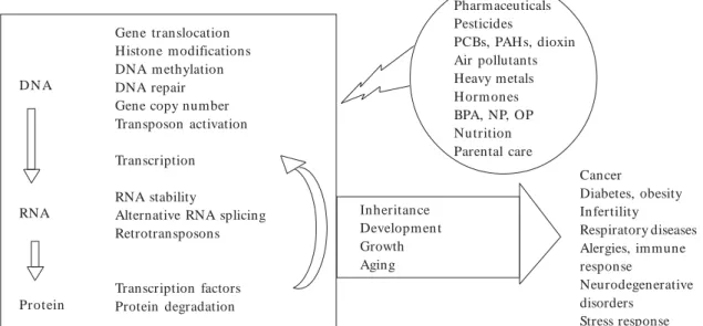
Yet, receptor-mediated changes in gene expres-sion are just the tip of the iceberg. There are many more mechanisms of gene regulation that are potentially susceptible to alteration by environ-mental influences. The effect of environenviron-mental contaminants on health is a major concern be-cause exposure is associated with a number of diseases, including cancer, diabetes, and infertility. The purpose of this review is to identify points of gene expression regulation, occurring along the process described by the central dogma (DNA . RNA . protein), that have been shown to be affected by environmental factors, particularly contaminants (Figure 1). We have drawn on re-search that
shows a strong connection between environ-m entally induced changes in gene regulatory mechanisms and disease etiology (Figure 1).
Pesticides, gene translocation, and non-Hodgkin lymphoma
A number of cancers, including childhood leuke-m ia an d follicu lar n on -H odgkin lyleuke-m ph oleuke-m a (NHL), are characterized by specific transloca-tions1,2 promoted by physical proximity of the
affected genes within the nucleus (reviewed by Verschure3). In follicular NHL, the
anti-apop-totic B-cell leukemia/ lymphoma 2 (bcl-2) gene, normally found on chromosome 18, translocates to the im m unoglobulin heavy chain locus on chromosome 14 4,2. This t(14;18) translocation
places bcl-2 under the control of the heavy chain enhancer, resulting in the overexpression of bcl-2 and, consequently, increased cell survival and lym-phomagenesis (reviewed by Roulland et al.4).
The t(14;18) translocation occurs in the lym-phocytes of healthy people and those with follic-ular NHL, and not all people with follicfollic-ular NHL possess the tran slocation5,4. Non etheless, in
-creased frequency of the translocation is a mark-er for increased lymphoma risk6. Epidemiologic
evidence indicates a positive association between t(14;18)-positive NHL and exposure to a variety of pesticides, including dieldrin, toxaphene,
lin-dane, atrazine, and fungicides6. Roulland et al.4
show that occupational exposure to pesticides can increase the frequency of the t(14;18) transloca-tion, both in terms of the number of people af-fected, and the number of affected lymphocytes within those people. Exposure to 2,3,7,8-tetra-chlorodibenzo-p-dioxin (TCDD) is also corre-lated with increased number of circulating t(14;18) positive lymphocytes5. It appears that t(14;18)
translocation frequency depends on how often pesticide exposure occurs, such that NHL risk increases with ongoing accumulation of genetic instability, acquired as a result of pesticide expo-sure4. Thus, variability in environmental
expo-sure, coupled with genetic events like transloca-tion, alters disease risk.
Environmental factors, DNA methylation, and fetal origins of adult disease
A growing number of animal studies show that parental diet and other exposures can influence fetal DNA methylation patterns and permanent-ly affect health outcomes in later life7,8,9.
More-over, there is evidence that environmentally in-duced changes in DNA methylation patterns are heritable across generations10,11,12,13,14,15. To
un-derstand how and when environmental factors m igh t ch an ge DNA m eth ylation , wh y th ese changes are potentially heritable, and how they can contribute to the fetal origins of adult dis-ease, it is helpful to consider examples in the con-text of ontogeny.
Ontogeny of DNA methylation and environmental influence
DNA methylation occurs in two modes: dy-namic methylation and theoretically permanent methylation, such as X chromosome inactiva-tion and genomic Gene-by-environment inter-actions and disease im printing16. In dynam ic
methylation, DNA methylation/demethylation reactions turn genes on or off throughout the life of an organism17. For exam ple, Wilks et al.18
showed active demethylation of the vitellogenin promoter in response to estradiol treatment in chickens. The more permanent, although not ir-reversible, methylation patterns are determined during early embryogenesis19,20,21,22,8 and
contin-ue to adjust through the neonatal period23,24.
aú
d
e C
o
le
tiv
a, 1
3
(1
):2
6
9
-2
8
1
, 2
0
0
8
not all the imprints and methylation patterns in his or her germ cells21. This ensures that the
par-ent passes on an imprinted pattern that exclusive-ly reflects his or her own sex. Between fertilization and implantation, the embryo demethylates most of its genes, with the exception of imprinted and some repeat genes25,21,26. The maintenance of
im-printed genes through the preimplantation peri-od is essential for normal embryonic develop-ment26,8. However, demethylation of other genes
is im portant for m aking the genom e broadly available to the undifferentiated and developing embryo. In addition, demethylation in the em-bryo may help to remove epigenetic modifications acquired during parental gametogenesis. Between cleavage and gastrulation, remethylation occurs throughout the embryo. X chromosome inacti-vation occurs over the period between implanta-tion and organogenesis22. At gastrulation, the
pri-mordial germ cells (PGCs) are newly formed and exhibit methylation similar to the surrounding somatic cells20. Some germ cells also exhibit X
in-activation28. During PGC proliferation and
mi-gration, X inactivation is completed28.
However, when the germ cells enter the geni-tal ridge, their DNA undergoes global demethy-lation, including loss of parental imprints, and reactivation of the inactive X20, 21,25,26,28. Whether
demethylation affects all genes is under debate. Remethylation and imprinting then occur in a sex-specific manner during gametogenesis21.
Af-ter birth, somatic cell methylation patAf-terns con-tinue to adjust, based on developmental and
en-vironmental factors24, 29. As an individual ages,
there is a gradual loss or gain of methylation, depending on the cell, tissue, or organ30, 31.
Even before conception, organisms are vul-nerable to environmentally altered methylation. This phenomenon has been observed in mice exposed preconceptionally to chrom ium (III) chloride, a carcinogen found in welding fumes. The 10-week-old offspring of male mice treated with chromium 2 weeks before conception ex-hibit significant increases in serum corticoster-one and glucose concentrations32, as well as
in-creased tumor risk and frequency of developmen-tal abnormalities, relative to controls9. Observed
tumors or anomalies include pheochromocyto-mas, thyroid follicular cell and Harderian gland tumors, ovarian cysts, uterine abnormalities, lung tum ors (fem ale offspring only), reproductive gland tumors (male offspring only), and renal nonneoplastic lesions (male offspring only). These effects result, in part, from chromium-induced epigenetic changes in the sperm that alter paren-tal imprinting. Specifically, the sperm of chromi-um-treated mice exhibit a significant increase in the number of undermethylated copies of the 45S ribosomal RNA gene (rRNA)33,34. 45S rRNA is
the precursor of other rRNAs that are part of the ribosome machinery and a control point for the number of ribosomes in a cell. Increased ribo-som e num ber and associated deregulation of protein synthesis could be one step in the pro-gression toward tissue growth, proliferation, and ultimately malignancy35, 34.
Figure 1. Summary of gene regulatory mechanisms affected by environmental exposures, with disease implications.
Abbreviations: BPA, bisphenol A; NP, 4-nonylphenol; PAHs, polycyclic aromatic hydrocarbons, PCBs, polychlorinated biphenlys; OP, 4-tert-octylphenol.
Gene translocation Histone modifications DNA m ethylation DNA repair Gene copy number Transposon activation
Transcription
RNA stability
Alternative RNA splicing Retrotransposons
Transcription factors Protein degradation D N A
RNA
Protein
Cancer
Diabetes, obesity Infertility
Respiratory diseases Alergies, immune response
Neurodegenerative disorders
Stress response Pharmaceuticals
Pesticides
PCBs, PAH s, dioxin Air pollutants Heavy metals H orm ones BPA, NP, OP Nutrition Parental care
E
d
w
a
rd
s,
T
. M
. &
M
After conception, between cleavage and gas-trulation, the timing and pattern of remethyla-tion is also subject to environmental influence. Am ino acid deficiency, for exam ple, causes a marked decrease in overall DNA methylation, along with abnormal expression of the normally silent, paternally imprinted and growth-related H19 allele in cultured mouse embryos36.
Con-versely, Wu Q et al.27 showed increased
methyla-tion of the H19/insulin-like growth factor 2 (Igf2) imprint control region, increased methyltrans-ferase activity, and decreased fetal growth after transfer to a recipient dam following in vitro ex-posure of mouse embryos to TCDD from the one-cell stage to the blastocyst stage. TCDD is a widespread and persistent environmental con-taminant that is formed during the production of paper, polyvinyl chloride (PVC) plastics and chlorinated pesticides, and during the incinera-tion of chlorine-containing products. Human exposure to TCDD is primarily dietary37.
Aber-rant H19/Igf2 imprinting and expression are as-sociated with development of a number of tu-mors, including Wilms tumor38, testicular germ
cell tumors39, ovarian tumors40, and
adrenocor-tical tumors41. Furthermore, alterations in fetal
growth related to H19/Igf2 imprinting are asso-ciated with metabolic disorders in adulthood, including obesity and diabetes42.
In addition to H19/Igf2 imprinting patterns, other genes that predispose for obesity can be af-fected by maternal diet, Dolinoy et al.7 observed
that dietary genistein (the major phytoestrogen in soy) during gestation in mice increased methy-lation of a retrotransposon located upstream of the Agouti gene, effectively reducing expression of the gene. Agouti transcription causes yellow fur, obesity, and tumorigenesis. Thus, Agouti expres-sion in the unexposed offspring predisposed them to obesity later in life. In addition, only 7% of the genistein-supplemented mice were yellow, com-pared with 21% of the unsupplemented animals. The degree of DNA methylation was similar in endodermal, mesodermal, and ectodermal tissues, suggesting genistein acts during early embryonic development.
After birth, somatic cell methylation patterns continue to adjust to developmental and environ-mental factors. Li et al.43 showed that neonatal
exposure to the synthetic estrogen diethylstil-bestrol (DES) caused abnormal demethylation of the CpG sites upstream of the estrogen-response element of the lactoferrin promoter. Lactoferrin is an important estrogen-inducible glycoprotein component of uterine secretions and is a useful
m arker of estrogen responsivity44. In another
study, Weaver et al.24 showed that increased
lick-ing, groomlick-ing, and arched-back nursing behav-ior of rat mothers reduced methylation of the glu-cocorticoid receptor (GR) promoter region in the hippocampus. Thus, rats that experienced high-quality (vs. low-high-quality) maternal behavior ex-hibited increased GR expression, greater gluco-corticoid feedback sensitivity, and a better response to stress later in life (lower plasma corticosterone concentrations after 20 min of restraint stress). The epigenetic alteration was noticeable in the first week after birth and persisted into adulthood. The effect could be reversed with cross-fostering, show-ing it to be a consequence of maternal behavior rather than gestation or genetic inheritance. Fur-thermore, the effect of high-quality maternal care on GR demethylation and reduced stress response could be reversed in adulthood with a central in-fusion of the methyl donor L-methionine29. These
studies by Weaver et al.24,29 emphasize the
poten-tial for DNA methylation patterns to respond to environmental influences throughout life.
Finally, as an individual ages, there is a gradu-al loss or gain of methylation, depending on the tissue, cell, or organ. The interaction between ab-errant methylation and age is recognized as a pos-sible early step in carcinogenesis (reviewed by Ri-chardson31). This is especially true of oncogenes
or tumor suppressor genes that become incor-rectly demethylated or methylated, respectively30.
In some cases these changes may be mediated by improperly regulated
DNA m ethyltransferase (Dnm t) enzym es. Gastric cancer cells are often characterized by over-expression of Dnmt1 with hypermethylation of genes relevant to the etiology of gastric cancer, including human MutL homologue 1 (hMLH1), thrombospondin-1 (THBS-1), and E-cadherin45.
This pattern is associated with Epstein-Barr virus infection, which potentially stimulates Dnmt1 over-expression45. In other cases, altered
methy-lation of oncogenes or tumor suppressor genes is mediated by dietary folate intake. Folate, found in fresh fruits and vegetables, is needed to make S-adenosylmethionine, the primary methyl donor for methylation46. Folate deficiency is associated
wit h h yp er m et h ylat io n o f t h e p 16IN K4A (CDKN2A) gene in human head and neck squa-mous cell carcinoma (HNSCC) and a rat model of hepatocellular carcinoma46, 47. In HNSCC,
aú
d
e C
o
le
tiv
a, 1
3
(1
):2
6
9
-2
8
1
, 2
0
0
8
Figure 2. Ontogenetic timeline of methylation in mammals. PGCs, primordial germ cells. From parental
gametogenesis, through fertilization, embryonic and neonatal development, and aging, the genome experiences stages of methylation and demethylation. Asymmetry of methylation is sometimes observed between maternal and paternal genomes, between somatic and germ cells, and among different tissues. Environmental factors have been shown to influence methylation patterns at multiple points in development. Please see “Ontogeny of DNA methylation and environmental influence” for details.
Female parent Male parent
X chrom osom e reactiveted before m eiosis ( Tam et al., 1994). De novo methylation and resetting of
im-prints (possibly incom plete) occurs during oocyte growth (postmeiotic arrest (Reik et al, 2001).
De novo methylation and resetting of imprints
(pos-sibly incomplete) occurs during prospermatogonial stage of gametogenesis (Reik et al., 2001).
W
W
W
W
W
W
W
W
At gastru lation , em bryon icgerm cells exhibit methylation and pren-tal imprinting patterns (Hakova et al., 2002). During proliferation and
migration of PGCs, X inactivation is com pleted ( Tam et al., 1994).
When PGCs reach the genital ridge, th ey are ch aracterized by global dem ethylation , in clu din g loss of paren tal im prin ts, repetitive ele-ments exhibit more protracted or incom plete dem ethylation (H
ajk-ova et al., 2002; Reik et al., 2001;
Surani, 2001).
Reactivation of the inactive X oc-cu r s before m eiosis ( Tam et al.,
1994). Remethylation and imprint-in g of germ cells imprint-in sex-specific manner takes place during game-togenesis (Reik et al., 2001).
In the zygote, enhancement of imprinting asym etry between paren tal gen om es oc-curs; paternal genome undergoes demeth-ylation, whereas maternal genome inder-goes de novo methylation (Surani, 2001).
Global dem ethylation of m ost genes, ex-cept imprinted and some repeat genes (Reik
et al., 2001; Surani, 2001).
Remethylation of somatic and germ cells; tim ing and pattern depends on species, tissue lineage, intrauterine environm ent and maternal nutrition (Guerrero-Bosa-gn a et al., 2005; H ajkova et al., 2002;
Reik et al., 2001; Wu G et al., 2004).
W
Tissue dependent, gradual loss or gain of methylation wirh aging; aberrant methyla-tion is an early step in carcinogenesis (re-viewed by Richardson, 2003).
W
Continued adjustment of methylation pat-terns (e.g.; altered methylation of the glu-cocorticoid receptor, induced by maternal lickin g an d froom in g beh avior in m ice (Weaver et al., 2004); altered methylation
patterns in the prostates of 8-week-old male mice fed a genistein diet (Day et al., 2002).
W
Fertilization
Cleavage
Blastocyst
Implantation
Gastrulation
Organogenesis
Neo natal development
Aging
E
d
w
a
rd
s,
T
. M
. &
M
Inheritance of methylation patterns
Inheritance of methylation patterns is of great in terest because it provides a m echan ism by which a parent’s acquired alterations in methyla-tion could be passed to offspring. Anway et al.10
showed that gestational exposure of male rats to vinclozolin (antiandrogenic fungicide) or meth-oxychlor (estrogenic organochlorine insecticide) during the time of gonadal sex determination caused decreased sperm count and viability and increased rates of infertility in adulthood. This loss of fertility was perpetuated through the male germ line for four generations and occurred in conjunction with altered, heritable methylation patterns.
Inheritance of altered methylation patterns could explain the transgenerational effects of DES exposure. DES is a synthetic estrogen given to pregnant women between 1938 and Gene-by-en-vironment interactions and disease 1971 to pre-vent miscarriages. The children and grandchil-dren of humans and mice exposed to DES in ute-ro exhibit increased rates of uterine sarcomas and adenocarcinomas, lymphomas, malignant repro-ductive tract tumors in both males and females, proliferative lesions of the rete testis, and benign ovarian tumors11, 12, 14, 15. Li et al.48 showed that
DES exposure alters methylation patterns asso-ciated with the promoters of many estrogen-re-sponsive genes that regulate reproductive organ development in both mice and humans. In addi-tion, Ruden et al.13 recently published a novel
hypothesis suggesting that the transgenerational effects of DES are associated with altered DNA methylation, possibly mediated through modi-fied WNT signaling.
Cadmium, DNA repair, and diabetes
In 2003 Schwartz et al.49 published a large
cross-sectional human study in which they re-ported a significant positive relationship between urinary cadmium, impaired fasting glucose, and diabetes, su ggestin g that cadm iu m exposu re plays a role in diabetes etiology. Using monkeys, Kurata et al.50showed that cadmium accumulates
in the pancreas and that chronic exposure ini-tiates degeneration of islet ß cells and induces the clinical signs of diabetes. Cadmium exposure also potentiates some diabetic complications related to renal tubular and glomerular function51.
Cadmium-induced diabetes may be a side effect of enhanced DNA repair. A variety of geno-toxic environmental factors, including cadmium
and several pesticides, cause DNA strand breaks or fragmentation52,53 54. Poly (ADP-ribose)
poly-merase-1 (PARP-1) recognizes the strand breaks and promotes break repair by loosening chro-matin structure (Benjamin and Gill55; reviewed
by Rouleau et al.56). Specifically, PARP-1 catalyzes
histone ribosylation—the addition of ADP-ribose molecules to histone lysine residues. The result-ing negative charge to the histones causes elec-trostatic repulsion away from the n egatively charged DNA, making it more accessible to re-pair enzymes57. Following DNA repair, the
ADP-ribose is freed, becoming briefly available for the stable glycation of oth er h iston es an d p r o-teins58,59. This mimics glycation caused by
hyper-glycemia60 and could explain why chronic
cad-mium exposure induces clinical signs of diabetes. In addition, sugar-modified histones can un-dergo other transformations (described by Cer-vantes-Laurean et al.58) to form advanced
glyco-sylation end products (AGEs). AGE accumula-tion associated with histones and other proteins is implicated in the progression of aging and age related diseases like diabetes and Alzheimer dis-ease58,60.
Air pollution, activation of inflammatory genes, and respiratory disease
Respiratory diseases such as chronic obstructive pulmonary disease, cystic fibrosis, interstitial lung disease, acute respiratory distress syndrome, and asthma are characterized by expression of inflam-mation genes, which can be activated or intensi-fied by exposure to air pollution.
Air pollution consists of tiny ambient parti-cles measuring < 10–15 µm in diameter (PM10) that come from dust, smoke, or aerosol liquids produced by vehicles, factories, construction sites, plowed fields, or burning wood. Air pollution can include residual oil fly ash (ROFA), an inor-ganic mixture of silicates and metal salts con-taining vanadium, zinc, iron, and nickel released during the combustion of low-grade oil61,62.
In vitro studies with human airway epithelial
cells show that exposure to diesel soot and other PM10 particles activates pro-inflammatory genes in a process mediated by free radical/oxidative stress mechanisms63, 64, 65, 66, 67. The oxidative stress
induces pro-inflammation transcription factors such as nuclear factor .B (NF-.B) and activator protein 1 (AP-1)64, which in turn promote
aú
d
e C
o
le
tiv
a, 1
3
(1
):2
6
9
-2
8
1
, 2
0
0
8
release (IL-8 is a marker of inflammation), and finally, expression of inflammatory genes65, 67.
ROFA exposure stimulates a similar cascade of events. Using a perfused rabbit lung model, Samet et al.62 showed that the vanadium
compo-nent of ROFA can inhibit tyrosine phosphatases, causin g phosphorylation of NF-.B an d other proinflammation transcription factors, includ-ing activatinclud-ing transcription factor 2 (ATF-2), c-Jun, and cAMP response element binding–pro-tein (CREB). Again, this leads to expression of inflammatory genes, potentially exacerbating res-piratory distress.
Environmental estrogens, transcription, and allergies
Bisphenol A (BPA) is a synthetic estrogen that was investigated for use in birth control pills but was instead favored as a plasticizer for use in polycarbonate plastics, dental sealants, and the lining of food cans. Both BPA and 4-nonylphe-nol (NP), a derived product of nonionic surfac-tants, have been shown to activate estrogen re-ceptor alpha (ER-a), induce estrogen-dependent gene expression, and stimulate growth in estro-gen responsive MCF7 breast cancer cells68.
The effects of these chemicals are not limited to classical estrogen signaling. Lee et al.69, 70 showed
that BPA, NP, and 4-tert-octylphenol (OP), used widely in detergents and wetting agents, increased the T-cell allergic response, measured as an in-crease in interleukin-4 (IL-4) mRNA levels, in an-tigen-stimulated mouse T cells. The IL-4 promoter in mice and humans contains multiple binding sites for a transcription factor called nuclear fac-tor of activated T cells (NF-AT)70. It appears that
BPA, OP, and NP enhance IL-4 production by stimulating the calcium-dependent calcineurin sig-naling pathway. This causes dephosphorylation of cytoplasmic NF-AT with subsequent translo-cation of the transcription factor to the nucleus.
Lee et al.69, 70 suggest that increased NF-AT
con-centrations in the nucleus up-regulate IL-4 tran-scription, causing the T-cell allergic response ob-served with BPA, NP, or OP exposure.
Environmental factors, RNA stability, and tumor development or
reproductive dysfunction
TCDD is a persistent and widespread environ-mental contaminant that is a potent carcinogen
in rodents and a suspected human carcinogen with multiple modes of action. One way that TCDD prom otes carcinogenesis is by stabilizing the mRNA of urokinase plasminogen activator (uPA), a serine protease that contributes to matrix turn-over and growth of tumor cells71, 72. High uPA
mRNA concentrations are found in tumors such as hepatocellular carcinoma but not in healthy tissues, and similarly, survival time is inversely related to uPA mRNA concentrations73. In rat
liv-er cells, the TCDD-induced stabilization of uPA mRNA is mediated by a 50-kDa cytoplasmic pro-tein (p50) that binds specifically to sites in the 3´ untranslated region of uPA mRNA72. Shimba et
al. 72 showed that p50 is activated rapidly (in 15
min) by TCDD-mediated phosphorylation. They suggest that p50 stabilizes uPA mRNA by protecting nuclease cleavage sites from attack. In a second TCDD study, Minegishi et al.74
showed that TCDD exposure reduced the half-life of lutein izin g horm on e receptor (LH -R) mRNA in rat granulosa cells. This could impact steroidogenesis, luteinization, and ovulation by reducing granulosa cell sensitivity to circulating LH. The authors speculate that TCDD affects production or activity of regulatory proteins that destabilize LH-R mRNA. For example, TCDD may facilitate the binding of appropriate pro-teins to the AU (adenylate/uridylate)-rich ele-ments (AREs) of LH-R, thus promoting degra-dation of the mRNA by exosomes. In addition to uPA, several other proteases are im portant in tumor development. Among these are the matrix metalloproteinases (MMPs), which promote tu-mor invasion. Bao et al.75 showed that vitamin D
(1a, 25-dihydroxyvitamin D3) reduced the mRNA stability of metalloproteinase 9 (MMP-9) in a human prostate cancer cell line. This, along with other actions of vitamin D, inhibited the invasive ability of the cancer cells.
The role of vitamin D in the prevention of prostate cancer could be applicable in other situ-ations as well. For example, Ritchie et al.76
report-ed that long-term, low-dose exposure to poly-chlorinated biphenyls (PCBs) could contribute to an increased risk of prostate cancer in the gen-eral human population. PCBs are no longer man-ufactured in the United States, but even after sev-eral decades of banning, PCBs remain a persis-tent environmental contaminant that people en-counter mostly through their diet77. Given the
study by Bao et al.75, which suggests that vitamin
E
d
w
a
rd
s,
T
. M
. &
M
and their offspring78. It is worth testing if PCBs
increase cancer risk via changes in vitamin D metabolism or efficacy with regard to processes like reduced MMP-9 mRNA stability.
Pesticides, Impaired protein degradation, and Parkinson disease
Parkinson disease (PD) is a neurodegenerative disease that affects more than 1 million people in the United States alone79. It is diagnosed by the
p r esen ce of in t r acyt op lasm ic in clu sion s in dopaminergic neurons. These inclusions, known as “Lewy bodies,” are composed primarily of a-synuclein protein aggregates80. Lewy body
for-mation also characterizes some forms of demen-tia and Alzheimer disease81.
PD can occur in families or sporadically and is associated with both genetic and environmen-tal causes82. A familial form of PD involves
ge-nomic triplication and consequent overexpres-sion of the a-synuclein gene83. In both familial
and sporadic forms of PD, the timely degrada-tion of a-synuclein is inhibited in part by protea-some dysfunction84, an effect that appears to be
exacerbated by a-synuclein overexpression79.
Using an im m ortalized rat m esencephalic dopaminergic cell line (N27 cells) transfected with the human a-synuclein gene, Sun et al.79 showed
that exposure to 30 µM dieldrin (an organochlo-rine pesticide) impaired protea-some function, resulting in a marked increase in a-synuclein pos-itive aggregates. This dose was deduced by the authors to represent the expected human expo-sure level after 50 years; dieldrin is lipophilic and bioaccumulates significantly in the central nervous system85. In addition, when a-synuclein was
over-expressed the dieldrin worked additively with the protein to impair proteasome function and trig-ger neuronal apoptosis. Sun et al.79 argue that
ex-posure to neurotoxic chemicals such as dieldrin increases risk for PD, particularly among individ-uals predisposed to a-synuclein accumulation for genetic or age-related reasons. Other pesticides, including the herbicide paraquat the fungicide maneb and the insecticide rotenone, are causally linked to a-synuclein accumulation and dopam-inergic cell degeneration and apoptosis (reviewed by Meredith et al.86). Rotenone is so effective that
it is used to generate a rodent model for PD 87.
Pharmaceuticals, gene amplification/muta-tion, and drug resistance
When faced with death, cells adapt individually and as a population. For example, in Escherichia coli, starvation of lac– bacteria on lactose medi-um induces lac+ revertants. The revertants exhib-it eexhib-ither gene amplification (20- to 100-fold) of the lac– allele, or a compensatory frameshift mu-tation that randomly produces the lac+ allele in association with wide-spread, stress-induced, hy-permutation 88, 89, 90. Amplification of the
lac–al-lele (increased gene copy number) eases the star-vation stimulus because the allele is leaky, confer-ring 1–2% of the wild-type ß-galactosidase activ-ity91. The revertants are apparently produced de
novo in response to starvation because they ap-pear more rapidly and at higher frequencies than would be predicted by selection-only models (re-viewed by Hersh et al.90). Hence, the
phenome-non is termed “adaptive amplification/mutation.” E. coli provide empirical evidence for the abil-ity of cells to enhance their survival in response to environmental pressures through genomic plas-ticity and adaptation. A major difficulty that af-fects cancer therapy is the progressive develop-ment of drug resistance observed in a subset of patients. As in the E. coli example, tumor cells can respond to treatment by amplifying and/or mu-tating genes that promote their survival. For ex-ample, androgen deprivation is a common thera-py for prostate cancer. However, some patients respond well initially to the therapy, but then slow-ly develop resistance resulting in improved tumor growth92. Koivisto et al. 92 report that this pattern
is associated with progressive amplification of the androgen receptor (AR) gene along with substan-tially increased AR mRNA expression.
A related observation was made by Copur et al.93 who showed that continuous exposure of
cultured human colon cancer cells to the colon can cer d r u g 5- flu o r o u r acil ( 5- FU) , cau ses thymidylate synthase (TS) gene amplification and overexpression of the TS protein. Overexpres-sion of TS conferred 5-FU resistance and pro-vides an explanation for the development of flu-oropyrimidine chemotherapy resistance among patients with colon cancer93.
aú
d
e C
o
le
tiv
a, 1
3
(1
):2
6
9
-2
8
1
, 2
0
0
8
In 1989, Prody et al.94 published a suggestive but
isolated study in which they reported de novo 100-fold amplification of the silent serum utyryl-cholinesterase (BtChoEase) gene in a farmer ex-posed chronically to organophosphate insecti-cides, which inhibit BtChoEase. The silent BtCh-oEase gene codes for a defective protein that makes homozygous individuals particularly sen-sitive to organophosphate poisoning. The am-plification was not present in the man’s parents but was inherited by his son, indicating that germ cells were also affected.
Insecticides, alternative RNA splicing, and pesticide resistance
Not all forms of resistance result from gene amplification or compensatory mutation. Glu-tathione S-transferases (GSTs) make up a family of multifunction enzymes that play an impor-tant role in detoxification of xenobiotic com-pounds95. GSTs contribute to insecticide resistance
among insects, including mosquitoes96, and to
multidrug resistance in tumor cell lines and can-cer patients97. In Anopheles mosquitoes, one of
the GST genes, adgst1AS1, codes for (at least) four RNA splice variants that vary in their bind-ing characteristics with regard to permethrin, a pyrethroid insecticide95. Alternative RNA
splic-ing could explain the rapid increase in permethrin resistance, associated with GST upregulation, observed among Culex mosquitoes selected for just one or three generations98. It is not known if
pyrethroid exposure causes changes in GST ac-tivity in humans.
Environmental contaminants, transposons, and carcinogenesis
Transposon derived sequences account for at least 45% of the human genome, a hefty proportion when compared with the 1% given over to pro-tein coding regions99. They are of great medical
interest for two reasons. First, LINE-1 (L1) ret-rotransposons, along with the 412 retrotranspo-son in Drosophila and the VL30 retrotransporetrotranspo-sons in mice and humans, are activated in the normal course of gonad and gamete development, and in fact, may play a regulatory role in these process-es100,101,102. Second, retrotransposons and reverse
transcriptase (RT) are activated in the tumors of several different cancers (reviewed by Sinibaldi-Vallebona et al.103). Sinibaldi-Vallebona et al.103
report that drug-mediated inhibition of RT
activ-ity or silencing of L1 retrotransposons by RNA interference (RNAi) reduces cell growth and stim-ulates differentiation of cancer cell lines. These observations suggest an active role for retrotrans-posons in carcinogenesis that could be related to their original developmental role becoming mis-regulated later in life.
Further, a small amount of evidence suggests that transposons can be activated by environmen-tal contaminants. Transposon activation has been observed in HeLa cells (human cervical cancer cell line) and vascular smooth muscle cells of mice and humans exposed in vitro to the carcinogen benzo[a]pyrene104, 105, 106 and in the livers of male
mummichogs (fish) exposed to pyrene, a com-mon PAH found ubiquitously in the environment and at several estuarine Superfund sites in the United States106. El-Sawy et al.108 reported that
nickel activates L1 retrotransposition in transfect-ed HeLa cells. In addition, Morales et al.109 showed
that serum, testosterone, dihydrotestosterone, and a mixture of 17 organochlorine pesticides stimu-lated transcription of the L1Hs retrotransposon promoter in human choriocarcinoma cells. These examples support the hypothesis that environ-mentally induced transposon activity could be important in the etiology of cancer and possibly other diseases.
Conclusions
The studies reviewed in this article show how en-vironmental factors influence a diverse array of molecular mechanisms and consequently alter disease risk. They emphasize the plasticity of the genome and its regulation, providing support for genomic reaction and adaptation in response to environmental stimuli. Further, they provide di-rect evidence that chemicals placed in the environ-ment by human activity can and do promote dis-ease by altering gene expression. Epigenetic stud-ies in particular provide insights that further our understanding of fetal origins of adult disease, while also offering new research avenues for the investigation of acquired, and potentially herita-ble, genetic variation and disease susceptibility.
var-E
d
w
a
rd
s,
T
. M
. &
M
References
Sm it h MT, McH ale CM, Wiem els JL, Zh an g LP, Wien cke JK, Zhen g S, Gun n L, Skibola CF, Ma X, Buffler PA. Molecular biom arkers for the study of childhood leukem ia. Toxicol Appl Pharm acol 2005;
206(2):237–245.
Tsujim oto Y, Gorham J, Cossm an J, Jaffe E, Croce CM. The T(14,18) chrom osom e translocations in-volved in B-cell neoplasm s result from m istakes in VDJ joining. Science 1985; 229(4720):1390–1392.
Verschu re PJ. Position in g th e gen om e with in th e nucleus. Biol Cell 2004; 96(8):569–577.
Roulland S, Lebailly P, Lecluse Y, Briand M, Pottier D, Gauduchon P. Characterization of the t(14;18) BCL2-IGH translocation in farm ers occupationally exposed to pesticides. Cancer Res 2004; 64(6):2264–
2269.
Baccarelli A, Hirt C, Pesatori AC, Consonni D, Patter-son DG, Bertazzi PA, Dolken G, Landi MT. t(14;18) tran slocation s in lym ph ocytes of h ealth y dioxin -exposed individuals from Seveso, Italy. Carcinogen-esis 2006; 27(10):2001–2007.
Schroeder JC, Olshan AF, Baric R, Dent GA, Wein-berg CR, Yount B, Cerhan JR, Lynch CF, Schuman Ie OM, Tolbert PE, Rothman N, K PC, Blair A. Agricul-tural risk factors for t(14;18) subtypes of non-Hodgkin’s lymphoma. Epidemiology 2001; 12(6):701–709.
Dolinoy DC, Weidman JR, Waterland RA, Jirtle RL. Matern al gen istein alters coat color an d protects A(vy) m ouse offspring from obesity by m odifying the fetal epigenom e. Environ Health Perspect 2006;
114:567–572.
Wu GY, Bazer FW, Cudd TA, Mein in ger CJ, Spen-cer TE. Maternal nutrition and fetal development. J Nutr 2004; 134(9): 2169–2172.
Yu W, Sip owicz MA, Hain es D C, Birely L, Diwan BA, Riggs CW, Kasp rzak KS, An derson LM. Pre-con ception u rethan e or chrom iu m (III) treatm en t of m ale m ice: m u ltip le n eop lastic an d n on - n eo-plastic changes in offspring. Toxicol Appl Pharmacol
1999; 158(2):161–176.
Anway MD, Cu p p AS, Uzu m cu M, Skin n er MK. Epigen etic tran sgen eration al action s of en docrin e d isr u p t o r s an d m at e fer t ilit y. Scien ce 2005;
308(5727):1466–1469.
Newbold RR, H an son RB, Jefferson WN, Bu llock BC, H asem an J, McLachlan JA. In creased tu m ors but uncom prom ised fertility in the fem ale descen-dants of m ice exposed developm entally to diethyl-stilbestrol. Carcinogenesis 1998; 19(9):1655–1663.
Newbold RR, H an son RB, Jefferson WN, Bu llock BC, Haseman J, McLachlan JA. Proliferative lesions and reproductive tract tumors in male descendants of m ice exp osed d evelop m en t ally t o d iet h ylst il-bestrol. Carcinogenesis 2000; 21(7):1355–1363.
Ruden DM, Xiao L, Garfin kel MD, Lu X. H SP90 and environm ental im pacts on epigenetic states: a m odel for the trans-generational effects of diethyl-stilbestrol on uterine development and cancer. Hum Mol Genet 2005; 14:R149–R155.
Turusov VS, Trukhanova LS, Parfenov YD, Tom atis L. Occurrence of tumors in the descendants of CBA male mice prenatally treated with diethylstilbestrol.
Int J Cancer 1992; 50(1): 131–135.
Walker BE, H aven MI. In ten sit y of m u ltigen er a-tional carcinogenesis from diethylstilbestrol in mice.
Carcinogenesis 1997; 18(4):791–793.
Bestor TH . The DNA m ethyltran sferases of m am -mals. Hum Mol Genet 2000; 9(16):2395–2402.
Cervoni N, Szyf M. Demethylase activity is directed by histone acetylation. J Biol Chem 2001; 276(44):
40778–40787.
Wilks A, Seldr an M, Jost JP. An estrogen depen -den t dem eth ylation at th e 5’ en d of th e ch icken vitellogenin gene is independent of DNA synthesis.
Nucleic Acids Res 1984; 12(2):1163–1177.
Guerrero-Bosagna C, Sabat P, Valladares L. Environ-m ental signaling and evolutionary change: can ex-posure of pregnant mammals to environmental es-trogens lead to epigenetically induced evolutionary changes in embryos? Evol Dev 2005; 7(4):341–350.
Hajkova P, Erhardt S, Lane N, Haaf T, El-Maarri O, Reik W, Walter J, Su r an i MA. Ep igen et ic r ep ro-gram m in g in m ou se prim ordial germ cells. M ech Dev 2002; 117(1–2):15–23.
Reik W, Dean W, Walter J. Epigenetic reprogram -m in g in -m a-m -m alian d evelop -m en t. Science 2001;
293(5532):1089–1093.
Tan SS, William s EA, Tam PPL. X- ch rom osom e in activation occu rs at differen t tim es in differen t tissu es of th e p ost- im p lan tation m ou se em br yo.
Nature Genet 1993; 3(2):170–174.
Day JK, Bau er AM, DesBordes C, Zhu an g Y, Kim BE, Newton LG, Nehra V, Forsee KM, MacDonald RS, Besch - Willifor d C, H u an g TH , Lu bah n DB. Genistein alters methylation patterns in mice. J Nutr
2002; 132(8):2419S–2423S.
Weaver ICG, Cervoni N, Champagne FA, D’Alessio AC, Sharma S, Seckl JR, Dymov S, Szyf M, Meaney MJ. Epigen etic program m in g by m atern al behav-ior. Nat Neurosci 2004; 7(8):847–854.
Acknowledments
We thank W. Hessler for her editorial suggestions. The John Merck Fund provided funding for this review.
1.
2.
3.
4.
5.
6.
7.
8.
9.
10.
11.
12.
13.
14.
15.
16.
17.
18.
19.
20.
21.
22.
23.
24.
aú
d
e C
o
le
tiv
a, 1
3
(1
):2
6
9
-2
8
1
, 2
0
0
8
Reik W, Walt er J. Gen o m ic im p r in tin g: p aren tal in flu en ce o n t h e gen o m e. N at Rev Gen et 2001;
2(1):21–32.
Su ran i MA. Reprogram m in g of gen om e fu n ction t h r o u gh ep igen et ic in h er it an ce. N at u re 2001;
414(6859):122–128.
Wu Q, Ohsako S, Ishimura R, Suzuki JS, Tohyam a C. Exposure of mouse preimplantation embryos to 2,3,7,8-tetrachlorodibenzo-p-dioxin (TCDD) alters the methylation status of imprinted genes H19 and Igf2. Biol Reprod 2004; 70(6):1790–1797.
Tam PPL, Zhou SX, Tan SS. X-Chrom osom e activ-ity of the m ouse prim ordial germ cells revealed by the expression of an X-linked LacZ transgene. De-velopment 1994; 120(10):2925–2932.
Weaver ICG, Champagne FA, Brown SE, Dym ov S, Sharm a S, Meaney MJ, Szyf M. Reversal of m ater-n al program m iater-n g of stress respoater-n ses iater-n adult off-sprin g throu gh m ethyl su pplem en tation : alterin g epigen etic m arkin g later in life. J N eurosci 2005;
25(47):11045–11054.
Ehrlich M. DNA methylation in cancer: too much, but also too little. Oncogene 2002; 21(35):5400–5413.
Richardson B. Im pact of agin g on DNA m ethyla-tion. Ageing Res Rev 2003; 2(3):245–261.
Chen g RYS, Alvord WG, Powell D, Kaspr zak KS, Anderson LM. Increased serum corticosterone and glucose in offspring of chromium(III)-treated male mice. Environ Health Perspect 2002; 110:801–804.
Ch en g RYS, H ockm an T, Cr awford E, An d er son LM , Sh iao YH . Ep igen et ic an d gen e exp r essio n changes related to trans-generational carcinogene-sis. Mol Carcinog 2004; 40(1): 1–11.
Shiao YH, Crawford EB, Anderson LM, Patel P, Ko K. Allele-specific germ cell epimutation in the spac-er prom otspac-er of the 45S ribosom al RNA gene aftspac-er Cr ( III) exp o su r e. Toxicol Appl Pharm acol 2005;
205(3):290–296.
Ruggero D, Pandolfi PP. Does the ribosom e trans-late cancer? N at Rev Cancer 2003; 3(3):179–192.
Doherty AS, Mann MRW, Tremblay KD, Bartolomei MS, Schultz RM. Differential effects of culture on im prin ted H 19 expression in the preim plan tation mouse embryo. Biol Reprod 2000; 62(6):1526–1535.
Centers for Disease Control and Prevention. Third National Report on Human Exposure to Environmental Chem icals. N at io n al Cen t er fo r En vir o n m en t al
H ealth Publ no 05-0570. Atlanta: Centers for Dis-ease Control and Prevention; 2005.
Taniguchi T, Sullivan MJ, Ogawa O, Reeve AE. Epi-gen etic chan ges en com passin g the Igf2/H 19 locus associated with relaxation of Igf2 im prin tin g an d silencing of H19 in Wilms tumor. Proc Natl Acad Sci USA 1995; 92(6):2159–2163.
Vangurp R, Oosterhuis JW, Kalscheuer V, Mariman ECM, Looijen ga LH J. Biallelic exp r ession of th e H 19 an d Igf2 gen es in hum an testicular germ cell tumors. J N atl Cancer Inst 1994; 86(14):1070–1075.
Kim H T, Choi BH , Niikawa N, Lee TS, Chan g SI. Frequent loss of imprinting of the H19 and IGF-II gen es in ovarian tu m ors. Am J M ed Genet 1998;
80(4):391–395.
Gao ZH, Suppola S, Liu J, Heikkila P, Janne J, Vou-tilainen R. Association of H 19 prom oter m ethyla-tion with the expression of H19 and IGF-II genes in adrenocortical tumors. J Clin Endocrinol Metab 2002;
87(3):1170–1176.
Sm it h FM , Gar field AS, War d A. Regu lat io n o f growth and m etabolism by im printed genes. Cyto-genet Genom e Res. 2006; 113(1–4):279–291.
Li S, Washburn KA, Moore R, Uno T, Teng C, New-bold RR, McLachlan JA, Negishi M. Developm en-tal exposure to diethylstilbestrol elicits dem ethyla-tion of estrogen-responsive lactoferrin gene in mouse uterus. Cancer Res 1997; 57(19):4356–4359.
Pentecost BT, Teng CT. Lactotransferrin is the m a-jor estr ogen in d u cible p r otein of m ou se u ter in e secretions. J Biol Chem 1987; 262(21):10134–10139.
Etoh T, Kan ai Y, Ushijim a S, Nakagawa T, Nakan -ishi Y, Sasako M, Kitano S, Hirohashi S. Increased DNA methyltransferase 1 (DNMT1) protein expres-sion correlates significantly with poorer tumor dif-feren tiation an d frequ en t DNA hyperm ethylation of m u ltiple CpG islan ds in gastric can cers. Am J Pathol 2004; 164(2):689–699.
Kraunz KS, Hsiung D, McClean MD, Liu M, Osany-ingbem i J, Nelson HH, Kelsey KT. Dietary folate is associated with p16INK4A methylation in head and neck squam ous cell carcinom a. Int J Cancer 2006;
119(7):1553–1557.
Po gr ib ny IP, Jam es SJ, Jer n igan S, Po gr ib n a M . Gen om ic hypom ethylation is specific for pren eo-plastic liver in folate/methyl deficient rats and does n ot occu r in n on -target tissu es. M utat Res 2004;
548:53–9.
Li SF, Hursting SD, Davis BJ, McLachlan JA, Barrett JC. Environmental exposure, DNA methylation, and gene regula-tion—lessons from diethylstilbesterol-induced cancers. In: Epigenetics in Cancer Preven-tion: Early Detection and Risk Assessment. New York: New York Academy of Sciences; 2003. p. 161–169. Schwartz GG, Il’yasova D, Ivanova A. Urinary cad-m iu cad-m , icad-m paired fastin g glu cose, an d diabetes in the NHANES III. Diabetes Care 2003; 26(2):468–470.
Kurata Y, Katsuta O, Doi T, Kawasuso T, Hiratsuka H , Tsu chitan i M, Um emu r a T. Chr on ic cadm ium treatm en t in duces islet B cell in jury in ovariecto-m ized Cyn oovariecto-m olgu s ovariecto-m o n keys. Jpn J Vet Res 2003;
50(4):175–183.
Åkesson A, Lundh T, Vahter M, Bjellerup P, Lidfeldt J, Nerbrand C, Samsioe G,. Stromberg U, Skerfving S. Tubular and glomerular kidney effects in Swedish women with low environmental cadmium exposure.
Environ Health Perspect 2005; 113:1627–1631.
Bolognesi C. Genotoxicity of pesticides: a review of h u m an biom on it or in g st u d ies. M u tat Res 2003;
543(3):251–272.
Palus J, Rydzynski K, Dziubaltowska E, Wyszynska K, Natarajan AT, Nilsson R. Gen otoxic effects of occupational exposure to lead and cadm ium . M u-t au-t Res Gen eu-t Tox icol En v iron . M u u-t agen . 2003;
540(1):19–28.
Yan g JM, Ar n u sh M, Ch en QY, Wu XD, Pan g B, Jian g XZ. Cadm iu m -in du ced dam age to prim ary cu ltu res of r at Leydig cells. Reprod Toxicol 2003;
17(5):553–560. 25.
26.
27.
28.
29.
30.
31.
32.
33.
34.
35.
36.
37.
38.
39.
40.
41.
42.
43.
44.
45.
46.
47.
48.
49.
50.
51.
52.
53.
E
d
w
a
rd
s,
T
. M
. &
M
Benjamin RC, Gill DM. Poly(ADP-ribose) synthesis in vitro programmed by damaged DNA—a compar-ison of DNA molecules containing different types of strand breaks. J Biol Chem 1980; 255(21):502–508.
Ro u leau M , Au b in RA, Po ir ier GG. Po ly( AD P-ribosyl)ated chrom atin dom ain s: access gran ted. J Cell Sci 2004; 117(6):815–825.
Virág L, Szabó C. The therapeutic potential of poly (ADPribose) polym erase inhibitors. Pharm acol Rev
2002; 54(3):375–429.
Cer van tes-Laurean D, Jacobson EL, Jacobson MK. Glycation an d glycoxidation of histon es by ADP-ribose. J Biol Chem 1996; 271(18):10461–10469.
Diefenbach J, Bürkle A. Introduction to poly(ADP-ribose) m etabolism . Cell M ol Life Sci 2005; 62(7–
8):721–730.
Gugliucci A, Bendayan M. H istones from diabetic rats contain increased levels of advanced glycation end products. Biochem Biophys Res Com m un 1995;
212(1):56–62.
Henr y WM, Knapp KT. Compound form s of fossil fu el fly-ash em ission s. Environ Sci Technol 1980;
14(4):450–456.
Sam et JM, Silbajor is R, Hu an g T, Jasp ers I. Tran -scription factor activation following exposure of an intact lung preparation to metallic particulate mat-ter. Environ Health Perspect 2002; 110:985–990.
Bolan d S, Bonvallot V, Four n ier T, Baeza-Squiban A, Aubier M, Maran o F. Mechan ism s of GM-CSF in crease by diesel exhaust particles in hum an air-way ep it h elial cells. Am J Physiol Lung Cell M ol Physiol 2000; 278(1):L25–L32.
D on ald so n K, Sto n e V, Bor m PJA, Jim en ez LA, Gilm our PS, Schins RPF, Knaapen AM, Rahm an I, Faux, SP, Brown DM, MacNee W. Oxidative stress and calcium signaling in the adverse effects of envi-ron m en tal particles (PM10). Free Radic Biol M ed
2003; 34(11):1369–1382.
Gilm o u r PS, Rah m an I, H ayash i S, H o gg JC, Don aldson K, MacNee W. Aden oviral E1A pr im es alveolar epithelial cells to PM10-induced transcrip-tion of in terleukin -8. Am J Physiol Lung Cell M ol Physiol 2001; 281(3):L598–L606.
Marano F, Boland S, Bonvallot V, Baulig A, Baeza-Squ iban A. H u m an airway epithelial cells in cu l-ture for studying the molecular mechanisms of the inflam m atory response triggered by diesel exhaust par ticles. Cell Biol Toxicol 2002; 18(5):315–320.
Gilm our PS, Rahm an I, Donaldson K, MacNee W. Histone acetylation regulates epithelial IL-8 release m ediated by oxidative stress from en viron m en tal particles. Am J Physiol Lung Cell Mol Physiol 2003;
284(3):L533–L540.
Vivacqua A, Recchia AG, Fasan ella G, Gabriele S, Carpino A, Rago V, Di Gioia ML, Leggio A, Bono-figlio D, Maggiolin i M, Ligu or i A. The food con-tam in an ts bisphen ol A an d 4-n on ylphen ol act as agonists for estrogen receptor alpha in MCF7 breast cancer cells. Endocrine 2003; 22(3):275–284.
Lee M H , Ch u n g SW, Kan g BY, Par k J, Lee CH , H wan g SY, Kim TS. En han ced in ter leu kin -4 p rduction in CD4(+ ) T cells an d elevated im m un o-globulin E levels in antigen-primed mice by bisphen ol A abisphen d bisphen obisphen ylphebisphen ol, ebisphen docribisphen e disruptors: ibisphen -volvem en t of nuclear factor-AT an d Ca2+ . Im m u-nology 2003; 109(1):76–86.
Lee MH , Kim E, Kim TS. Exposu re to 4-tert-oc-tylphenol, an environm entally persistent alkylphe-n ol, ealkylphe-n haalkylphe-n ces ialkylphe-n terleu kialkylphe-n -4 produ ctioalkylphe-n ialkylphe-n T cells via NF-AT activation. Toxicol Appl Pharm acol 2004;
197(1):19–28.
Gaido KW, Maness SC. Posttranscr iptional stabili-zation of urokinase plasminogen-activator messen-ger RNA by 2,3,7,8-tetrachlorodibenzo-p-dioxin in a hum an keratin ocyte cell lin e. Toxicol Appl Phar-macol 1995; 133(1):34–42.
Shimba S, Hayashi M, Sone H, Yonemoto J, Tezuka M. 2,3,7,8- tetr ach lor od iben zo- p - d ioxin ( TCDD) in d u ces bin d in g o f a 50 kDa p ro tein o n t h e 3 ‘ untranslated region of urokinase-type plasminogen act ivat or m RN A. Biochem Biophys Res Com m u n
2000; 272(2):441–448.
De Petro G, Tavian D, Copeta A, Portolani N, Giulini SM, Barlati S. Expression of urokinase-type plasmi-n o geplasmi-n activator (u PA), u PA receptor, aplasmi-n d t issu e-type PA m essenger RNAs in hum an hepatocellular carcinoma. Cancer Res 1998; 58(10): 2234–2239.
Minegishi T, Hirakawa T, Abe K, Kishi H, Miyamoto K. Effect of IGF-1 and 2,3,7,8-tetrachlorodibenzo-p-dioxin (TCDD) on the expression of LH receptors during cell differentiation in cultured granulosa cells.
Mol Cell Endocrinol 2003; 202(1–2): 123–131.
Bao BY, Yeh SD, Lee YF. 1 á, 25-dihydroxyvitam in D-3 inhibits prostate cancer cell invasion via mod-ulation of selective proteases. Carcinogenesis 2006;
27(1):32–42.
Ritchie JM, Vial SL, Fuortes LJ, Guo HJ, Reedy VE, Sm ith EM. Organ och lorin es an d risk of prostate cancer. J Occup Environ Med 2003; 45(7):692–702.
Ritchie JM, Vial SL, Fuortes LJ, Robertson LW, Guo H, Reedy VE, Smith EM. Comparison of proposed fram eworks for grouping polychlorinated biphenyl congener data applied to a case-control pilot study of prostate cancer. Environ Res 2005; 98(1):104–113.
Lilienthal H , Fastabend A, H any J, Kaya H , Roth-Harer A, Dunemann L, Winneke G. Reduced levels of 1,25-dihydroxyvitamin D-3 in rat dams and off-spring after exposure to a reconstituted PCB m ix-ture. Toxicol Sci 2000; 57(2):292–301.
Sun F, Anantharam V, Latchoumycandane C, Kan-thasamy A, KanKan-thasamy AG. Dieldrin induces ubiq-uitin-proteasom e dysfunction in a-synuclein overexp r essin g d op am in er gic n eu r on al cells an d en -hances susceptibility to apoptotic cell death. J Phar-macol Exp Ther 2005; 315(1):69–79.
Baba M, Nakajo S, Tu PS, Tomita T, Nakaya K, Lee VMY, Trojan owski JQ, Iwatsubo T. Aggregation of a-syn uclein in Lewy bodies of sporadic Parkinson’s disease and dementia with Lewy bodies. Am J Pathol
1998; 152(4):879–884.
Tu PH ,Galvin JE, Baba M, Giasson B, Tom ita T, Leight S,. Nakajo S, Iwatsubo T, Trojan owski JQ, Lee VM-Y. Glial cytop lasm ic in clu sion s in wh ite m atter oligoden drocytes of m u ltiple system atro-phy brains contain insoluble a-synuclein. Ann Neu-rol 1998; 44(3):415–422.
Mo or e D J, West AB, D awso n VL, D awso n TM . Molecular pathophysiology of Parkin son’s disease.
Annu Rev N eurosci 2005; 28:57–87.
55.
56.
57.
58.
59.
60.
61.
62.
63.
64.
65.
66.
67.
68.
69.
70.
71.
72.
73.
74.
75.
76.
77.
78.
79.
80.
81.
aú
d
e C
o
le
tiv
a, 1
3
(1
):2
6
9
-2
8
1
, 2
0
0
8
Sin gleton AB, Farrer M, Joh n son J, Sin gleton A, H agu e S, Kachergu s J, Hu lih an M, Peu r alin n a T, Dutra A, Nussbaum R, Lincoln S, Crawley A, Han-so n M , Mar agan o r e D, Ad ler C, Co o kHan-so n M R, Muenter M, Baptista M, Miller D, Blancato J, Har-dy J, Gwin n -H arHar-dy K. a-Syn u clein locu s triplica-t io n cau ses Par kin so n’s d isease. Scien ce 2003;
302(5646):841–841.
Myt ilin eo u C, McNau gh t KSP, Sh ash id h ar an P, Yabut J, Baptiste RJ, Parnandi A, Olanow CW. Inhibition of proteasom e activity sen sitizes dopam -in e n eu ron s to prote-in alteration s an d oxidative stress. J Neural Transm 2004; 111(10–11):1237–1251.
Fleming L, Mann JB, Bean J, Briggle T, Sanchezramos JR. Parkinson’s disease and brain levels of organochlo-rine pesticides. Ann Neurol 1994; 36(1):100–103.
Meredith GE, Halliday GM, Totterdell S. A cr itical review of the development and importance of pro-teinaceous aggregates in anim al m odels of Parkin-son’s disease: new insights into Lewy body form a-tion. Parkinsonism Relat Disord 2004; 10(4):191–202.
Betarbet R, Sherer TB, MacKenzie G, Garcia-Osuna M, Panov AV, Greenamyre JT. Chronic systemic pes-ticide exposure reprodu ces features of Par kin son’s disease. Nat Neurosci 2000; 3(12):1301–1306.
Cairns J, Foster PL. Adaptive reversion of a frame-shift m u tation in Escherichia coli. Genetics 1991;
128(4):695–701.
Hastin gs PJ, Slack A, Petrosin o JF, Rosenberg SM. Adaptive am plification and point m utation are in-dependent mechanisms: evidence for various stress-in ducible m utation m echan ism s. PLOS Biol 2004;
2(12):2220–2233.
Hersh MN, Ponder RG, Hastings PJ, Rosenberg SM. Adaptive mutation and amplification in Escherichia coli: two pathways of genome adaptation under stress.
Res Microbiol 2004; 155(5):352–359.
Foster PL. Population dyn am ics of a lac(–) strain of Escherichia coli during selection for lactose uti-lization. Genetics 1994; 138(2):253–261.
Koivisto P, Kon on en J, Palm ber g C, H yyt in en E, Tam m ela T, Tr apm an J, Isola J, Visakor pi T, Kal-lion iem i O-P. An drogen receptor gen e am plifica-tion : A possible m olecular m echan ism for an dro-gen d eprivation therapy failure in prostate cancer.
Cancer Res 1997; 57(2):314–319.
Cop u r S, Aiba K, Dr ake JC, Allegr a CJ, Ch u E. Thymidylate synthase gene amplification in human colon can cer cell lin es resistan t to 5-fluorouracil.
Biochem Pharm acol 1995; 49(10): 1419–1426.
Prody CA, Dreyfus P, Zam ir R, Zakut H , Soreq H . De novo am plification within a silent hum an cho-linesterase gene in a family subjected to prolonged exposure to organ ophosphorous in secticides. Proc Natl Acad Sci USA 1989; 86(2):690–694.
Jirajaroenrat K, Pongjaroenkit S, Krittanai C, Pra-panthadara LA, Ketterman AJ. Heterologous expres-sion and characterization of alternatively spliced glu-tathione S-transferases from a single Anopheles gene.
Insect Biochem Mol Biol 2001; 31(9):867–875.
Brown AWA. Insecticide resistance in mosquitoes— a pragmatic review. J Am Mosq Control Assoc 1986;
2(2):123–140.
H ayes JD, Pu lfor d DJ. Th e glu tath io n e S- tr an s-ferase su pergen e fam ily: Regu lation of GST an d t h e co n t r ib u t io n o f t h e iso en zym es t o can cer chemoprotection and drug resistance. Crit Rev Bio-chem Mol Biol 1995; 30(6):445–600.
Xu Q, Liu HQ, Zhang L, Liu NN. Resistance in the m osqu ito, Cu lex qu in qu e-fasciatu s, an d possible m echan ism s for resistan ce. Pest M anag Sci 2005;
61(11):1096–1102.
Jord an IK, Rogozin IB, Glazko GV, Koon in EV. Origin of a substantial fraction of human regulato-r y sequen ces fregulato-rom tregulato-ran sposable elem en ts. Trends Genet 2003; 19(2):68–72.
Brookm an JJ, Toosy AT, Sh ash id h ar a LS, Wh it e RAH . The 412 retrotran sposon an d the develop-m ent of gonadal develop-m esoderdevelop-m in Drosophila. Devel-opment 1992; 116(4):1185–1192.
Schiff R, Itin A, Keshet E. Tran scription al activa-tion of m ouse retrotran sposon s in vivo—specific expr ession in ster oidogen ic cells in r espon se to trophic hormones. Genes Dev 1991; 5(4):521–532.
Trelogan SA, Mar tin SL. Tightly regulated, devel-opmentally specific expression of the first open read-ing frame from Line-1 durread-ing mouse embryogene-sis. Proc Natl Acad Sci USA 1995; 92(5):1520–1524.
Sinibaldi-Vallebona P, Lavia P, Gar aci E, Spadafo-ra C. A role for endogenous reverse tSpadafo-ranscriptase in tum origenesis and as a target in differentiating can cer therapy. Genes Chrom osom es Cancer 2006;
45(1):1–10.
Lu KP, H allb er g LH , Tom lin son J, Ram o s KS. Benzo(a)pyrene activates L1Md retrotransposon and inhibits DNA repair in vascular smooth muscle cells.
Mutat Res 2000; 454(1–2):35–44.
Lu KP, Ramos KS. Identification of genes differen-tially expressed in vascu lar sm ooth m u scle cells followin g ben zo[a]pyren e challen ge: im plication s for chem ical atherogen esis. Biochem Biophys Res Commun 1998; 253(3):828–833.
Stribinskis V, Ramos KS. Activation of human long interspersed nuclear elem ent 1 retrotransposition by ben zo( a) pyren e, a u biqu itou s en viron m en tal carcinogen. Cancer Res 2006; 66(5):2616–2620.
Roling JA, Bain LJ, Baldwin WS. Differential gene expression in m um m ichogs (Fun dulus heterocli-tus) following treatm ent with pyrene: com parison to a creosote contam inated site. M ar Environ Res
2004; 57(5):377–395.
El-Sawy M, Kale S, Dugan C, Nguyen TQ, Belancio V, Bruch H , Roy-Engel AM, Deininger PL. Nickel stimulates L1 retrotransposition by a post-transcrip-tional mechanism. J Mol Biol 2005; 354(2):246–257.
Morales JF, Snow ET, Murnane JP. Environm ental factors affecting transcription of the human L1 re-trotransposon. I. Steroid hormone-like agents. Mu-tagenesis 2002; 17(3):193–200.
83.
84.
85.
86.
87.
88.
89.
90.
91.
92.
93.
94.
95.
96.
97.
98.
99.
100.
101.
102.
103.
104.
105.
106.
107.
108.