RESUMO
Amorim CG, Malbouisson LMS, Saraiva BM, Pedro FMS, Martins MA, Carmona MJC - Avaliação do Óxido Nítrico Exalado em Pacientes Submetidos à Revascularização do Miocárdio com Circulação Extracorpórea.
JUSTIFICATIVA E OBJETIVOS: A circulação extracorpórea (CEC) pode causar disfunção pulmonar. As alterações inflamatórias po-dem afetar a liberação de óxido nítrico (NO). Objetivou-se avaliar o NO exalado em pacientes submetidos à revascularização do miocárdio (RM) com CEC.
MÉTODO: Foram estudados prospectivamente nove pacientes adultos submetidos à RM com CEC. Inicialmente, foi coletada amostra de ar para análise de NO no sistema que alimenta o aparelho de anestesia. A seguir, anestesia iniciada por via venosa com etomidato (0,3 mg.kg-1), sufentanil (0,3 µg.kg-1), pancurônio (0,08 mg.kg-1) e mantida com isoflurano (0,5 a 1,0 CAM) e sufentanil (0,5 µg.kg-1.h-1). O volume corrente fixado a 8 mL.kg-1,com FIO2 de 0,6, exceto durante a CEC. Trinta minutos depois da indução, e trin-ta após a CEC, três amostras sequenciais de ar exalado foram co-lhidas para análise de NO, por quimioluminescência. Os dados foram analisados por meio do teste t de Student.
RESULTADOS: O valor do NO do ar ambiente foi de 5,05 ± 3,37 ppb. O NO exalado decresceu após a CEC, variando de 11,25 ± 5,65 ppb para 8,37 ± 3,17 ppb (p = 0,031).
CONCLUSÕES: A redução do NO exalado pós-CEC observada nesse estudo não permite confirmar o papel dessa molécula como marcador de lesão pulmonar. Entretanto, os variados graus de co-lapso do parênquima pulmonar, o método de obtenção dos dados, os fármacos utilizados, dentre outros, podem ter contribuído para a redução.
Unitermos: CIRURGIA CARDÍACA: revascularização do miocárdio, circulação extracorpórea; COMPLICAÇÕES: hipoxemia, lesão endotelial, lesão epitelial; VENTILAÇÃO: controlada mecânica, óxido nítrico exalado.
SUMMARY
Amorim CG, Malbouisson LMS, Saraiva BM, Pedro FMS, Martins MA, Carmona MJC – Evaluation of Exhaled Nitric Oxide in Patients Undergoing Myocardial Revascularization with Cardiopulmonary Bypass.
BACKGROUND AND OBJECTIVES: Cardiopulmonary bypass (CPB) can cause pulmonary dysfunction. Inflammatory changes may affect the release of nitric oxide (NO). The objective of this study was to evaluate exhaled NO in patients undergoing myocardial revascularization (MR) with CPB.
METHODS: This is a prospective study with nine adult patients undergoing MR with CPB. Initially, air samples were collected to analyze the presence of NO in the system that feeds the anesthesia equipment. Intravenous anesthesia was then initiated with ethomidate (0.3 mg.kg-1), sufentanil (0.3 µg.kg-1), and pancuronium (0.08 mg.kg-1), and maintained with isoflurane (MAC from 0.5 to 1.0) and sufentanil (5 µg.kg-1.h-1). Tidal volume was fixed at 8 mL.kg-1 and FiO2 0.6, except during CPB. Thirty minutes after induction and 30 minutes after CPB, three sequential samples of exhaled air were collected for NO analysis by chemiluminescence. Data were analyzed by the Student t test.
RESULTS: The level of NO in room air was 5.05 ± 3.37 ppb. Levels of exhaled NO decreased after CPB, varying from 11.25 ± 5.65 ppb to 8.37 ± 3.71 ppb (p = 0.031).
CONCLUSIONS: The reduction of exhaled NO after CPB observed in this study does not confirm the role of this molecule as a marker of pulmonary lesion. However, the different degrees of pulmonary parenchymal collapse, the method used to collect the data, and the drugs, among others, could have contributed for this reduction.
Keywords: COMPLICATIONS: hypoxemia, endothelial lesion, epithelial lesion; SURGERY, Cardiac: myocardial revascularization, cardiopulmonary bypass; VENTILATION: controlled mechanical, ex-haled nitric oxide.
Avaliação do Óxido Nítrico Exalado em Pacientes Submetidos
à Revascularização do Miocárdio com Circulação
Extracorpórea
*
Evaluation of Exhaled Nitric Oxide in Patients Undergoing Myocardial
Revascularization with Cardiopulmonary Bypass
Célio Gomes de Amorim1, Luiz Marcelo Sá Malbouisson, TSA2, Beatriz Mangueira Saraiva3, Fernanda Maria da Silva Pedro4,
Milton Arruda Martins5, Maria José Carvalho Carmona, TSA6
* Recebido da (Received from) Divisão de Anestesiologia do Instituto Cen-tral do Hospital das Clínicas da Faculdade de Medicina da Universidade de São Paulo (HC-FM/USP)
1. Médico Assistente da Divisão de Anestesia do Instituto Central do HC-FM/ USP; Médico Assistente do HC-FM da Universidade Federal de Uberlândia; Pós-Graduando Nível de Doutorado pelo Programa de Pós-graduação em Anestesiologia da FM/USP
2. Doutor em Ciências pela USP; Especialista em Medicina Intensiva – AMIB; Médico Supervisor da UTI da Disciplina de Anestesiologia do Instituto Cen-tral do HC-FM/USP
3. Doutora em Ciências pela Fisiopatologia da FM/USP 4. ME2 do CET/SBA da FCM/UNICAMP
5. Professor Titular pela Disciplina de Clínica Médica da FM/USP; Chefe do Laboratório de Terapêutica Experimental 1 da FM/USP
6. Professora Livre Docente Associada da Disciplina de Anestesiologia da FMUSP; Diretora da Divisão de Anestesiologia do Instituto Central do HC-FM/ USP
Apresentado (Submitted) em 02 de agosto de 2007 Aceito (Accepted) para publicação em 20 de janeiro de 2009
Endereço para correspondência (Correspondence to): Dra. Maria José Carvalho Carmona
Divisão de Anestesia do ICHC
Av. Enéas Carvalho de Aguiar, 255 – 8o andar. Cerqueira César
quando foi submetida ao teste pareado com 8 graus de li-berdade, encontrou-se um baixo poder (α < 0.800),
denotan-do a impossibilidade de se detectar diferenças realmente existentes, devido ao tamanho da mesma.
Em conclusão, a alteração do NO exalado após a circulação extracorpórea é multifatorial, devendo-se considerar as con-dições pré-operatórias dos pacientes, os efeitos da CEC propriamente dita sobre a atividade inflamatória, os variados graus de colapso do parênquima pulmonar e o método uti-lizado na coleta de dados.
Evaluation of Exhaled Nitric Oxide in
Patients Undergoing Myocardial
Revascularization with Cardiopulmonary
Bypass (CPB)
Célio Gomes de Amorim, M.D.; Luiz Marcelo Sá Malbouisson, TSA, M.D.; Beatriz Mangueira Saraiva, M.D.; Fernanda Maria da Silva Pedro, M.D.; Milton Arruda Martins, M.D.; Maria José Carvalho Carmona, TSA, M.D.
INTRODUCTION
In cardiac surgery, the inflammatory reaction produced by cardiopulmonary bypass (CPB) can lead to postoperative organic dysfunction1. Changes in pulmonary capillary
permeability may vary from subtle changes in oxygenation to acute respiratory distress syndrome, with pulmonary vascular resistance increase and refractory hypoxemia, partly attributable to the loss of hypoxic pulmonary vasoconstriction (HPV) 2. Ischemia and reperfusion seen during CPB can lead
to endothelial damage and change in the production and release of nitric oxide (NO) 3,4.
Nitric oxide is a potent vasodilator and bronchodilator, and it interferes on the stages of inflammatory reactions by inhibiting platelet aggregation, besides other actions 5. It
cau-ses vasodilation through the activation of soluble guanylate cyclase by binding with the iron in the heme group, which cau-ses an increase in the intracellular production of cyclic 3,5-guanosine monophosphate (cGMP), and relaxing the vascular musculature 6. Nitric oxide-induced vasodilation can avoid the
accumulation of vascular damage mediators and inactivate free superoxide radicals generated by activated leukocytes 7.
In the lungs, NO is produced by the bronchial epithelium, vas-cular endothelium, interstitial macrophages, and bacteria in the bronchial tree 2. However, there are controversies on how
much of the exhaled NO comes from the endothelium or epithelium and the contribution of the upper airways (rhino-pharynx), which can contribute with large amounts 8. Some
authors suggest that the amount of exhaled NO can be up to 50% higher in males 8,9.
Studies have associated exhaled NO to possible endothelial or epithelial lesions and the consequent postoperative
pul-monary dysfunction, but that would be different in the post-CPB period, with some authors referring an increase in its concentration while others report a reduction 10-13. The reasons
for such discrepancies may be related with the different methods of collection used. However, studies evaluating the levels of exhaled NO in patients undergoing myocardial re-vascularization, considering possible interferences asso-ciated with variations in respiratory flow during mechanical ventilation and NO concentrations in the gas network of hos-pitals are lacking. The objective of this study was to evaluate possible changes in the concentration of exhaled NO during myocardial revascularization with CPB.
METHODS
The study was approved by the Scientific Commission of the Instituto do Coração (InCor) and by the Ethics Commission for Analysis of Research Projects (CAPPesq) of the Hospi-tal das Clínicas da Faculdade de Medicina da Universidade de São Paulo.
Nine patients with indication of elective myocardial revas-cularization (MR) with CPB, weighing from 40 to 90 kg, ages 15 to 70 years, height ranging from 150 to 180 centimeters, physical status ASA II and III, according to the criteria of the American Society of Anesthesiologists (ASA), participated in this prospective study. Individuals with clinical signs suggestive of congestive heart failure (CHF) greater than gra-de three according to the classification of the New York Heart Association, as well as those with physical status ASA greater than IV, with moderate surgical risk, greater than four according to Higgins 14 stratification criteria, or body mass
index (BMI) greater than 35, were excluded from the study. All patients received intramuscular midazolam, 0.1 to 0.3 mg. kg-1, with a maximal dose of 15 mg, 30 minutes before
in-duction of anesthesia.
Samples of compressed air from the hospital gas-delivery system were collected to analyze the levels of NO before beginning the procedure. Compressed air in this system is delivered by a compressor system and stored under high pressure in a cylinder whose final contents are free of oil and humidity, connected to a pressure regulator that supplies a fixed outlet of 4 kgf.cm2.
In the operating room, patients were monitored with elec-trocardiogram at DII and V5 derivations, pulse oximeter, and invasive blood pressure.
Anesthetic induction was accomplished with the intravenous administration of 0.3 mg.kg-1 of ethomidate, 0.3 µg.kg-1 of
sufentanil, and 0.08 mg.kg-1 of pancuronium, followed by
manual ventilation with a unidirectional circuit with carbon dioxide absorber, and afterwards patients were intubated with a tracheal tube adjusted for the diameter of the trachea. Isoflurane, at an inhalational concentration adjusted for MAC of 0.5 to 1.0 with the aid of a gas analyzer, whose sample was collected close to the junction with the tracheal tube and 0.5 µg.kg-1.h-1 of sufentanil administered by an infusion pump
Additional monitoring was instituted after anesthetic in-duction, such as PETCO2 by continuous capnography, eso-phageal temperature, urine output, and right atrial pressure (CVP) by catheterizing the right internal jugular vein.
Patients were ventilated with a circular valve system with carbon dioxide absorber, according to resolution NBR/ABNT nº 10012, using a microprocessed electronic ventilator from the set of the Cícero® anesthesia equipment (Cícero; Dräger
and Siemens Company, Lübeck, Germany) with a tidal volume (VT) of 8 mL.kg-1, FiO
2 0.6, 1:2 I:E ratio , respiratory rate (RR)
12 bpm, positive end-expiratory pressure (PEEP) 5 cmH2O, adjusted to maintain PETCO2 between 35 and 40 mmHg. Measurements were interrupted during CPB.
Sequential samples of exhaled air were collected in three balloons, which were appropriate for the task, 30 minutes after institution of controlled mechanical ventilation, to deter-mine mean NO. Samples of nitric oxide were collected in a three-way system such that, at the beginning of inspiration the flow to the reservoir balloon used to collect the samples was interrupted and released at the beginning of expiration. When the balloons were filled, they were sent for deter-mination of NO concentration by chemiluminescence on a Sievers® equipment (Sievers NOA 280 model, American
Thoracic Society, 1999). To determine the exhaled flow, expi-ratory time, number of exhalations, and the time necessary to fill the 1.5 L balloon were considered. Nitric oxide was collected from the gas delivery system at a flow of 1 L.min-1.
Results of NO concentrations are expressed in nanoliter per liter or parts per billion (ppb).
Membrane oxygenator and non-pulsating roller flow (Braile, São José do Rio Preto, Brazil), perfusate with 1,500 mL of Ringer’s lactate with 0.8 g.kg-1 of mannitol, and heparin were
used during the CPB in the study population. Blood or blood products were not added.
Before cannulating the ascending aorta and right atrium, 500 IU.kg-1 of heparin were administered intravenously. Perfusion
was maintained between 2 and 4 L.min-1 during controlled
hypothermia, with esophageal temperature varying from 30° to 32° C. Mean arterial pressure was maintained between 60 and 70 mmHg during CPB. At the time of disconnection from CPB, patients were treated with dobutamine, 3 to 5 µg.kg-1.min-1; nitroglycerin, 10 to 80 µg.min-1, or sodium
nitro-prusside, 0.5 to 1.0 µg.kg-1.min-1.
After supplementing the perfusate in the CPB circuit, any re-sidual heparin was neutralized with 1 mg of protamine/100 IU of heparin.
Thirty minutes after reinstitution of controlled mechanical ventilation, after the end of CPB, other three samples of ex-haled air were collected and all balloons, properly identified, were sent for NO analysis.
The sample size was calculated assuming that after CPB, exhaled NO concentrations would be reduced by 5 ppb, with a standard deviation of three ppb, α error of 0.05, and study
po-wer of 80% in the Student t test for bicaudal paired measure-ments. A sample size of at least six patients was proposed.
Demographic data were presented descriptively, and NO values before and after CPB were compared using means and standard deviation.
RESULTS
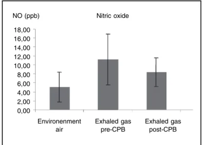
Tables I and II show the demographic data of patients and the characteristics of exhaled NO collection, respectively. Figure 1 shows the results of NO analysis in the compres-sed air network pre- and post-CPB. The level of NO in the compressed air of the gas network was 5.05 ± 3.37 ppb. The concentration of exhaled NO decreased after CPB, va-rying from 11.25 ± 5.65 ppb pre-CPB to 8.37 ± 3.17 post-CPB (p = 0.031) when extraction of environmental NO for analysis was not considered.
Table I – Demographic Data of the Study Patients
Mean Standard Deviation Minimal Maximal
Age (years) 61.8 9.5 47 76
Weight (kg) 79.5 16.3 41.7 98
Height (cm) 164.5 8.6 148 173
Table II – Characteristics of the Collection of Exhaled Nitric Oxide
Mean SD Minimal Maximal
Number of 23.66 2.44 20 29
exhalations
Total time (sec) 73.1 7.21 68 98
Exhalation flow 0.020 0.002 0.015 0.022 (L.sec-1)
Figure 1 – Distribution of Mean Values in the Air, Pre-, and Post-CPB.
NO (ppb) Nitric oxide
Environenment air
Exhaled gas post-CPB Exhaled gas
pre-CPB 18,00
DISCUSSION
The results of the study showed that the compressed air used had a variable concentration of NO, besides a statistically significant reduction in exhaled NO when pre-and post-CPB levels were compared (p < 0.05). When in-cluding the analysis of NO in the gas delivery system for the anesthesia device, some measurements of exhaled NO, pre- or post-CPB, presented variable results. This observa-tion generated negative means and elevated the standard deviations. Although the final calculation of NO is not simply the result of this subtraction, both analyses (with and without subtracting NO levels in compressed air) demonstrated a tendency for a reduction in exhaled NO levels after CPB (p < 0.05). On the other hand, it is possible that the differences found in the levels of the gas delivery network might have not been properly evaluated, since the conditions of flow and time of collection were not the same and were not the objective of this study. Those considerations indicate perspectives for new studies.
Since the molecule of NO is an important element related with the physiology and pathophysiology of microcirculatory mechanisms of the respiratory system, it has been extensi-vely studied 7,10,15-17. In many studies it is considered an
inflam-matory marker of the airways, since patients with asthma and other lung diseases have elevated levels of exhaled NO 2,18. In asthma patients, it seems to indicate both
disea-se exacerbation and the dodisea-se-respondisea-se relationship in the treatment with corticosteroids, especially inhaled 2,18,19. When
the maintenance dose is not adequate, the patient becomes refractory to treatment, or in the case of a fast reduction in the maintenance dose of patients who remain stable during treatment, substantial changes in exhaled NO levels are seen, indicating its interference in endothelial tonus 19.
Ni-tric oxide also seems to be elevated in patients with
bronchiectasis or decompensated chronic obstructive pulmonary disease (COPD) 20-22. However, regarding
myo-cardial revascularization, the literature does not agree on the behavior observed after CPB, or on the definitive mecha-nisms that would be implicated in those changes 10,11,16,23-25.
It is known that ventilation patterns in patients on mechani-cal ventilation are related with the levels of exhaled NO 16.
Changes in minute ventilation (Vm), secondary to changes in respiratory rate (RR) or tidal volume (VT), lead to non-linear changes in exhaled NO levels. Tornberg et al. 10 observed a
reduction in post-CPB exhaled NO, both on what they considered as NO output (NO concentration × exhaled airflow × 60) and mean NO peaks (mNO) extracted from reading the curve during the expiratory flow. Unlike the present study, in which off-line measurements were used, those authors used on-line measurements and obtained mNO of 3.2 ppb.L-1 and
output of 12.2 ppb.min-1, as can be seen in table III, which
compares collection methods among this and other studies. Using the same proposal as Tonberg et al. 10, if the pre-CPB
level obtained here (11.3 ppb) were considered equivalent to the output, 9.4 ppb.L-1 would correspond to the mean study
level (versus 3.2 ppb.L-1). This difference could possibly be
secondary to the exhaled flow, which in the present case was 0.020 L.sec-1, while theirs was 0.063 L.sec-1, obtained
using the same equation as before where maintaining the output, mean NO levels would be inversely proportional to the flow. In addition, comparing ventilation data, one observes that the tidal volume (VT) in the present study was greater than in their study (Table III). On the other hand, they used a higher respiratory rate (RR). Thus, the higher VT justifies according to Harefield’s 16 group lower levels of exhaled NO
or in this case lower output, corroborating this line of thought. Similarly, the higher RR used by those authors should also lead to lower exhaled NO levels when compared with the present study, what was not the case. Even with their higher Table III – Comparison among Methods of Collection of Exhaled Nitric Oxide
Present study Tonberg et al. Ishibe et al. Sheppard et al. Beghetti et al.
(2004) (2000) (2004) (1998)
Analysis Off-line On-line On-line On-line On-line
VT (mL.kg-1) 8 4 12 n n
RR (bpm) 12 20 8-10 n n
Ventilation Volume-controlled Volume-controlled Volume-controlled Pressure-controlled n
Post-CPB moment 30 min 2 h 6 h n 30 min
Parameters [NO] in 1.5 L pNO for 30 sec and mNO, pNO, VNO in a prolonged [NO] collected
balloon NO output VNO and Q exhalation in 20 mL
Values [NO] =11.3 ppb pNO = 3.2 ppb mNO = 5.7 ppb VNO = 2.58 ppb.sec-1 [NO] = 7.0 ppb Output = 12.2 pNO = 15.4 ppb and
(ppb.min-1) VNO=29.16 ppb.min-1
RR and the higher VT of the present study, the equation showed higher output (12.2 ppb.min-1 versus the hypothetic
11.3 ppb), which should also be due to the flow.
Certainly, the variability of the collection methods as shown in table III translates into different results. According to this viewpoint, and using the same line of thought applied in the comparison with the study of Tonberg et al. 10, considering the
mean level of 5.7 ppb (mppb) obtained by Ishibe et al. 11 the
flow in this case would be 0.085 L.sec-1. Analyzing those
results, one observes that the higher NO output (VNO corrected for one minute) – 29.16 ppb.min-1 – could also be related
with the expiratory flow used for the reading, which was higher than in the present study and in the study of Tonberg et al. 10. Differences in mean values are probably secondary
to the VNO/flow ratio and, therefore, the values obtained by Ishibe et al. 11 were between those of Tonberg et al. 10 and the
present study.
Although Sheppard et al. 12 did not specify the ventilation
parameters used, they mentioned the use of pressure-con-trolled ventilation (PCV), through which they collected the samples based on a 30-second ventilator pause followed by a prolonged expiration (Table III). The technique of interrup-ting ventilation followed by a prolonged exhalation increases the exhaled NO curve and maintains it elevated throughout the observation period and, therefore, peak NO levels could be as high as 35 to 40 ppb, which does not reflect the true NO concentration as a normal exhalation does 16,26.
Nitric oxide concentrations in the airways seem to increase when RR or VT is decreased or when the I:E ratio is increa-sed. During prolonged inspiration or exhalation, it is possible to measure mean and peak concentrations, and also the area under the NO release curve. Besides, the plateau of the NO curve seems to occur approximately 20 to 30 seconds, during the ventilator pause, indicating higher values during sustained expiration when compared to inspiration 16.
This detection during inspiration seems to suggest its con-tinuous production/release also observed in the bronchial epithelium 8.
Several studies have emphasized the importance of the flow-dependency mechanism for collection and analysis of exhaled NO concentrations. The European Respiratory So-ciety (ERS) and the American Thoracic SoSo-ciety (ATS) recom-mend constant expiratory flow and decontamination of NO from the upper airways in patients with spontaneous ven-tilation 26. In patients in controlled ventilation, ventilator
para-meters (type of ventilation, I:E ratio, PEEP, tidal volume, and contamination with inspired air) should be considered 16,26.
On the other hand, evaluation of the behavior of NO in pa-tients undergoing CPB, especially diabetics or papa-tients using the so-called NO donors (nitroglycerine, sodium nitroprus-side, and nitrates), should consider those aspects as rele-vant. In diabetics, the basal level of microcirculatory stress that produces damage mediators, which combine with supe-roxide anions to form peroxynitrite, leads to NO consump-tion 27,28. Both nitroglycerine and sodium nitroprusside,
routinely used, are exogenous NO donors, behaving as an-tioxidants of radicals produced during CPB, and inhibiting the endogenous production of NO 27,28. Tonberg et al. 10 observed
a reduction in the response of exhaled NO to nitroglycerin infusion, attributing this response either to a reduction in the conversion of nitroglycerin to NO or to an increase in the con-sumption of NO by free radicals produced during CPB. Some authors have also considered that the type of flow produced by the CPB equipment may interfere with basal NO relea-se 29,30. During non pulsating CPB, terminal capillaries tend
to close as a result of the reduction of the contact with the vessel walls 29,30. The endothelial release of NO, considered
physiological and produced by the stimulation of eNOS (en-dothelial nitric oxide synthetase), seems to be a function of the frequency and amplitude of the pulsating flow 29,30.
Variable degrees of post-CPB atelectasis are related with different pulmonary conditions with the consequent change in gas exchange 31-33, which are also relevant in the evaluation
of NO. The reduction in post-CPB exhaled NO can be asso-ciated with a reduction in the PaO2/FiO2 ratio, as well as with an increase in the alveolo-arterial gradient (P(A-a)O2) 10.
Chan-ges in pulmonary compliance have also been associated with this post-CPB reduction 11. However, inferences on the
effects of different degrees of post-CPB atelectasis on va-riations in exhalation flow and levels of exhaled NO cannot be found in the literature. Those considerations need to be better understood. If changes in ventilation pattern are rela-ted with the curve of exhaled NO, they could become relevant not only during its observation in the post-CPB phase, but also in any major surgery in which precise ventilator moni-toring is fundamental, making NO relevant for the detection of sudden changes, may they be epithelial, endothelial, or purely mechanical.
From what was exposed, there is an unquestionable re-lationship between the production and basal release of NO and the rupture of the physiological mechanisms of gas exchange caused by CPB. Once established, this connection will show its relevance as an online marker of perioperative pulmonary function. However, since more conclusive data are not available, this inference is hasty and certainly NO will continue to be the focus of extensive studies.
The limitations of the present study are related to the lack of distinction between groups of diabetic and non-diabetic patients, more judicious investigation of pulmonary function or inflammatory response parameters, and the lack of quan-tification of drugs used that have the potential to interfere with basal NO release. Although the study population was consi-dered statistically normal, the paired test with 8 degrees of freedom revealed a low power (α < 0.800), indicating the
REFERÊNCIAS – REFERENCES
01. Laffey JG, Boylan JF, Cheng DC - The systemic inflammatory response to cardiac surgery: implications for the anesthesio-logist. Anesthesiology 2002;97:215-252.
02. Barnes PJ, Liew FY - Nitric oxide and asthmatic inflammation. Immunol Today 1995;16:128-130.
03. Engelman DT, Watanabe M, Maulik N et al. - L-arginine reduces endothelial inflammation and myocardial stunning during ische-mia/reperfusion. Ann Thorac Surg 1995;60:1275-1281. 04. Tsao PS, Lewis NP, Alpert S et al. - Exposure to shear stress
alters endothelial adhesiveness. Role of nitric oxide. Circulation 1995;92:3513-3519.
05. Moncada S, Higgs A - The L-arginine-nitric oxide pathway. N Engl J Med 1993;329:2002-2012.
06. Negri MLS, Camargo EA - Óxido nítrico: uma alternativa no tra-tamento da hipertensão pulmonar. J Bras Med 2003;85:15-23. 07. Rubanyi GM, Ho EH, Cantor EH et al. - Cytoprotective function
of nitric oxide: inactivation of superoxide radicals produced by human leukocytes. Biochem Biophys Res Commun 1991;181: 1392-1397.
08. Sartori C, Lepori M, Busch T et al. - Exhaled nitric oxide does not provide a marker of vascular endothelial function in healthy hu-mans. Am J Respir Crit Care Med 1999;160:879-882.
09. Gustafsson LE, Leone AM, Persson MG et al. - Endogenous ni-tric oxide is present in the exhaled air of rabbits, guinea pigs and humans. Biochem Biophys Res Commun 1991;181:852-857. 10. Tornberg DC, Angdin M, Settergen G et al. - Exhaled nitric oxide
before and after cardiac surgery with cardiopulmonary bypass: response to acetylcholine and nitroglycerin. Br J Anaesth 2005; 94:174-180.
11. Ishibe Y, Liu R, Hirosawa J et al. - Exhaled nitric oxide level de-creases after cardiopulmonary bypass in adult patients. Crit Care Med 2000;28:3823-3827.
12. Sheppard SV, Gibbs RV, Smith DC - Does the use of leucocyte depletion during cardiopulmonary bypass affect exhaled nitric oxide production? Perfusion 2004;19:7-10.
13. Beghetti M, Black SM, Fineman JR - Endothelin-1 in congenital heart disease. Pediatr Res, 2005;57:16R-20R.
14. Higgins TL, Estafanous FG, Loop FD et al. - Stratification of morbidity and mortality outcome by preoperative risk factors in coronary artery bypass patients. A clinical severity score. JAMA 1992;267:2344-2348.
15. Beghetti M, Silkoff PE, Caramori M et al. - Decreased exhaled nitric oxide may be a marker of cardiopulmonary bypass-induced injury. Ann Thorac Surg 1998;66:532-534.
16. Marczin N, Kovesi T, Royston D - Exhaled nitric oxide as a marker of lung injury in coronary artery bypass surgery. Br J Anaesth 2003;90:101-105.
17. Wilkins MR, Zhao L, al-Tubuly R - The regulation of pulmonary vascular tone. Br J Clin Pharmacol 1996;42:127-131.
18. Kharitonov SA, Barnes PJ - Effects of corticosteroids on noninvasive biomarkers of inflammation in asthma and chronic obstructive pulmonary disease. Proc Am Thoracic Soc 2004;1: 191-199.
19. Kharitonov SA, Barnes PJ - Clinical aspects of exhaled nitric oxi-de. Eur Respir J 2000;16:781-792.
20. Barnes PJ, Chowdhury B, Kharitonov SA et al. - Pulmonary bio-markers in chronic obstructive pulmonary disease. Am J Respir Crit Care Med 2006;174:6-14.
21. Horvath I, Donnelly LE, Kiss A et al. - Exhaled nitric oxide and hydrogen peroxide concentrations in asthmatic smokers. Res-piration 2004;71:463-468.
22. Brindicci C, Ito K, Resta O et al. - Exhaled nitric oxide from lung periphery is increased in COPD. Eur Respir J 2005;26:52-59.
23. Zegdi R, Fabre O, Cambillau M et al. - Exhaled nitric oxide and acute lung injury in a rat model of extracorporeal circulation. Shock 2003;20:569-574.
24. Zegdi R, Fabre O, Cambillau M et al. - Exhaled nitric oxide does not reflect the severity of acute lung injury: an experimental study in a rat model of extracorporeal circulation. Crit Care Med 2002;30:2096-2102.
25. Beghetti M, Adatia I - Inhaled nitric oxide and congenital cardiac disease. Cardiol Young 2001;11:142-152.
26. American Thoracic Sosicety. European Respiratory Society. ATS/ ERS recommendations for standardized procedures for the on-line and offon-line measurement of exhaled lower respiratory nitric oxide and nasal nitric oxide, 2005. Am J Respir Crit Care Med 2005;171:912-930.
27. Matata BM, Galinanes M - Effect of diabetes on nitric oxide metabolism during cardiac surgery. Diabetes 2001;50:2603-2610.
28. Matata BM, Galinanes M - Cardiopulmonary bypass exacerba-tes oxidative stress but does not increase proinflammatory cy-tokine release in patients with diabetes compared with patients without diabetes: regulatory effects of exogenous nitric oxide. J Thorac Cardiovasc Surg 2000;120:1-11.
29. Hutcheson IR, Griffith TM - Release of endothelium-derived relaxing factor is modulated both by frequency and amplitude of pulsatile flow. Am J Physiol 1991;261:H257-262.
30. Macha M, Yamazaki K, Gordon LM et al. - The vasoregulatory role of endothelium derived nitric oxide during pulsatile cardiopul-monary bypass. Asaio J 1996;42:M800-804.
31. Hachenberg T, Tenling A, Nystrom SO et al. - Ventilation-perfu-sion inequality in patients undergoing cardiac surgery. Anesthe-siology 1994;80:509-519.
32. Magnusson L, Zemgulis V, Wicky S et al. - Atelectasis is a major cause of hypoxemia and shunt after cardiopulmonary bypass: an experimental study. Anesthesiology 1997;87:1153-1163. 33. Tenling A, Hachenberg T, Tyden H et al. - Atelectasis and gas
exchange after cardiac surgery. Anesthesiology 1998;89:371-378.
RESUMEN
Amorim CG, Malbouisson LMS, Saraiva BM, Pedro FMS, Martins MA, Carmona MJC - Evaluación del Óxido Nítrico Exhalado en Pacientes Sometidos a la Revascularización del Miocardio con Circulación Extracorpórea.
JUSTIFICATIVA Y OBJETIVOS: La circulación extracorpórea
(CEC), puede causar una disfunción pulmonar. Las alteraciones inflamatorias pueden afectar la liberación de óxido nítrico (NO). Se buscó evaluar el NO exhalado en pacientes sometidos a la revascularización del miocardio (RM) con CEC.
MÉTODO: Se estudiaron prospectivamente nueve pacientes
adul-tos sometidos a la RM con CEC. Inicialmente fue recolectada una muestra de aire para el análisis de NO en el sistema que alimenta el aparato de anestesia. A continuación, se inició la anestesia por vía venosa con etomidato (0,3 mg.kg-1), sufentanil (0,3 µg.kg-1),
pancuronio (0,08 mg.kg-1) y se mantiene con isoflurano (0,5 a 1,0
CAM) y sufentanil (0,5 µg.kg-1.h-1). El volumen corriente fijado fue
8 mL.kg-1,con FiO2 de 0,6 excepto durante la CEC. Treinta
RESULTADOS: El valor del NO del aire ambiente fue de 5,05 ± 3,37 ppmm. El NO exhalado se redujo después de la CEC, variando de 11,25 ± 5,65 ppmm para 8,37 ± 3,17 ppmm (p = 0,031).
CONCLUSIONES: La reducción del NO exhalado pos-CEC,
obser-vada en este estudio, no permite confirmar el papel de esta