Iranian Journal of Basic Medical Sciences
ijbms.mums.ac.ir
Oxidative stress and cytotoxic potential of anticholinesterase
insecticide, malathion in reproductive toxicology of male
adolescent mice after acute exposure
Selmi Slimen
1*, El Fazaa Saloua
1, Gharbi Najoua
11 Laboratory of Animal Physiology, Department of Biology, Faculty of Sciences, University Tunis El Manar, Tunis, Tunisia
A R T I C L E I N F O A B S T R A C T Article type:
Original article
Objective(s):The present study was undertaken to determine the effects of acute exposure to malathion on oxidative stress and cytotoxic potential of anticholinesterase insecticide, malathion in reproductive toxicology of adolescent male mice.
Materials and Methods: Thirty two adolescent male mice at pubertal age were treated with 500 mg/kg body weight (BW) of malathion for three days. After exposure, biochemical markers and sperm analysis were evaluated and finally histological modifications of testis and sperm were assessed.
Results: Our data showed that treatment of male mice with malathion (500 mg/kg, BW) could lead to oxidative stress. Induced oxidative stress status can be assessed due to increased malondialdhyde (MDA) content, decreased thiol group content, as well as increased antioxidant enzyme activities. On the other hand, exposure to malathion at the pubertal age led to alteration of semen parameters; sperm production and percentage of motile sperm were decreased in the treated groups compared to the control. Furthermore, exposure of male mice to malathion led to a decrease of testosterone level, inhibition of acetylcholinesterase, and decrease of the reproductive performance of male mice after three days of treatment at the age of puberty.
Conclusion: The importance to carry out in vitro reproductive toxicology assays lies on the need of knowing the alterations these insecticides may cause at cellular level, since they are endocrine disruptors that interfere with reproductive functions.
Article history: Received: Sep 3, 2013 Accepted: May 2, 2014
Keywords:
Acetylcholinestrase Malathion Oxidative stress Testosterone
►
Please cite this paper as:Slimen S, Saloua EF, Najoua Gh. Oxidative stress and cytotoxic potential of anticholinesterase insecticide, malathion in reproductive toxicology of male adolescent mice after acute exposure. Iran J Basic Med Sci 2014; 17:522-530.
Introduction
Organophosphate insecticides (OPI) constitute one of the most widely used classes of pesticides being employed for both agricultural and pest control. However, the uncontrolled use of insectici-des in agriculture and public health operation importantly increased the scope of ecological imbalance and thus many non-target organisms have become victims (1). Malathion was one of the most important OPI used extensively on a large number of crops against several pests. Although the toxicity of organophosphorus compounds in humans and experimental animals are well demonstrated, the underlying mechanisms of their action remain unclear (2-4). The most plausible mechanism that may be operative in vivo is the generation of reactive oxygen species (ROS), which may initiate lipid peroxidation (LPO), oxidative damage to critical macromolecules such as proteins or DNA, and cell damage or death. LPO constitutes a free radical oxidation process in which poly-unsaturated fatty
acids of the cell membrane decompose to yield, among others, highly reactive lipid hydroperoxides, H2O2, hydroxyl radicals, and malondialdehyde (MDA) (5). MDA has been demonstrated to cause
cross-linking and polymerization of membrane
components and may contribute to mutagenic, genotoxic, and carcinogenic effects (6-8). In fact, it has been reported that malathion induces toxicity through the inhibition of acetylcholinesterase (AchE) and subsequent activation of cholinergic receptors (9, 10). Moreover, the lipophilic nature of malathion facilitates its interaction with cell membrane and leads to perturbations of the phospholipids bilayer structure of most visceral organs (11). In addition, there is now considerable evidence that male reproductive function is declining in human and wildlife populations (12); the mechanism of action may be disturbed testicular apoptosis and altered hepatic biotransformation of steroids. In rats, exposure to malathion results in reduction of repro-
Iran J Basic Med Sci, Vol. 17, No. 7, Jul 2014 523 ductive organs weights and spermatozoa parameters
with increase in sperm death and abnormalities. In the same context, Pina- Guzmán et al (13) demons-trated that malathion can affect late stages of spermatogenic cells maturation in mice causing damaged DNA and reduced chromatin in spermato-gonia and spermatids. The present study aimed to determine the effect of acute exposure to malathion on sperm function, induction of oxidative stress, and reproductive performance to elucidate the mechani-sm of reproductive toxicology of organophosphorus compounds.
Materials and Methods
ChemicalsMalathion (98% purity) (fyfanon 50 EC 500 g/l),
acetylthiocholine iodide, . ’
-dithiobis-(2-nitroben-zoic acid) (DTNB), Triton X-100, eosin stain, and RPMI (Roswell Park Memorial Institute), were purchased from SIGMA and Invitrogen.
Animals and dosage exposures
Twenty five adult male and female Swiss mice obtained from the Laboratory Animal Breeding Center of Pasteur, Tunis, Tunisia were housed in cages with constant temperature and humidity, with food and water available ad-labium. Animals were allowed to adjust to their new surroundings for 1 week before treatment and then were cohoused one to one/box. In the age of puberty (45 days after), male Swiss mice were separated and then randomly divided into two groups: control (n=16) and malathion treated mice (n=16). Malathion treated group received malathion, at a dose of 500 mg/kg of body weight (BW) in corn oil (1 ml per animal) for 3 days, which was given through gavages to mice in the experiment group. Control group received equal amount of corn oil for 3 days.
Body and reproductive organ weights
Initial (weight at start point) and final (weight at end point) BW were recorded. Male mice from each group were killed by decapitation after three days of the treatment. The reproductive organs were stripped from fatty tissues and blood vessels, blotted, and their absolute weights were determined. Clinical signs of body and reproductive organs were evaluated for toxicological criteria. To normalize the data for statistical analysis, organ weights were expressed per 100 g BW.
Plasma acetylcholinesterase activity (AChE) The cholinesterase activities were assayed in plasma by the method of Ellman (14) using acetylthi-ocholine iodide as a substrate. The rate of hydrolysis of acetylthiocholine was measured at 405 nm by the reaction of thiocholine with DTNB to give the yellow 5-thio-2-nitrobenzoate anion with spectrophoto-meter. The enzyme activity was expressed as nmol of
substrate hydrolyzed/min/mg protein.
Evaluations of reproductive hormone in serum Immediately after the animals reached the mating period, peripheral blood samples were harvested. The serum was separated and stored at − °C for subsequent hormone assays. Blood serum prepara-tions were used for measurement of steroid hormone. The concentration of testosterone was measured by commercial enzyme-linked immune-absorbent assays (ELISA) purchased from demeditec (Ref, DEV 9911), radioimmunoassay, using comer-cial kits as per manufacturer’s instructions.
Evaluation of sperm characteristics Sperm collection
After the test period, the laparotomy was conducted for all exposed and control male mice following weighing. Testes and epididymides were carefully excised. The sperm count was assessed from right cauda epididymides while sperm motility and morphology were analyzed from the left one. Epididymis was excised and minced in 1ml of RPMI to obtain sperm suspension. The other testes and epididymides were frozen in - 20°C until usage.
Sperm count
The cauda epididymal sperm count was performed according to the method of Vega et al
(15). Epididymal sperm counts were expressed as number of sperms per epididymis. To minimize the error, the count was repeated three times on each sample.
Sperm motility
Ten μl of sperm suspension was layered onto a warmed microscope slide. Sperm motility was assessed by counting all progressive motile, non-progressive motile and immotile spermatozoa. The number of motile sperm cells in each field was divided by the total number, and the average of the fields was assayed (16). The percentage of motile spermatozoa was determined.
Sperm viability
Sperm viability was assessed using the eosin stain. The staining was performed with one drop of freshly collected semen (10 μl and two drops μl of eosin. The live sperms were unstained (intact cell membrane) and dead sperms revealed purple to red-stained head with damaged membranes. The dye exclusion was evaluated in 100 spermatozoa. Sperm viability was defined as the percentage of dead sperm cells.
Sperm morphology
Iran J Basic Med Sci, Vol. 17, No. 7, Jul 2014 524
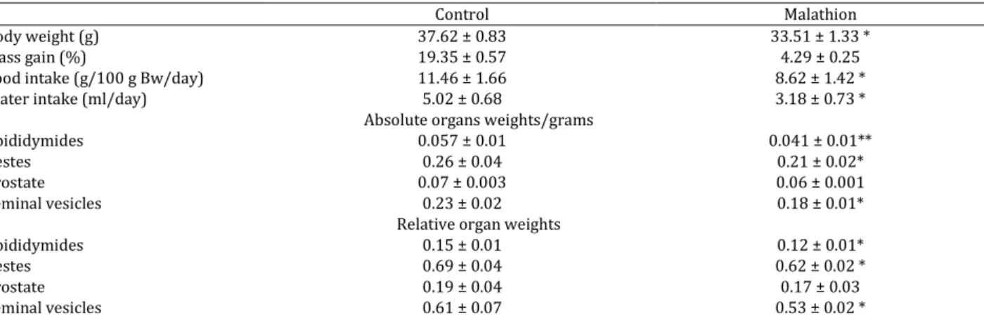
Table 1. Body weight, mass gain, food intake and water intake during acute exposure to malathion to male mice for 3 days
Control Malathion
Body weight (g) 37.62 ± 0.83 33.51 ± 1.33 *
Mass gain (%) 19.35 ± 0.57 4.29 ± 0.25
Food intake (g/100 g Bw/day) 11.46 ± 1.66 8.62 ± 1.42 *
Water intake (ml/day) 5.02 ± 0.68 3.18 ± 0.73 *
Absolute organs weights/grams
Epididymides 0.057 ± 0.01 0.041 ± 0.01**
Testes 0.26 ± 0.04 0.21 ± 0.02*
Prostate 0.07 ± 0.003 0.06 ± 0.001
Seminal vesicles 0.23 ± 0.02 0.18 ± 0.01*
Relative organ weights
Epididymides 0.15 ± 0.01 0.12 ± 0.01*
Testes 0.69 ± 0.04 0.62 ± 0.02 *
Prostate 0.19 ± 0.04 0.17 ± 0.03
Seminal vesicles 0.61 ± 0.07 0.53 ± 0.02 *
Results were expressed as the mean ± SD of three independent assays. n=16. Values significantly different with respect to control group were indicated as *P < 0.05 or **P < 0.01
sperm defects were evaluated and examined on optical microscope. At least one hundred spermato-zoa from different fields in each slide were examined and classified for criteria of morphological abnorm-alities (head, tail, and tail-head). Abnormal sperm cells were counted and the percentage was calculated (17).
Assessment of sperm production
Sperm content per g was determined using slight modifications on the method described previously.
(
)دیهدب سنرفرBriefly, after thawing at temperature
(25-27°C), the whole epididymal and the testicular tissues were homogenized for 5 min in 5 ml of physiological saline (0.9% NaCl) containing 0.05% (v/v) Triton X-100 using a manual homogenizer. The homogenates were diluted with 1.5 ml of the saline solution; spermato-zoa and spermatid were counted. Three counts per sample were averaged. These count values were used to obtain total number of spermatids per testis and sperm per epididymis; it was then divided by the testis or epididymis weight to determine the number per g of testis or epididymis.
Lipid peroxidation
LPO which is detected by the determination of MDA production was determined in this study. Homogenates of testis and epididymis were centrifuged at 1000 g for 10 min at 4°C to sediment cell debris and mitochondrial samples. Supernatants were suspended in phosphate buffered saline (PBS), pH=7.4, mixed with trichltoracetic acid (TCA), buthyl-hydroxytoluen (BHT-TCA solution ‰ w/v BHT dissolved in 20% TCA) and centrifuged at 1000 g for 35 min. Then, supernatant was mixed with 0,5 NaCl and 120 mM thiobarbituric acid (TBA) in 26 mM Tris and heated in water bath at 80°C for 10 min. After cooling, we determined the absorbance of the resulting chromophore at 532 nm. MDA levels were determined by using an extinction coefficient for MDA-TBA complex of 1.56 105 M-1 cm-1.
Determination of the concentration of the thiol groups
Homogenates of testis and epididymis were added to 0.25 M Trizma-Base and 20 mM EDTA (Ethylen diamine tetraacetic acid) pH 8.2. Then, the mixture was vortexed and its absorbance was determined at 412 nm. The first value was noted A1. After that, 10 mM DTNB was added, incubated for 15 min, and a new value A2 was determined. The white tube of DTNB contains only DTNB and buffer; its absorbance value was noted B.
We calculated the concentration of thiol groups per tube using this expression: (A2-A1-B)×1.57 mM.
Intracellular mediator
H2O2 was measured according to Kakinuma et al
(18) using commercial kits from Biomaghreb (Tunisia).
Antioxidant activities assays
The activity of SOD and its isoformes in testis and epididymis of control and treated mice was assessed by spectrophotometric method. Manganese super-oxide dismutase (Mn-SOD) activity was measured in the presence of 2.0 mM sodium cyanide, an inhibitor of copper and zinc-containing SOD (Cu/Zn-SOD). Cu/Zn-SOD activity was calculated from the total activity minus the activity observed in the presence of sodium cyanide. CAT activity was measured by the modified method of Aeibi (19).
Protein determination
Protein concentration in the supernatant of each tissue (epididymis and testis) was determined by the method of Bradford (20), using bovine serum albumin (BSA) as standard.
Histological examination
Iran J Basic Med Sci, Vol. 17, No. 7, Jul 2014 525 Figure 1. plasmatic Acetylcholinesterase activity after 3 days
exposure to 500 mg/kg Bw (n= 16 for each groups)
Statistical analysis
All data were expressed as the mean ± SD for each group. Differences in group means were assessed via overall one-way analysis of variance (ANOVA), and
unpaired Student’s t-test. For all comparisons,
statistical significance was set at P<0.05. All analyses were performed with SPSS version 10.0.1. *: P<0.05; **: P<0.01.
Results
The findings depict the indices of reproductive toxicology in adolescent male mice after adminis-tration of anticholinesterase insecticide, malathion for 3 days at pubertal age.
Mortality and macroscopic symptoms of toxicity In the present study, although all animals survived (both treated and control groups animals) until the end of experimental period, cholinergic signs of adverse toxicological effects were observed such as sluggishness, muscular tremors, irregular movement, and abdominal tremble. The progression of these signs proceeds during experiment of such treated male mice. At necropsy, no macroscopic alteration that could be attributed to treatment with malathion was found in the testes and/or reproductive accessories.
Body, reproductive organ weights, and food and water intake
There was a significant correlation between
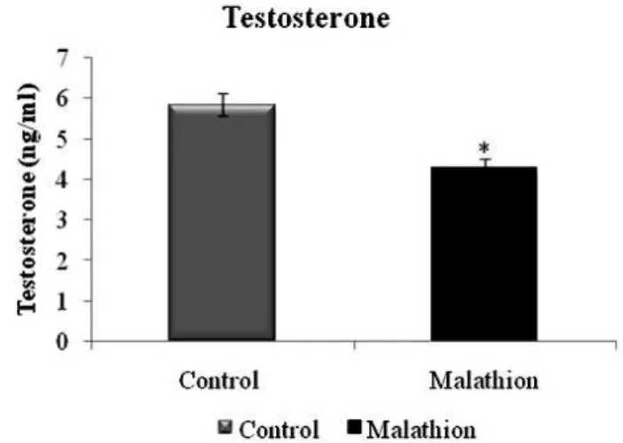
Figure 2. means± SD of the plasma concentration of testosterone in pubertal male mice after oral administration of 500 mg/kg Bw of malathion for 3 days (n=16)
symptoms of toxicity of both BW and reproductive organs weights of malathion treated group (500 mg/kg BW/day) (P<0.05) (Table 1). Furthermore, exposure to 500 mg/kg BW of malathion for 3 days caused a significant decrease in BW, absolute and relative weight of testis, epididymis, prostate, and seminal vesicle which was comparable to control. Furthermore, our results showed a decrease of food and water intake and mass gain of mice treated with malathion (Table 1).
Evaluations of biochemical findings
After three days of the treatment with malathion, a significant inhibition of plasma AChE activity (P<0.05) was observed in adolescent male mice treated with 500 mg/kg BW/day of malathion, as compared with control (Figure 1). Furthermore, serum testosterone levels were drastically reduced in the animals treated with 500 mg/kg BW/day of malathion (P<0.05) (Figure 2).
Evaluations of reproductive performance quality Administration to male mice of 500 mg/kg bw /day malathion resulted in paucity of epididymal spermatozoa counts and testicular spermatid enumeration (Table 2) and reduction of sperm motility (Table 2), while the number of dead sperms was increased. As shown in Table 2, a significant decrease was observed in the percentage of the total
Table 2. Effect of acute exposure to malathion on sperm parameters in treated male mice and control groups (n= 16/group)
Control groups Treated groups
Sperm concentration, 106/mL 6.8 ± 0.9 4.62 ± 0.6*
Motility, % 74 ± 7.16 61 ± 4.32*
Viability, % 83 ± 3.15 59 ± 5.68**
Morphology, normal forms, % 90.3 ± 2.31 73 ± 3.27**
Abnormal head, % 2.2 ± 0.58 9 ± 1.29**
Abnormal tail, % 7.6 ± 1.41 18 ± 3.82**
Sperm count/(g) epididymis (mean ×106) 117 ± 16 98 ± 13*
Spermatid count/testis (mean ×106) 51 ± 7 38 ± 5*
Spermatid count/(g) testis (mean ×106) 196 ± 29 181 ± 21
Iran J Basic Med Sci, Vol. 17, No. 7, Jul 2014 526
Figure 3. Patterns of sperm abnormalities. (a and b) Normal sperm with acrosome (arrow), (c,d) abnormal head (*), (e) clumped head with normal tail (*), (f) normal head with looped tail (*)
Figure 4. (A) Testis from control mice showing circumscribed somniferous tubules with intact basement membranes (Mb). Note the spermatogonia (Sg), spermatids (Sd), sperm (S), interstitial cells of leydig (L) and interstitial space (I). (B) Testis from mice treated with malathion 500 mg/kg/day showing widening of the interstitial spaces (I) and a small number of interstitial cells of leydig (L)
sperm viability. Morphological abnormalities of spermatozoa, categorized by head or tail (Table 2, Figures 3 a, b, c, d, e, and f) were significantly increased as shown in monitored smears of mice administrated 500 mg/kg BW/day of malathion.
Histological examination
No histological changes appeared in the testis of control group (Figure 4A). In the treated group with
500 mg/kg BW/day of malathion, the somniferous tubules showed wide lumen with ill-define sperma -togenesis. In addition, malathion led to widening of interstitial spaces with a small number of interstitial cells (Figure 4B). Testis showed malathion- associated degenerative changes in cells lining the somniferous tubules.
S Sd Sg
I L Mb
Sg
L Mb
I
Iran J Basic Med Sci, Vol. 17, No. 7, Jul 2014 527 Table 3. Changes in testis lipid peroxidation, total thiol groups and antioxidant enzyme activities after acute exposure of male mice to malathion during 3 days at pubertal age
Control Malathion
MDA (nmol/ mg protein) 3.58 ± 0.4 4.63 ± 0.31 *
Total thiol groups (mM) 0.53 ± 0.02 0.42 ± 0.01 *
H2O2 (µmol/mgprotein) 0.82 ± 0.13 1.48 ± 0.21 **
SOD (U/mg protein) Cu/zn-SOD (U/mg protein) Mn-SOD (U/mg protein) Fe-SOD (U/mg protein)
8. 31 ± 0.42 4.48 ± 0.29 2.57 ± 0.30 1.14 ± 0.13
10.67 ± 0.69 ** 5.46 ± 0.42*
3.6 ± 0.22* 1.21 ± 0.1
CAT (nmol/min/mg protein) 508 ± 18 762 ± 21 **
Values are means ± S.D. of 16 mice in each group. * and ** represent the statistical difference between control and treated groups respectively at P < 0.05 and P < 0.01
Lipid peroxidation and thiol group’s contents
We further looked at the testis and epididymis MDA and thiol-groups contents (Table 3 and 4). Our results showed that malathion exposure of male mice significantly increased the MDA level and thiol-groups content.
Intracellular mediators
Our results showed an increase of H2O2 content (Table 3 and 4).
Antioxidant enzyme activities
Results of the present study showed that malathion treatment significantly increased testis and epididymis anti-oxidant enzyme activities (SOD, its isoformes, and CAT) (Table 3 and 4).
Discussion
In our previous studies, we suggested that lactational exposure to malathion induces abnormal hyperglycemia, oxidative stress, and alternation of biochemical markers in liver and kidney of rat pups. Thus, malathion compound induces oxidative stress and cholinesterase inhibition in plasma, erythrocyte, and brain of rats pups (2, 3). Consequently, we suggested that continued repetition of this loop exhausts metabolic and endocrine control and can result in reproductive toxicology induction during the acute subchronic and long-term exposure.
Organophosphate compounds are primarily
recognized for their ability to induce toxicity in mammals through the inhibition of AChE, leading to accumulation of acetylcholine, and subsequent activation of cholinergic, muscarinic, and nicotinic receptors (21). On the other hand, malathion like dimethoate has no direct inhibitory action on the AChE activity prior to biotransformation. However, the mammalian toxicity (cholinergic crisis) of organophosphorus compounds like malathion depends on mixed function oxidase (MFO) catalyzed activation to its corresponding oxygen analogue, which is a direct inhibitor of AChE (22). Therefore, malathion may exert its biological effects through electrophilic attack on the cellular constituents of organ and tissues with simultaneous generation of reactive oxygen species (23). So, the adverse male-mediated reproductive effects of malathion may reflect its electrophilic attack on the mice cellular constituents. Furthermore, Spermatogenesis and fertility are critically dependent upon the maintenance of adequate levels of testosterone (24). In the present study, histological examination revealed that malathion caused testicular lesions characterized by markedly decreased testes weight with moderate to severe widening of interstitial spaces and partial arrest of spermatogenesis. Testicular spermatid and epididymal sperm counts indicated that spermatogenesis was partially arrested at malathion treated group during the experimental period. It has been shown that
Table 4. Changes in epididymis lipid peroxidation, total thiol groups and antioxidant enzyme activities after acute exposure of male mice to malathion during 3 days at pubertal age
Control Malathion
MDA (nmol/ mg protein) 3.14 ± 0.03 3.93 ± 0.05 *
Total thiol groups (mM) 0.28 ± 0.03 0.22 ± 0.03 *
H2O2 (µmol/mgprotein) 0.79 ± 0.18 1.74 ± 0.19 **
SOD (U/mg protein) Cu/zn-SOD (U/mg protein) Mn-SOD (U/mg protein) Fe-SOD (U/mg protein)
7. 21 ± 0.72 3.74 ± 0.28 2.91 ± 0.31 0.61 ± 0.07
9.67 ± 0.81 ** 5.06 ± 0.37* 4.23 ± 0.46* 0.64 ± 0.09
CAT (nmol/min/mg protein) 209.38 ± 24 357.41 ± 19**
Iran J Basic Med Sci, Vol. 17, No. 7, Jul 2014 528
pesticides can cause various histopathological and cytopathological changes in the reproduction system of male mammals (25). These changes include decreased spermatogenesis and sperm counts. For example, acephate, an OP insecticide, decreases the number of spermatogenic cells in the testes (26), while Khan et al (27) reported that phosphoro-thionate, an OP insecticide, inhibits spermatogenesis. In addition, Xu et al (28) found that male rats exposed to phoxim, an OP insecticide, along with fenvalerate, a pyrethroid insecticide, would show decreased daily sperm production. In addition, OP insecticides not only decrease sperm counts, they also reduce sperm motility (25-27). Moreover, pesticide exposed experimental animals produce significantly higher numbers of dead or abnormal sperm (25, 29, 30). These changes are concentration- dependent and will be intensified the longer the animals are exposed. Therefore, the effects of malathion on the fertility can be attributed to its ability to reduce serum testosterone levels (31, 32). Many studies suggested an alteration of the mecha-nism of steroidogenic hormones production. It is probable that the production of gonadotropins has been affected by malathion in male mice due to the disruption of the hypothalamic–pituitary–gonadal axis (33). Our findings indicated that sub-chronique doses of OPs in laboratory animals caused alterations in hormone levels.
In addition, the reported data indicated that oral malathion exposure caused adverse reproductive effects in male mice following subchronic exposure to 27 mg/kg/day for 30 days, decreased testicular weight, sperm motility, and increased the percentage of dead and abnormal sperm (34).
In agreement with previous results, the current study showed that malathion exposure impaired the motility and viability of mature spermatozoa collected at 3 days (35). It is likely that these effects of malathion and other OPs relate, at least in part, to their ability to cross the blood–testis barrier (25), induced oxidative stress, and lipid peroxidation damaging the biological membranes in the testes. This in turn may cause the degeneration of the spermatogenic and Leydig cells, which disrupts spermatogenesis and reduces sperm counts. Supporting this fact we found that acute exposure to malathion induced histopathological changes in the seminiferous tubules, namely, necrosis and oedema in the seminiferous tubules and interstitial tissue (36). The sperms themselves may also be damaged by the oxidative effects of OPs, which affect the activities of mitochondrial enzymes and the structure of the sperms microtubules. This in turn reduces their motility and reactive oxygen species which may contribute to infertility caused by defective sperm function. Importantly, acute exposure of male mice to malathion during three days, induced oxidative stress in testis and
epididymis as assessed by increased lipopero-xidation, decreased thiol groups level as well as an increase of antioxidant enzyme activities such as SOD and its isoformes Cu/Zn-SOD and Mn-SOD and CAT. Our data clearly corroborated other investigations which evidenced that organophosphorus administra-tion to male mice provoked an alteraadministra-tion in antioxi-dant enzymes activities in testis and epididymis tissues. However, it has been reported that hepatic and testis SOD and CAT activities pass by two phases during the sub-chronic treatment with malathion (34, 37, 38). However, it is likely that these effects of malathion and other OPs relate (39), at least in part, to their ability to cross the blood–testis barrier (25), after which they induce oxidative stress and lipid peroxidation causing damages in the biological membranes in the testes. This in turn may cause the degeneration of the spermatogenic and Leydig cells, which disrupts spermatogenesis and reduces sperm counts. Supporting this is the fact that we found that subacute exposure to malathion-induced histopatho-logical changes in the seminiferous tubules, namely, necrosis and edema in the seminiferous tubules and interstitial tissue. The sperm themselves may also be damaged by the oxidative effects of OPs, which affect the activities of mitochondrial enyzmes and the structure of the microtubulus in the sperm. This in turn reduces their motility. That reactive oxygen species may contribute to infertility caused by defective sperm function. OPs can also affect male reproductive function by damaging the DNA. Increases in abnormal sperm counts and the disruption of spermatogenesis are important indicators of genetic damage in pesticide-exposed mammals. Since sperm morphology is controlled by various autosomal and Y-specific genes, DNA damage may also reduce sperm motility.
Conclusion
The present study data demonstrate that malathion can produce adverse effects on fertility, reproductive performance, and sperm parameters in male mice. This pesticide can also inhibit acetylcholinesterase activity and testosterone secretion with changing cytotoxic effect and inducing oxidative stress in testis and epididymis tissues.
Acknowledgment
Financial support of the Tunisian Ministry of Enseignement Supérieur et Recherche Scientifique is appreciatively acknowledged. This study is a part of student thesis Financial disclosures: none declared.
Conflict of interest statement
The authors declared no conflict of interest.
References
Iran J Basic Med Sci, Vol. 17, No. 7, Jul 2014 529 subjected to dimethoate intoxication. Toxicology
2007; 231:137–146.
2. Selmi S, El-Fazaa S, Gharbi N. Oxidative stress and cholinesterase inhibition in plasma, erythrocyte and brain of rats’ pups following lactational exposure to malathion. Environ Toxicol Pharmacol 2012; 34:753-760.
3. Selmi S, El-Fazaa S, Gharbi N. Oxidative stress and alteration of biochemical markers in liver and kidney by malathion in rat pups. Toxicol Ind Health 2013; 1-7. 4. Fortunato JJ, Feier G, Vitali AM, Petronilho FC, Dal-Pizzol F, Quevedo J. Malathion-induced oxidative stress in rat brain regions. Neurochem Res 2006;
31:671–678.
5. Debnath D, Mandal TK. Study of quinalphos (an environmental oestrogenic insecticide) formulation (Ekalux 25 E.C.)-induced damage of the testicular
tissues and antioxidant defense systems in Sprague–
Dawley albino rats. J Appl Toxicol 2000; 20:197–204.
6. Padungtod C, Savitz DA, Overstreet JW, Christiani DC, Ryan LM, Xu X. Occupational pesticide exposure and semen quality among Chinese workers. J Occup
Environ Med 2000; 42:982–992.
7. Ramezani A, Goudarzi I, Lashkarboluki T, Ghorbanian MT, Abrari K, Elahdadi Salmani M. Role of oxidative stress in ethanol-induced neurotoxicity in the developing cerebellum. Iran J Basic Med Sci 2012; 15:965-974.
8. Khastar H, Kadkhodaee M, Sadeghipour HR, Seifi B,
Hadjati J, Najafi A, et al. Liver oxidative stress after
renal ischemia-reperfusion injury is leukocyte dependent in inbred mice. Iran J Basic Med Sci 2011; 14:534-539.
9. da Silva AP, Meotti FC. Lactational exposure to malathion inhibits brain acetylcholinesterase in mice.
Neurotoxicology 2006; 27:1101–1105.
10. Lasram MM, Alya BA, El Elj N, Selmi S, El Fazaa S,
Gharbi N. Effect of short-time malathion
administration on glucose homeostasis in Wistar rat.
Pestic Biochem Physiol 2008; 92:114–119.
11. Videira RA, Antunes-Madeira MC. Changes induced by malathion methylparathion and parathion on membrane lipid physicochemical properties correlate with their toxicity. Biochem Biophys Acta
2001; 1511:360–368.
12. Petrelli G, Mantovani A. Environmental risk factors and male fertility and reproduction.
Contraception 2002; 65:297–300.
13. Pina-Guzman B, Solıs-Heredia MJ
Quintanilla-Vega B. Diazinon alters sperm chromatin structure in mice by phosphorylating nuclear protamines. Toxicol
Appl Pharmacol 2005; 202: 189–198.
14. Ellman GA. New and rapid colorimetric
determination of acetylcholinesterase activity.
Biochem Pharmacol 1961; 7:88-95.
15. Vega SG, Guzman P, Garcia L, Espinosa J, De Nava CC. Sperm shape abnormality and urine mutagenicity in mice treated with niclosamide. Mutat Res 1988;
204:269–276.
16. Tardif S, Laforest JP, Comier N, Bailey JL. The importance of porcine sperm parameters on fertility
in vivo. Theriogenology 1999; 52:447–459.
17. Lobet JM, Colomina MT, Sivent JJ. Reproductive toxicology of aluminium in male mice. Fund Appl Toxicol 1995; 25:45-51.
18. Kakinuma K, Yamaguchi T, Kaneda M, Shimada K, Tomita Y, Chance B. determination of H2O2 release by the treatment of human blood polymorphonuclear
leucocytes with myristate. J Biochem1979; 86:87-95.
19. Aebi H. Catalase in vitro. Methods Enzymol 1984;
105:121–126.
20. Bradford MM. A rapid and sensitive method for the quantitation of µg quantities of protein utilizing the principal of protein-dye binding. Anal Biochem
1976; 72:248–254.
21. Rubin C, Esteban E, Kieszak S, Hill Jr, RH, Dunlop
B, Yacovac R, et al. Assessment of human exposure
and human health effects after indoor application of
methyl parathion in Lorain County, Ohio, 1995–1996.
Environ Health Perspect 2002; 110:1047–1051.
22. Pope CN. Organophosphorus pesticides: do they all have the same mechanism of toxicity? J Toxicol
Environ Health B Crit Rev 1999; 2:161–181.
23. Maroni M, Colosio C, Feridi A, Fait A. Organophosphorus pesticides. Toxicology 2000;
143:9–37.
24. Sharma Y, Somia B, Irshadb M, Datta G, Dograa TD. Effects of acute dimethoate administration on antioxidant status of liver and brain of experimental
rats. Toxicology 2005; 206:49–57.
25. Kidd H, James DR. The agrochemicals handbook. 3rd ed. Cambridge, UK: Royal Society of Chemistry Information Services; 1991.
26. Uzunhisarcikli M, Kalender Y, Dirican K, Kalender S, Ogutcu A, Buyukkomurcu F. Acute, subacute and subchronic administration of methyl parathion- induced testicular damage in male rats and protective role of vitamins C and E. Pestic Biochem
Physiol 2007; 87: 115–122.
27. Farag AT, Ewediah MH, El-Okazy AM.
Reproductive toxicology of acephate in male mice.
Reprod Toxicol 2000; 14:457–462.
28. Khan IA, Reddy BV, Mahboob M, Rahman MF, Jamil K. Effects of phosphorothionate on the reproductive system of male rats. J Environ Sci Health
B 2001; 36:445–456.
29. Xu L, Zhan N, Liu R, Song L, Wang X. Joint action of phoxim and fenvalerate on reproduction in male rats.
Asian J Androl 2004; 6:337–341.
30. Burruel VR, Raabe OG, Overstreet JW, Wilson BM, Wiley LM. Paternal effects from methamidophos administration in mice. Toxicol Appl Pharm 2000;
165:148–157.
31. Contreras HR, Bustos-Obregón E. Morphological alterations in mouse testis by a single dose of
malathion. J Exp Zool 1999; 284:355–359.
32. Griswold MD. The central role of Sertoli cells in
spermatogenesis, Semin. Cell Dev Biol 1998; 9:411–416.
33. Roohbakhsh A, Moghaddam AH, Delfan KM. Anxiolytic-like effect of testosterone in male rats: GABAC receptors are not involved. Iran J Basic Med Sci 2011; 14:376-382.
34. Lafuente A, Cabaleiro T, Caride A, Esquifino AI.
Toxic effects of methoxychlor administered
subcutaneously on the
hypothalamic-pituitary-testicular axis in adult rats. Food Chem Toxicol 2008;
46:1570–1575.
Iran J Basic Med Sci, Vol. 17, No. 7, Jul 2014 530
effect of vitamins C and E. Food Chem Toxicol 2009;
47:1903–1908.
36. Piña-Guzmán B, Solís-Heredia MJ, Rojas-García AE, Urióstegui-Acosta M, Quintanilla-Vega B. Genetic
damage caused bymethyl-parathion inmouse
spermatozoa is related to oxidative stress. Toxicol
Appl Pharmacol 2006; 216:216–224.
37. Kianifard D, Sadrkhanlou RA, Hasanzadeh S.The ultrastructural changes of the sertoli and leydig cells following streptozotocin induced diabetes. Iran J Basic Med Sci 2012; 15:623-635.
38. Rezg R, Mornagui B, El-Fazaa S, Gharbi N. Biochemical evaluation of hepatic damage in subchronic exposure to malathion in rats: effect on superoxide dismutase and catalase activities using
native PAGE. C R Biol 2008; 331:655–662.
39. Rezg R, Mornagui B, El-Fazaa S, Gharbi N. Caffeic
acid attenuates malathion induced metabolic
disruption in rat liver, involvement of
acetylcholinesterase activity. Toxicology 2008;